
Kidney nontumor / medical renal
Infection
Acute pyelonephritis
Authors: Dre Barnachea, M.D., Jian-Hua Qiao, M.D.
Editorial Board Member: Nicole K. Andeen, M.D.


Last author update: 31 March 2021
Last staff update: 31 March 2021
Copyright: 2003-2025, PathologyOutlines.com, Inc.
PubMed Search: Acute pyelonephritis [TIAB] free full text [SB] review [PT]

Table of Contents
Definition / general | Essential features | Terminology | ICD coding | Epidemiology | Sites | Pathophysiology | Etiology | Clinical features | Diagnosis | Laboratory | Radiology images | Complications | Prognostic factors | Case reports | Treatment | Gross description | Gross images | Microscopic (histologic) description | Microscopic (histologic) images | Sample pathology report | Differential diagnosis | Additional references | Practice question #1 | Practice answer #1 | Practice question #2 | Practice answer #2Cite this page: Barnachea S, Qiao JH. Acute pyelonephritis. PathologyOutlines.com website. https://www.pathologyoutlines.com/topic/kidneyacutepyelo.html. Accessed August 22nd, 2025.
Definition / general
- Acute suppurative (pus forming) infection of kidney collecting system as well as renal parenchyma
- Affects infants and young children with congenital lesions, women of childbearing age, men and women age 60+ years (due to nodular hyperplasia of prostate, cystoceles in women, cervical carcinoma, nephrolithiasis)
- Also associated with diabetes or immunocompromise
Essential features
- Acute suppurative (pus forming) infection of kidney collecting system as well as renal parenchyma
- E. coli is the most common uropathogen isolated from urine or pus cultures in almost 70% of cases (BJU Int 2011;107:1474)
- Early, mild cases treated with antibiotics and percutaneous drainage (PCD) have a good outcome
- Histology shows patchy suppurative inflammation, primarily cortical with edema, neutrophils in interstitium and tubular lumina and tubular necrosis; also cortical abscesses and necrosis
Terminology
- Acute pyelonephritis, papillary necrosis, perinephric abscess, pyonephrosis, emphysematous pyelonephritis
ICD coding
- ICD-10: N10 - acute pyelonephritis
Epidemiology
- More common in women and in patients with diabetes
Sites
- Kidney (left, right or bilateral)
Pathophysiology
- Impaired tissue perfusion and impaired immune response (J Urol 1994;151:125)
Etiology
- E. coli is the most common uropathogen isolated from urine or pus cultures in almost 70% of cases (BJU Int 2011;107:1474)
Clinical features
- Women, diabetics and immunosuppressed individuals are high risk (Urol Case Rep 2020;33:101390)
- E. coli is the most common pathogen (BJU Int 2011;107:1474)
- Leukocytosis was seen in 70 - 80% of cases (BJU Int 2011;107:1474)
- Causes: urinary tract infection (UTI), instrumentation, obstruction, pregnancy (4 - 6% have bacteriuria, 20% have symptoms if untreated), vesicoureteral reflux
- Bacterial UTI: due to colonization of distal urethra and introitus by coliform bacteria with adhesins on P fimbriae (pili), plus upward spread via instrumentation or catheterization; more common in women than men due to short urethra, no antibacterial prostatic fluid, hormonal changes, sex related trauma
- Usually gram negative rods from fecal flora (E. coli, Enterobacter, Klebsiella, Proteus, Streptococcus faecalis) or staph
- Ascending route most common; also hematogenous spread of bacteria to kidney
- Intrarenal reflux: via open ducts at tips of papillae; most common at poles of kidney where papillae have flat or concave tips; demonstrate via voiding cystourethrogram (seen in 50% of children with UTI)
- Vesicoureteral reflux: due to short intravesical ureter, spinal cord injuries; some UTIs; independent risk factor for renal scar formation after acute pyelonephritis in infants (J Urol 2012;187:1032)
- Signs / symptoms: sudden onset of costovertebral angle pain, symptoms of systemic infection or urinary tract infection, pyuria or white blood cell casts
Diagnosis
- Best imaging study for diagnosis is CT of kidneys, ureters and bladder (Arch Intern Med 2000;160:797)
Laboratory
- Bacterial culture of urine and renal pelvis most commonly positive for E. coli
Radiology images
Images hosted on other servers:

(a) CT: gas in
psoas muscle
(b) disappearance of
gas after treatment

CT: gas in renal parenchyma, and pararenal space

Ultrasound: "dirty" acoustic shadows (gas)

CT: gas in kidney and IVC
Complications
- Papillary necrosis: more common with diabetes and urinary tract obstruction; usually bilateral; variable number of pyramids involved; coagulative necrosis of tubules; usually limited white blood cell response
- Perinephric abscess: extension of pus through renal capsule into adjacent tissue
- Pyonephrosis: total or almost complete obstruction prevents drainage of pus
- Emphysematous pyelonephritis: gas in renal papillae, renal cortex, pararenal space and inferior vena cava; it is a medical emergency
Prognostic factors
- Early, mild cases treated with antibiotics and percutaneous drainage (PCD) have a good outcome
- In advanced cases, treatment with nephrectomy has the best outcome (Arch Intern Med 2000;160:797)
- Diabetes mellitus is a common risk factor for acute pyelonephritis; it is not associated with increased mortality (J Urol 2007;178:880)
Case reports
- 36 year old woman with uncontrolled diabetes with emphysematous pyelonephritis (Urol Case Rep 2020;33:101390)
- 56 year old man with diabetes, emphysematous cystitis and bilateral emphysematous pyelonephritis (CEN Case Rep 2020;9:313)
- 64 year old man with diabetes and bilateral emphysematous pyelonephritis (Diabet Med 2001;18:68)
- 65 year old woman with hypertension and fever (Urol Case Rep 2020;31:101172)
- Man with undiagnosed diabetes and symptoms of pyelonephritis (Hippokratia 2013;17:373)
Treatment
- Antibiotics usually eliminate symptoms in a few days (Am Fam Physician 2011;84:519, Lancet 2012;380:484)
- Persistent bacteriuria is usually associated with obstruction, diabetes and immunocompromise
- In emphysematous pyelonephritis, nephrectomy is usually the choice of treatment
Gross description
- Focal abscesses or wedge shaped areas of suppuration
- In emphysematous pyelonephritis, the kidney is smaller than usual with hemorrhage, necrosis and crepitus on palpation; gas filled cysts are present in renal cortex and renal papillae; renal capsule is detached and filled with subcapsular blood / blood clots
- Reference: Medscape: Acute Pyelonephritis [Accessed 22 March 2021]
Gross images
Contributed by Jian-Hua Qiao, M.D.
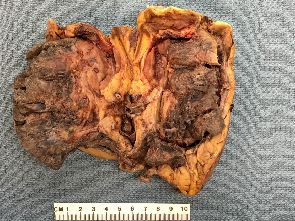
Bisected kidney
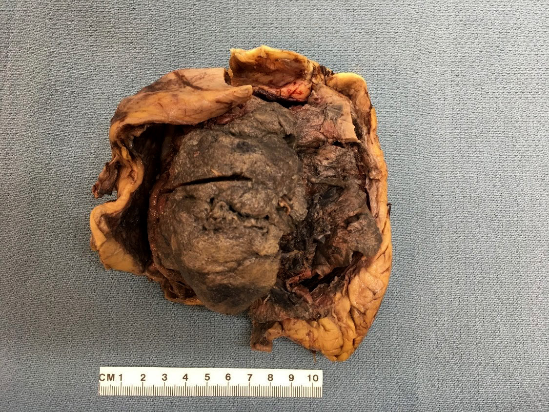
Detached renal capsule
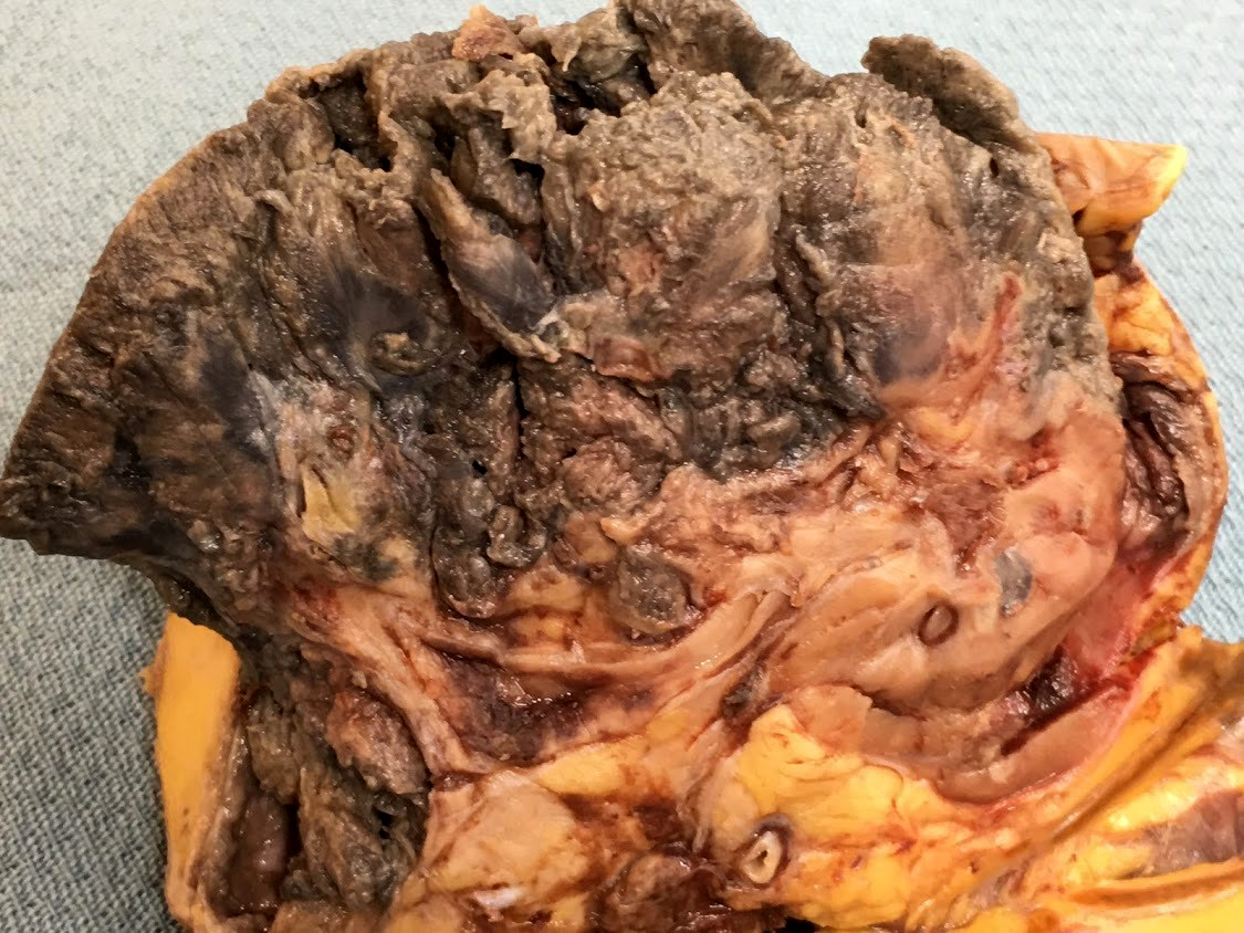
Close view
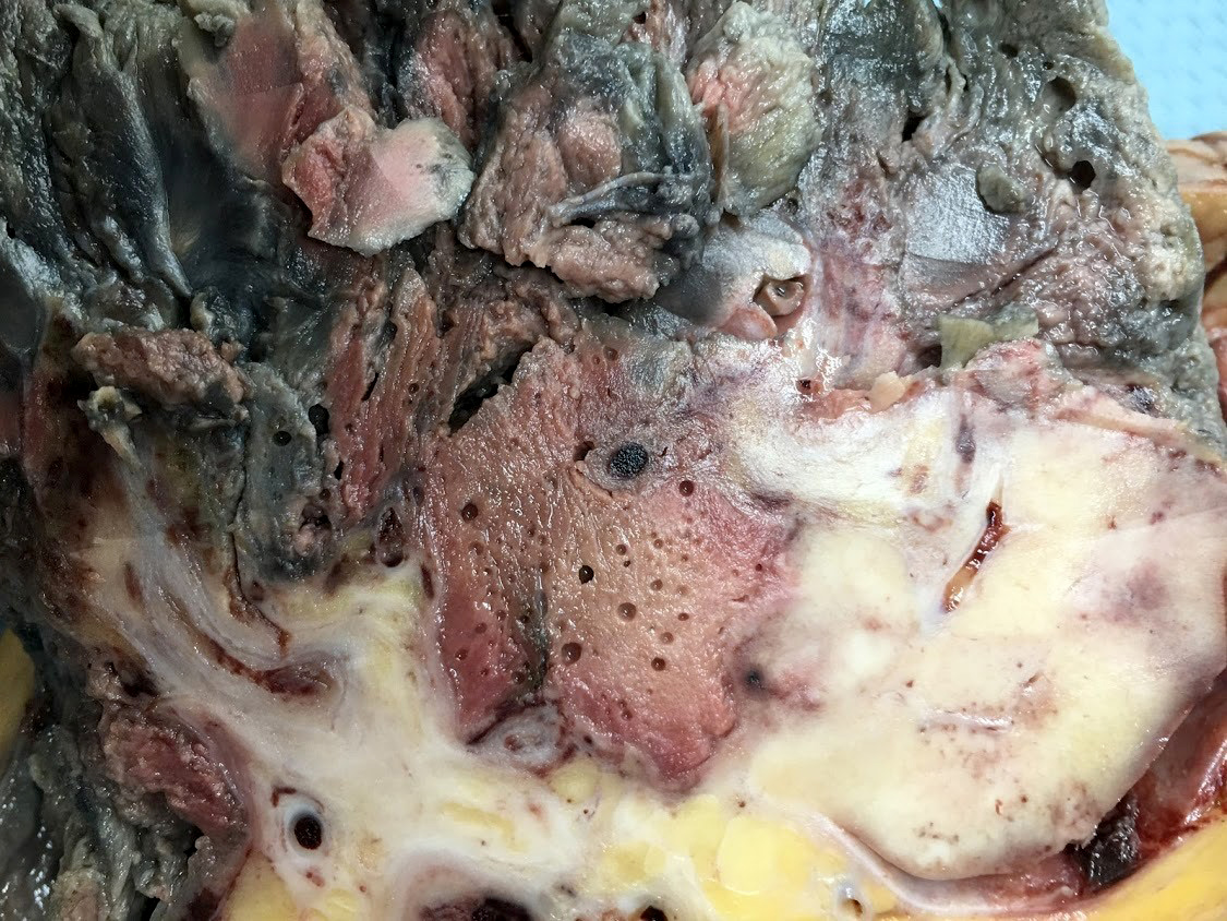
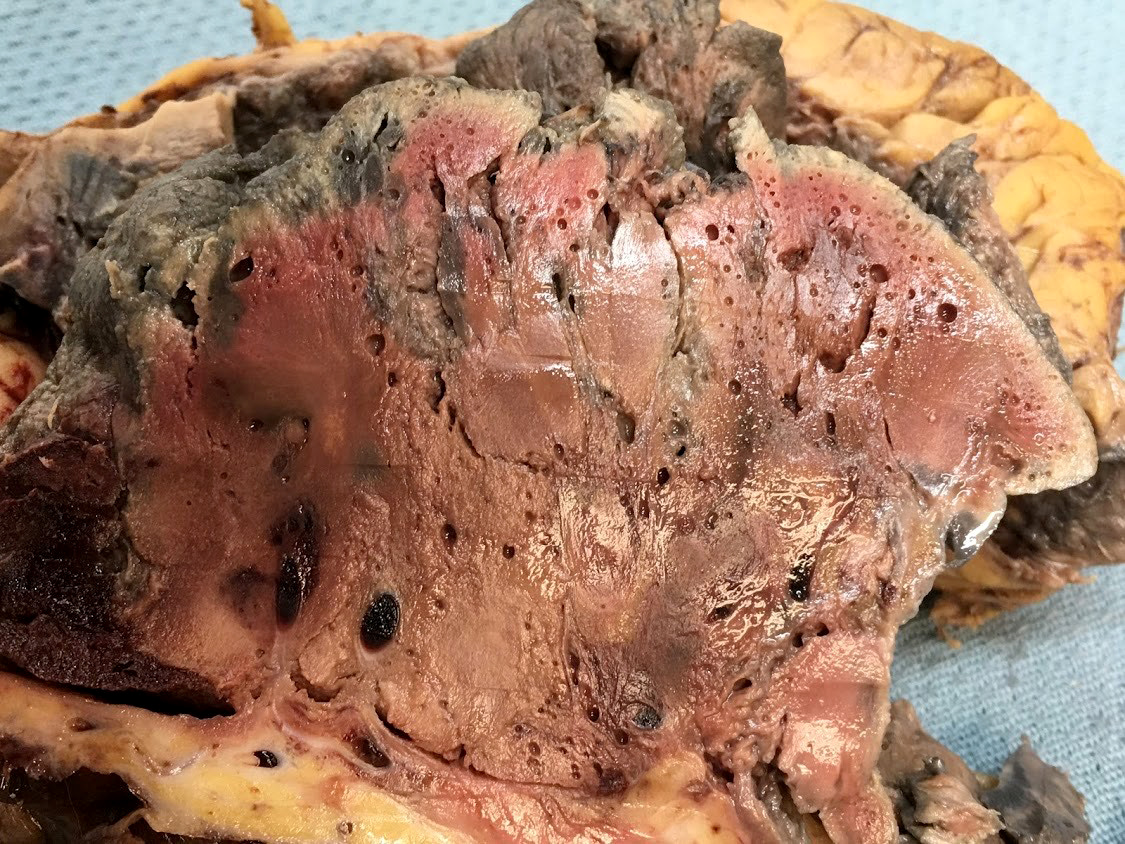
Gas filled cysts
Images hosted on other servers:

Areas of papillary necrosis
Microscopic (histologic) description
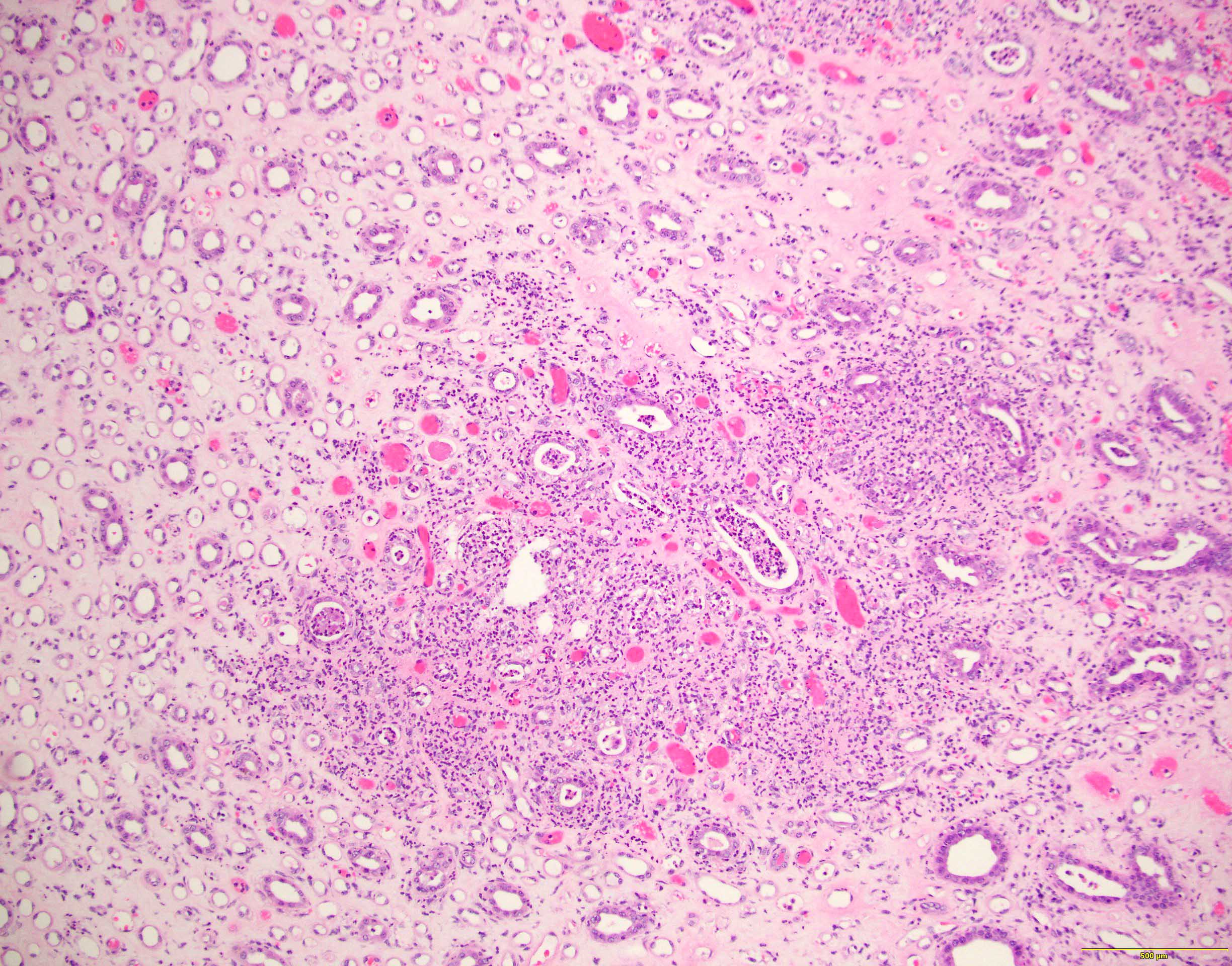
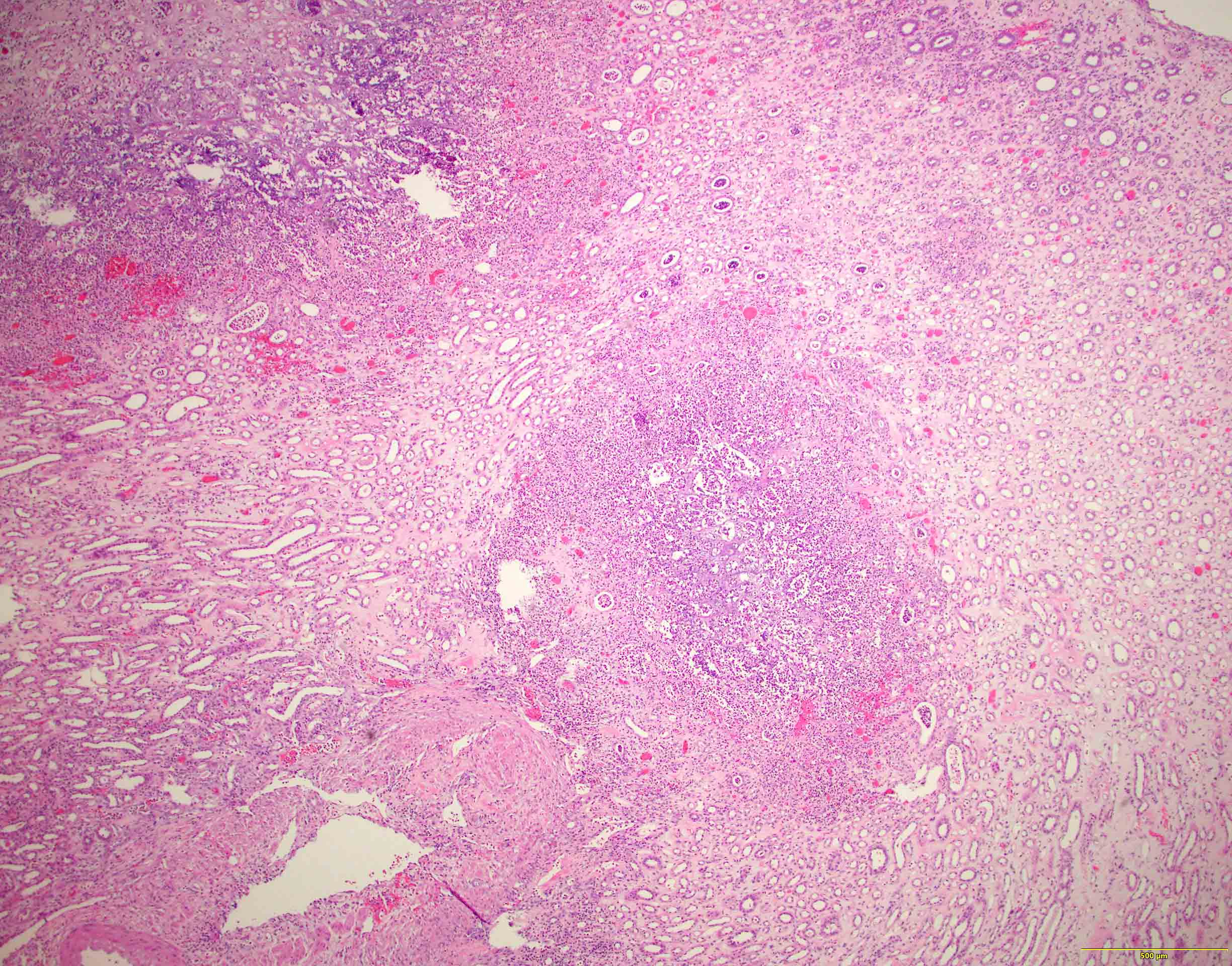
- Patchy suppurative inflammation, primarily cortical with edema, neutrophils in interstitium and tubular lumina and tubular necrosis
- Cortical abscesses and necrosis
- In emphysematous pyelonephritis, hemorrhagic and necrotic renal parenchyma with scattered gas filled cysts
- Reference: Medscape: Acute Pyelonephritis [Accessed 22 March 2021]
Microscopic (histologic) images
Contributed by Jian-Hua Qiao, M.D.
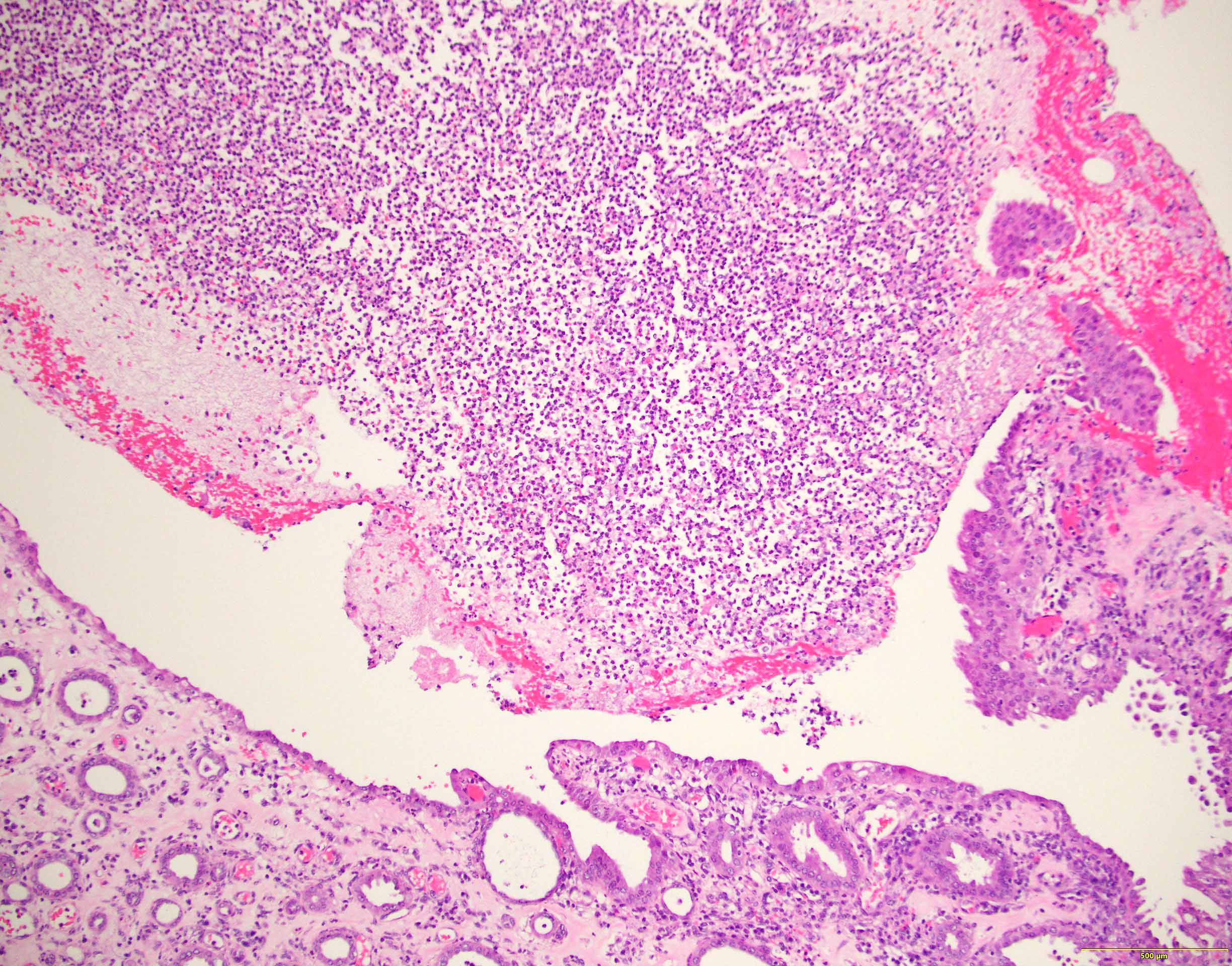
Acute inflammation
Neutrophil casts
Abscess
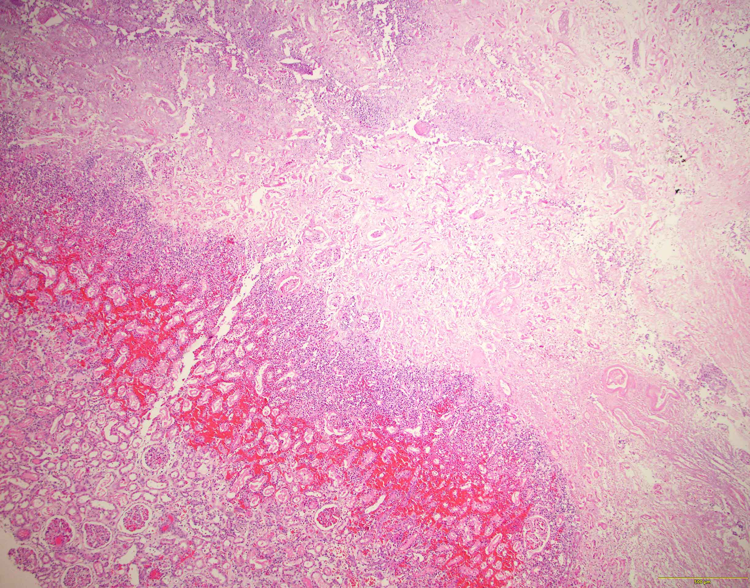
Necrotic renal cortex
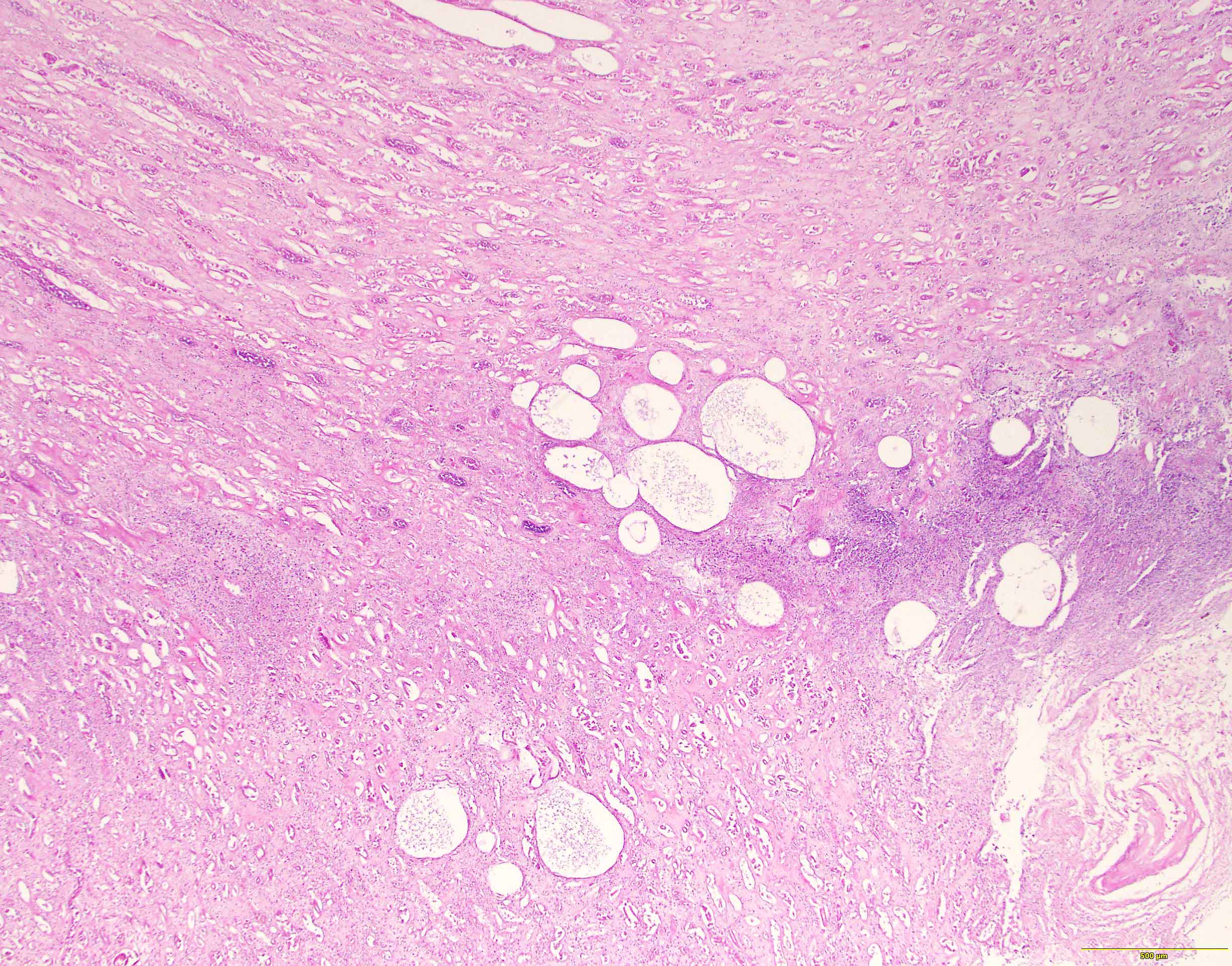
Gas filled cysts
Sample pathology report
- Left kidney, radical nephrectomy:
- Dissected renal capsule with massive subcapsular hematoma
- Acute pyelonephritis involving renal calyces and renal pelvis with abundant acute inflammatory exudates and abscess formation
- Necrotic renal cortex and renal papillae with formation of numerous gas filled cysts (emphysematous pyelonephritis)
- Culture of renal pelvis pus material positive for E. coli
- Negative for renal stone or malignancy
- One benign hilar lymph node, negative for malignancy (0/1)
Differential diagnosis
- Xanthogranulomatous pyelonephritis:
- In rare cases, emphysematous pyelonephritis may occur as a complication from xanthogranulomatous pyelonephritis (Urol Case Rep 2020;33:101299)
- Acute cystitis
- Viral infections, including BK nephropathy, adenovirus and CMV
Additional references
Practice question #1
A 41 year old woman presents with complaints of pain in the left side of her back, dysuria and urinary urgency for the past 3 days. She has a significant past medical history of diabetes mellitus. Abdominal CT shows renal fat stranding and emphysematous destruction confined to the parenchyma of the right kidney. What is the best course of management?
- Antibiotics only and fluids only
- Antibiotics, fluids, electrolytes and glucose control
- Bilateral nephrectomy
- Fluids and electrolytes only
- Right nephrectomy
Practice answer #1
B. Antibiotics, fluids, electrolytes and glucose control
Comment Here
Reference: Acute pyelonephritis
Comment Here
Reference: Acute pyelonephritis
Practice question #2
A 44 year old woman presents to the emergency room with fever, nausea, vomiting and dysuria for the past 4 days. Her past medical history is significant for uncontrolled diabetes mellitus. Ultrasound of the right kidney shows intense reflections and prominent acoustic shadows. Nephrectomy of the right kidney is performed. What is the most likely diagnosis?
- Autosomal polycystic kidney disease
- Diabetic nephropathy
- Emphysematous pyelonephritis
- Renal cell carcinoma
- Xanthogranulomatous pyelonephritis
Practice answer #2
