

Ovary
Seromucinous tumors
Seromucinous borderline tumor
Editorial Board Member: Stephanie L. Skala, M.D.
Deputy Editor-in-Chief: Gulisa Turashvili, M.D., Ph.D.


Last author update: 12 October 2023
Last staff update: 13 December 2023
Copyright: 2021-2024, PathologyOutlines.com, Inc.
PubMed Search: Seromucinous borderline tumor


Table of Contents
Definition / general | Essential features | Terminology | ICD coding | Epidemiology | Sites | Pathophysiology | Etiology | Clinical features | Diagnosis | Laboratory | Radiology description | Prognostic factors | Case reports | Treatment | Gross description | Frozen section description | Microscopic (histologic) description | Microscopic (histologic) images | Positive stains | Negative stains | Videos | Sample pathology report | Differential diagnosis | Additional references | Board review style question #1 | Board review style answer #1 | Board review style question #2 | Board review style answer #2Cite this page: Welter SM, Khalifa MA. Seromucinous borderline tumor. PathologyOutlines.com website. https://www.pathologyoutlines.com/topic/ovaryseromucinousborderline.html. Accessed May 13th, 2024.
Definition / general
- Ovarian seromucinous borderline tumor is a papillary neoplasm of the ovary that is associated with endometriosis and has an excellent prognosis
Essential features
- Papilla with hierarchical branching lined by a mix of Müllerian cell types
- No evidence of stromal invasion
Terminology
- Historic terms that are no longer used: endocervical type mucinous borderline tumor, Müllerian mucinous borderline tumor, Müllerian mucinous papillary borderline tumor, mixed epithelial papillary borderline tumor of Müllerian type
ICD coding
- ICD-O: 8474/1 - seromucinous borderline tumor
- ICD-11: 2C73.Y & XH0RB9 - other specified malignant neoplasms of the ovary & seromucinous borderline tumor
Epidemiology
- Uncommon (Am J Surg Pathol 2002;26:1529)
- Associated with endometriosis (Am J Surg Pathol 2002;26:1529, Am J Surg Pathol 2004;28:1311)
- Presents over a wide age range (mean age is 34 - 39 years) (Cancer 1988;61:546, Cancer 1988;61:340, Am J Surg Pathol 2004;28:1311)
Sites
- Ovary
Pathophysiology
- Subset with loss of ARID1A expression (Int J Gynecol Pathol 2012;31:297)
Etiology
- Associated with endometriosis
- Many arise from endometriotic cysts
Clinical features
- Incidental finding on imaging or may present with signs and symptoms of pelvic mass
- Considered to be endometriosis related ovarian neoplasm, along with endometrioid and clear cell tumors
Diagnosis
- Tumor can be identified via ultrasound or other imaging studies
- Removal of the ovary is then needed for histologic evaluation
Laboratory
- Generally not useful
Radiology description
- Cystic or solid mass
Prognostic factors
- Generally excellent prognosis
Case reports
- 23 year old woman with no history of pregnancy presented to the emergency room with lower abdominal pain (Gynecol Oncol Rep 2021;37:100839)
- 25 year old woman with complex cyst in the left ovary suggesting an endometriotic origin (Int J Womens Health 2020;12:601)
- 39 year old woman who was initially thought to have malignant transformation of endometriosis (Medicine (Baltimore) 2019;98:e15707)
- 65 year old woman with a previous history of bilateral salpingo-oophorectomy had peritoneal cysts increasing in size over 15 years and an increasing CA 19-9 level (BMJ Case Rep 2020;13:e234692)
Treatment
- Surgical management
- Treatment is surgical with the extent of surgery dependent on the stage
- Depending on patient's desire to preserve fertility, a conservative approach may be taken in younger patients (Ann Oncol 2016;27:i20)
Gross description
- Tumors range in size from 1.8 - 18 cm; mean size is 9 cm (Am J Surg Pathol 2015;39:983, Am J Surg Pathol 2002;26:1529)
- Up to 33% of cases are bilateral (Am J Surg Pathol 2002;26:1529)
- Typically a cyst with smooth outer lining and internal papillary excrescences
Frozen section description
- Frozen sections of ovarian tumors are common
- Frozen section will usually show architecture consistent with a borderline tumor with epithelium showing mixed cell types
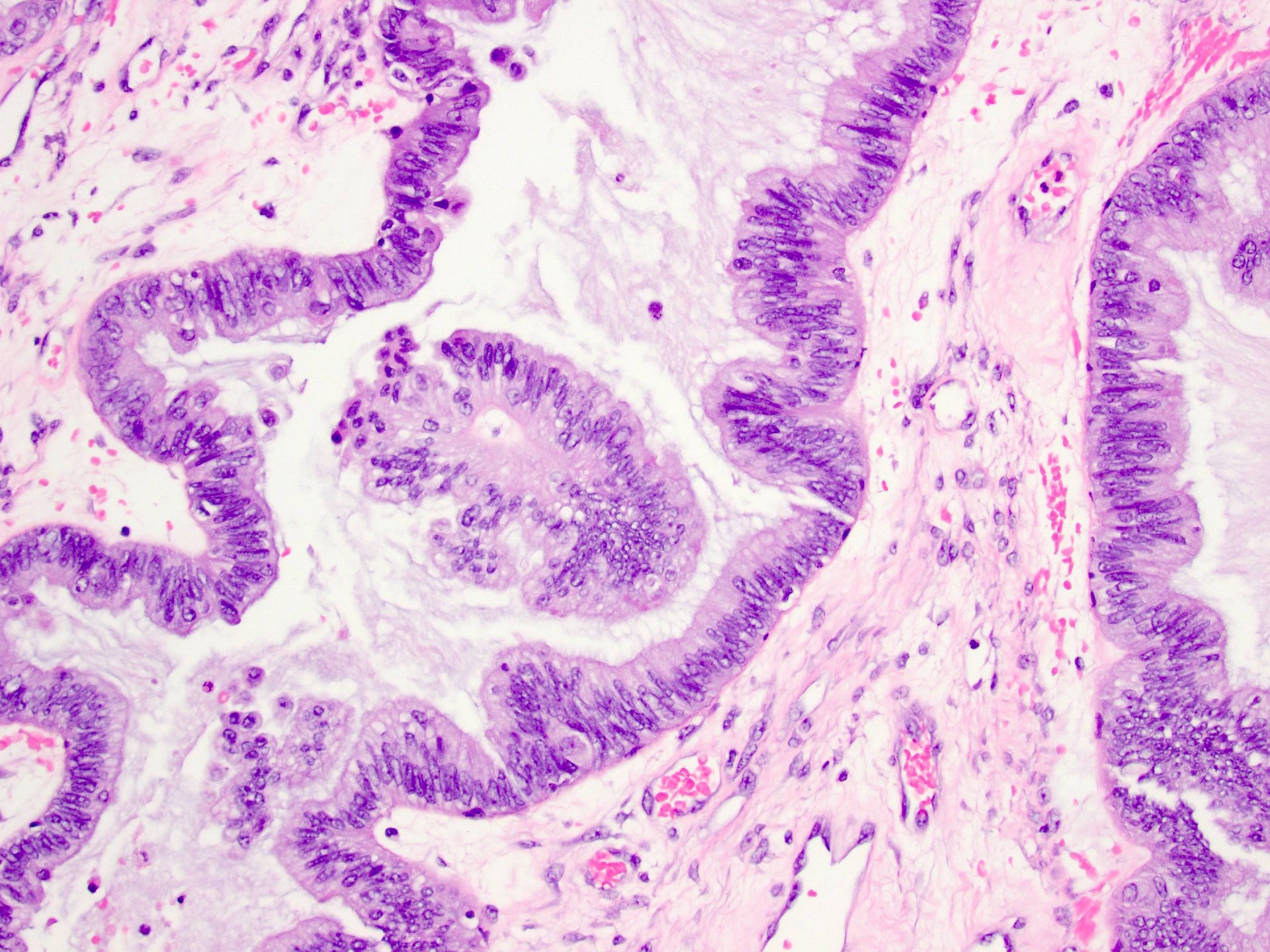
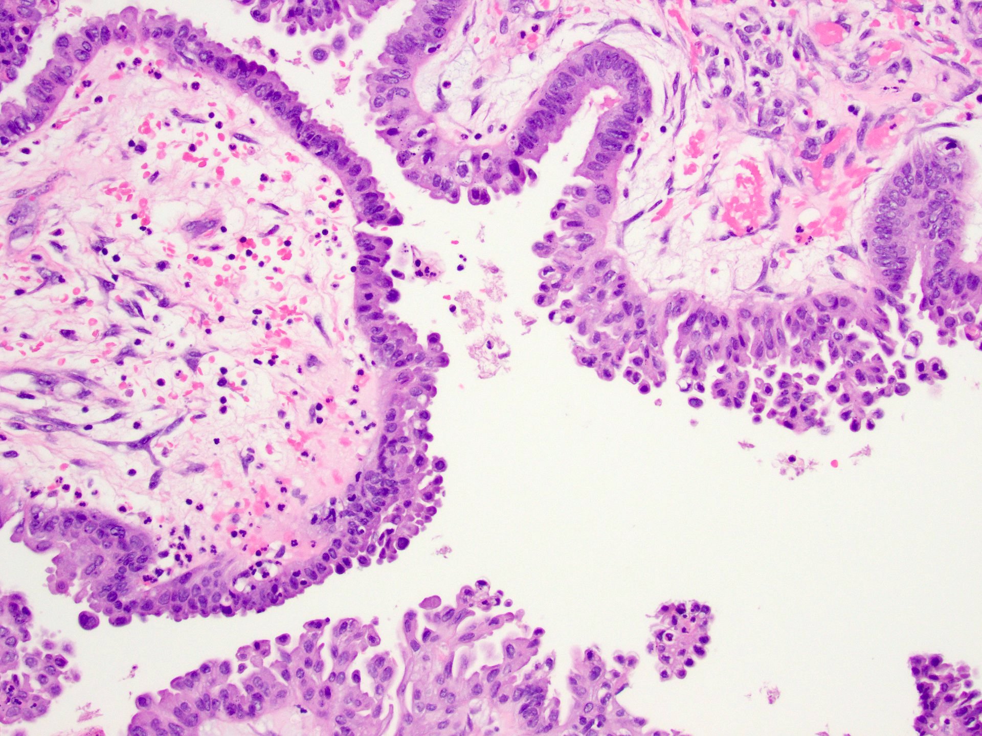
Microscopic (histologic) description
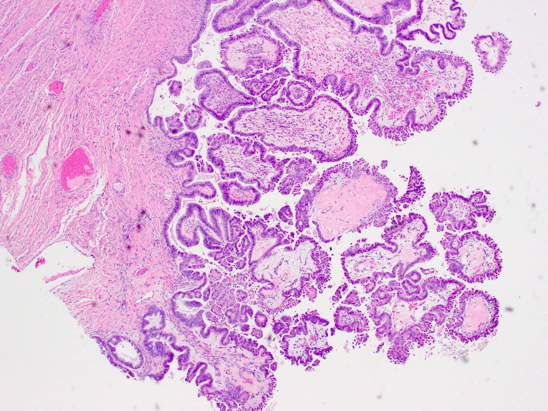
- Papillary tumor with hierarchical branching
- Epithelial lining composed of cytologically bland cells of mixed Müllerian types: endometrioid, squamous, endocervical type, ciliated, hobnail, clear and indifferent (nondescript) eosinophilic cells
- Stromal cores with edema or fibrosis with conspicuous inflammatory cells (neutrophils or eosinophils)
- Background endometriosis or endometriotic cyst
- Microinvasion has been described
- References: Cancer 1988;61:546, Cancer 1988;61:340, Am J Surg Pathol 2002;26:1529, Am J Surg Pathol 2004;28:1311
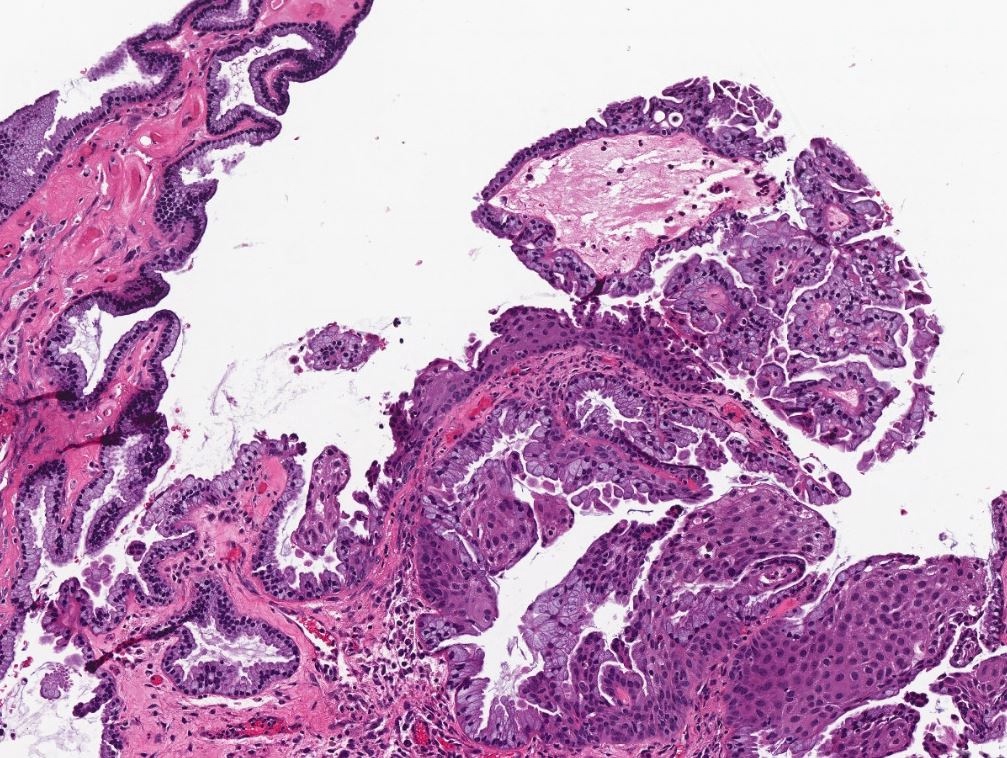
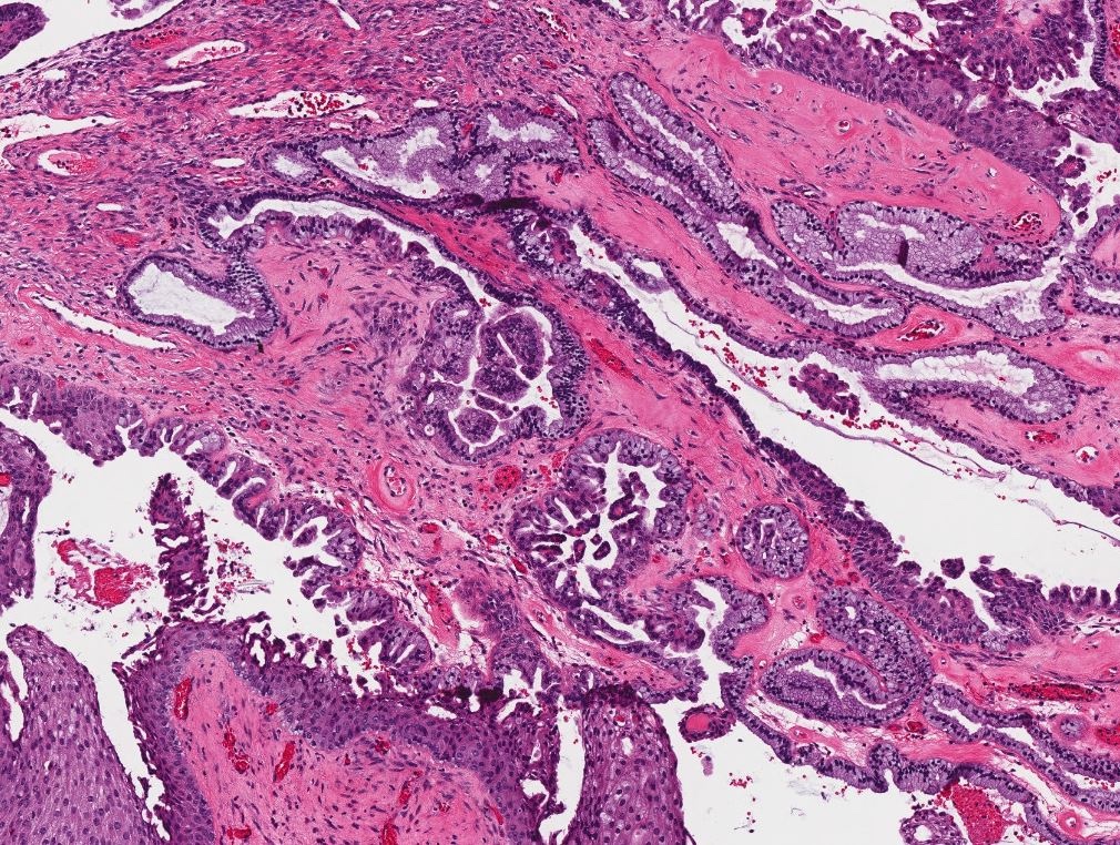
Microscopic (histologic) images
Contributed by Shannon Mingo Welter, M.D.
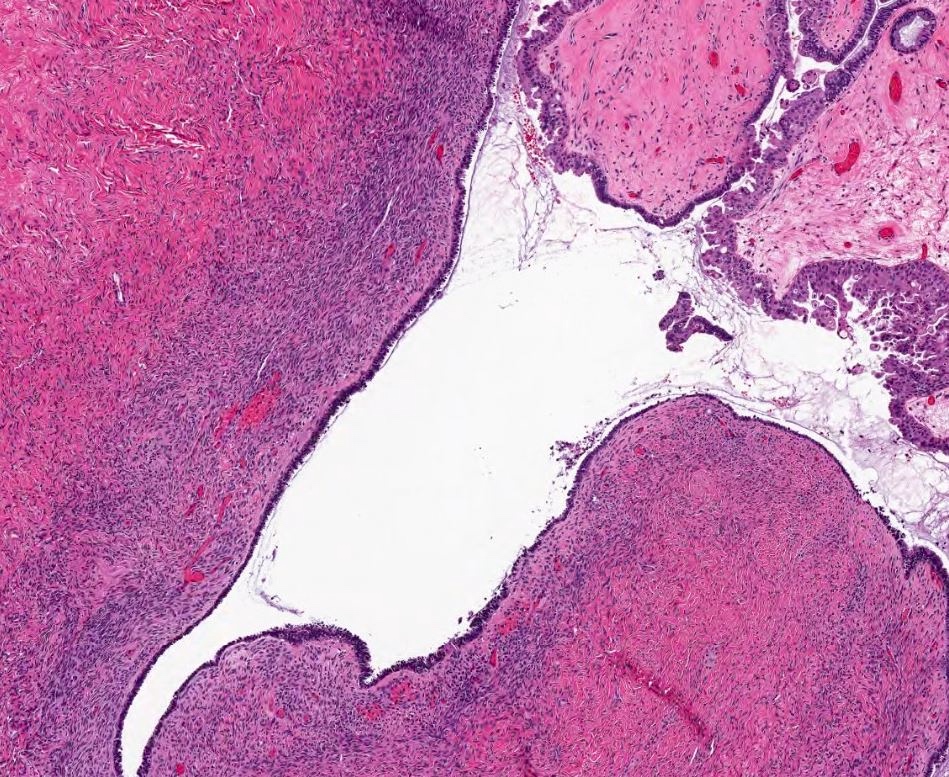
Endometriotic cyst
Squamous and endocervical
Stromal cores with edema or fibrosis
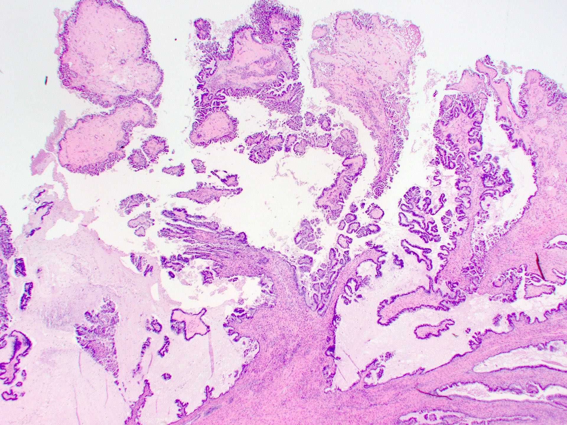
Papillary tumor
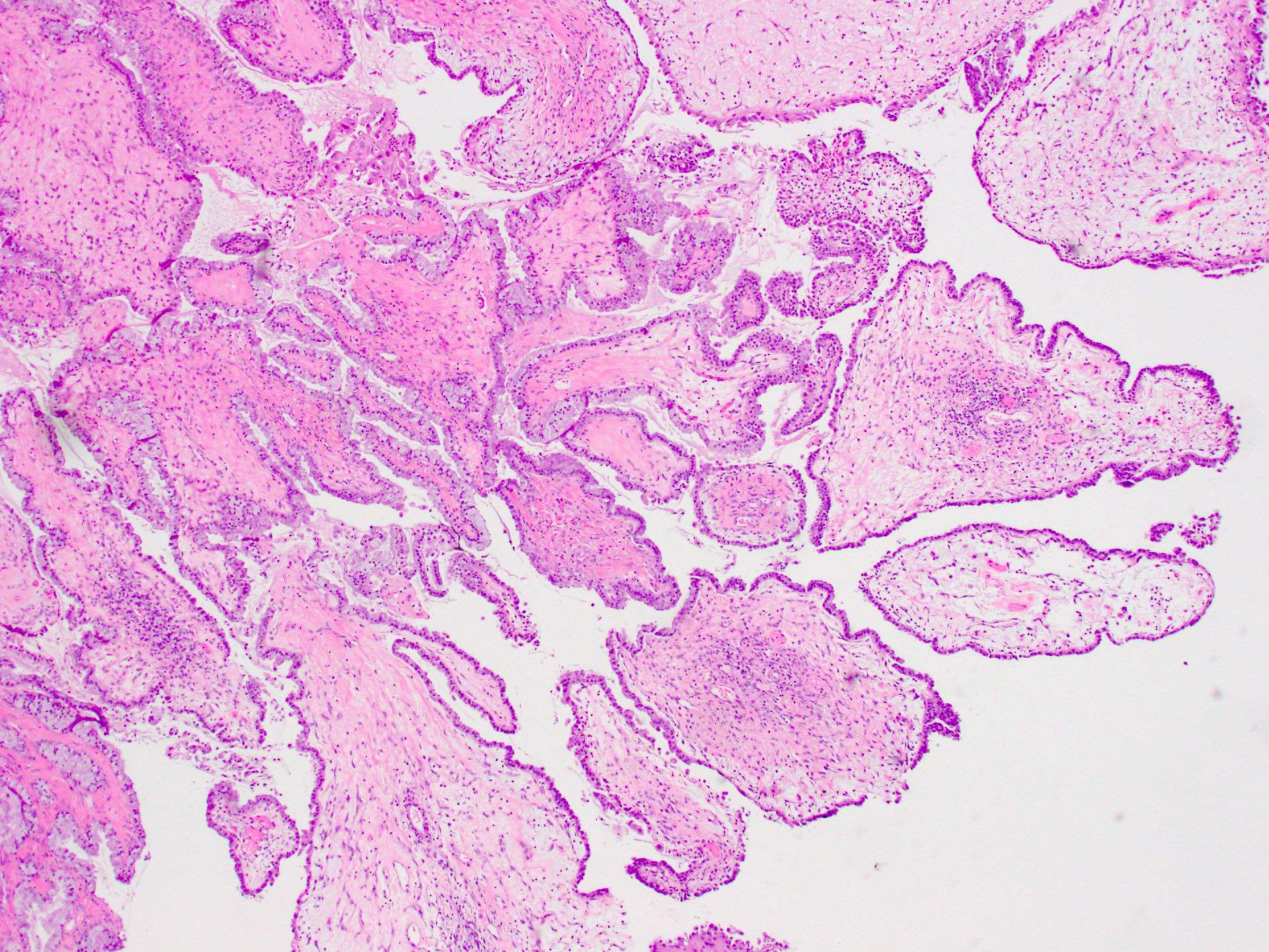
Fibrous cores with inflammation
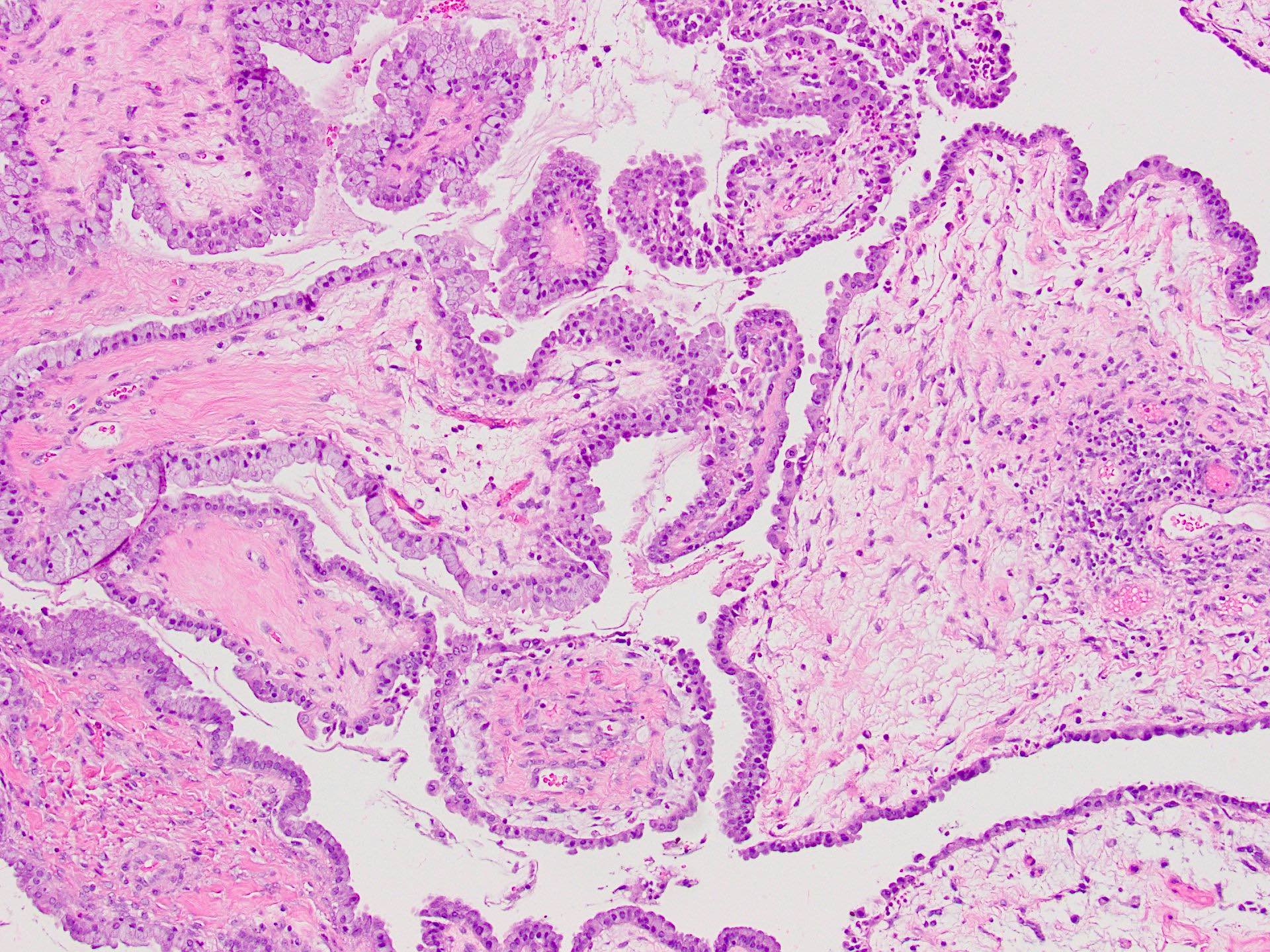
Variable cell types
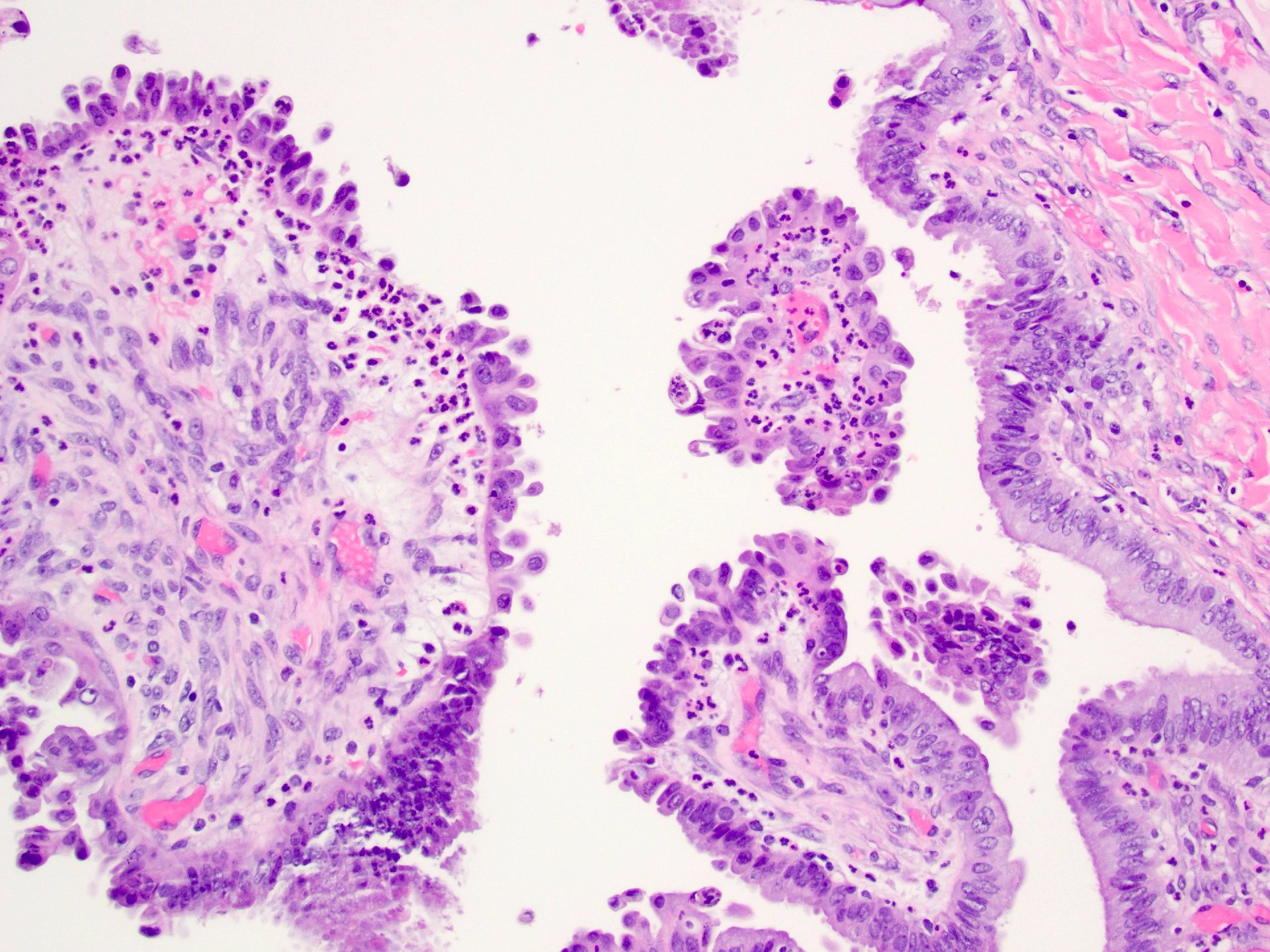
Hobnail and endocervical
Endocervical cell lining
Nondescript eosinophilic cells
Positive stains
Negative stains
- WT1 (can be positive patchy but generally not diffuse)
- CK20, CDX2 (Int J Gynecol Pathol 2016;35:78, In Vivo 2020;34:1341)
Videos
Ovary tumors associated with endometriosis
Sample pathology report
- Right ovary, oophorectomy:
- Seromucinous borderline tumor, size 9 cm (see synoptic report)
Differential diagnosis
- Serous borderline tumor:
- Similar morphology as papillary architecture but has single cell type and lacks other defined Müllerian cell types
- Usually WT1 positive
- Mucinous borderline tumor:
- Gastrointestinal differentiation (Int J Gynecol Pathol 2006;25:83)
- Older age, larger size, less likely to be bilateral, not associated with endometriosis (In Vivo 2020;34:1341)
Additional references
Board review style question #1
A 37 year old woman is found to have a 13.5 cm left ovarian cystic mass with smooth outer surface and internal papillary excrescences. On frozen section, the tumor has papillary, hierarchical branching. The epithelial lining is composed of cytologically bland cells of mixed Müllerian types. Endometriosis background is present but there is no stromal invasion. What is the most likely immunohistochemical profile of this tumor?
- CK7+, CK20-, PAX8-, WT1-
- CK7+, CK20+, PAX8-, WT1-
- CK7+, CK20-, PAX8+, WT1-
- CK7-, CK20+, PAX8-, WT1+
- CK7-, CK20-, PAX8+, WT1-
Board review style answer #1
C. CK7+, CK20-, PAX8+, WT1-. Answers A, B, D and E are incorrect because seromucinous borderline tumors are positive for CK7 and PAX8. They are negative for CK20 and usually negative for WT1. Serous tumors of the ovary are usually positive for WT1.
Comment Here
Reference: Seromucinous borderline tumor
Comment Here
Reference: Seromucinous borderline tumor
Board review style question #2
A 35 year old woman is found to have an 8.2 cm right ovarian mass. Histologic sections show a cyst lined by bland columnar epithelium with underlying spindle stroma. What tumors are known to be associated with this type of benign cyst?
- Clear cell carcinoma, mucinous carcinoma, low grade serous carcinoma
- Endometrioid carcinoma, seromucinous borderline tumor, clear cell carcinoma
- Seromucinous borderline tumor, clear cell tumor, mucinous borderline tumor
- Serous borderline tumor, mucinous borderline tumor, endometrioid carcinoma
Board review style answer #2
B. Endometrioid carcinoma, seromucinous borderline tumor, clear cell carcinoma. The picture shows an endometriotic cyst. Endometriosis associated malignancies include clear cell carcinoma, endometrioid carcinoma and seromucinous borderline tumors. Answers A, C and D are incorrect because serous and mucinous tumors are not associated with endometriosis.
Comment Here
Reference: Seromucinous borderline tumor
Comment Here
Reference: Seromucinous borderline tumor
