Penis & scrotum
General
Anatomy & histology-penis
Editorial Board Member: Maria Tretiakova, M.D., Ph.D.
Editor-in-Chief: Debra L. Zynger, M.D.


Last author update: 31 July 2020
Last staff update: 19 May 2021
Copyright: 2002-2025, PathologyOutlines.com, Inc.
PubMed Search: Anatomy[TI] penis pathology
Table of Contents
Definition / general | Essential features | Terminology | Physiology | Diagrams / tables | Clinical features | Gross description | Gross images | Microscopic (histologic) description | Microscopic (histologic) images | Virtual slides | Positive stains | Negative stains | Additional references | Practice question #1 | Practice answer #1 | Practice question #2 | Practice answer #2Cite this page: Sanchez DF, Cubilla AL. Anatomy & histology-penis. PathologyOutlines.com website. https://www.pathologyoutlines.com/topic/penscrotumanat.html. Accessed September 16th, 2025.
Definition / general
- Cylindric organ suspended from front and sides of pubic arch
- Mainly composed of erectile corpora
- Contains majority of urethra
- Orientation: the upper surface is termed dorsal, the undersurface is termed ventral
Essential features
- Glans and foreskin are the most important anatomic sections of the penis for clinical practice
- 3 erectile tissues: 2 corpora cavernosa (dorsal), 1 corpus spongiosum (ventral)
- Foreskin lacks skin adnexa
Terminology
- There are 3 main parts
- Proximal root
- Middle body (corpus or shaft)
- Distal glans (head)
- Root
- Erectile tissues: bulb, crura
- Muscles: ischiocavernosus and bulbospongiosus
- Penile shaft / middle body
- 3 cylindrical masses of erectile tissue
- Specialized venous sinuses of variable diameter and widely interconnected
- Bound together by fibrous tunica albuginea
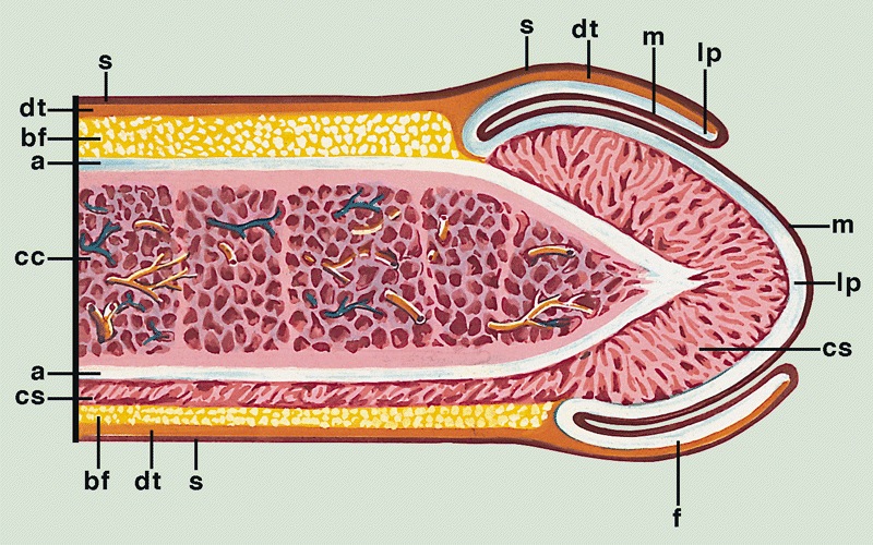
- Penile (Buck) fascia:
- Loose connective tissue between dartos layer and tunica albuginea
- From penile root to coronal sulcus
- Dartos layer:
- Smooth discontinuous muscle layer from homologous scrotal layer
- Throughout entire shaft between dermis and penile fascia
- Reflects itself over the coronal sulcus before continuing to foreskin (~50%)
- Continues directly to foreskin (~50%)
- Tunica albuginea:
- Dense fibrous membrane
- Encasing and separating corpora cavernosa from corpus spongiosum
- From penile root to tips of corpora cavernosa
- Slits containing small vessels, nerves and adipose tissue
- Considered route for cancer spread (Am J Surg Pathol 2017;41:1542)
- Corpora cavernosa:
- 2 lateral masses of erectile tissue that form the bulk of penis; posterior portions are called crura and connect to pubic arch
- Corpus spongiosum:
- Median ventral mass of erectile tissue
- Contains most of urethra
- Male urethra: see Anatomy & histology-male urethra
- Distal penis
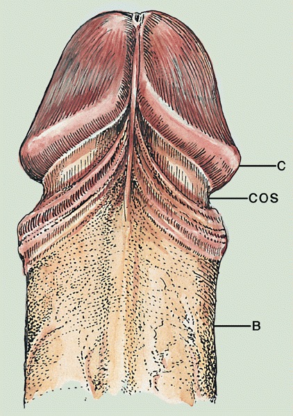
- Glans:
- Conical cup covering distal end of penile shaft
- Portion distal to coronal sulcus
- Glans corona:
- At base of glans
- Slightly elevated circumferential rim
- May contain small papillae over its free border (mistaken for Tyson glands, which are absent in humans), especially in sexually active males
- Meatus urethralis:
- Urethral opening
- Usually at central ventral glans penis
- Vertical cleft, related to frenulum (BJU Int 2007;100:161)
- Fossa navicularis:
- Terminal dilated portion of penile urethra
- Stratified, nonkeratinized squamous epithelium with clear cytoplasm
- Frenulum:
- Fibrous band of tissue attaching foreskin to ventral glans
- Coronal sulcus:
- Narrow and circumferential cul de sac (in noncircumcised) behind glans corona
- Area of insertion of dartos and Buck fascia
- Foreskin:
- Skin folded on itself covering the glans (clitoris in females)
- 3 types of foreskin (Am J Surg Pathol 2003;27:994)
- Long (orifice covers the glans)
- Medium (orifice is between meatus and glans corona)
- Short (orifice is between corona and sulcus)
- Layers are inner squamous epithelium, lamina propria, dartos layer and preputial skin
- Incidence of completely retractile foreskin increased from 0% at birth to 42% in adolescence
- Phimosis rate decreased with age from 99.7% to 7% (World J Pediatr 2009;5:312)
- Glans:
Physiology
- 2 main functions
- Urination
- Sexual intercourse
- Erection via parasympathetic innervation
- Muscles compression in the penis root prevent veins from draining erectile corpora
- Enlargement is obtained by erectile corpora filled and pressing against tunica albuginea
- Ejaculation via sympathetic innervation
Diagrams / tables
AFIP images

Transverse section
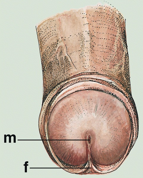
Meatus
Glans
Cut section
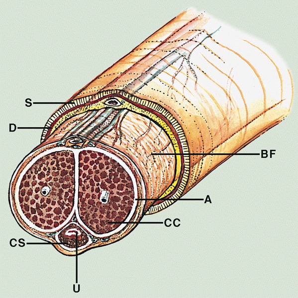
Shaft cross section
Images hosted on other servers:

Compartments


Local anatomy

Arteries

Veins

Urethra

Corpora cavernosa

Transverse sections
Clinical features
- Regional lymph nodes
- Superficial inguinal nodes (site of 1 - 3 sentinel nodes)
- Deep inguinal
- External iliac
- Internal iliac (pelvic nodes)
- Periurethral glands
- Cowper (bulbourethral) glands: mucinous acinar structures deep at level of membranous urethra
- Intraepithelial glands (Morgagni lacunae): 1 layer cylindrical intraepithelial glands
- Littre glands: tubuloacinar mucinous glands present along entire length of corpus spongiosum
- Miscellaneous
- Penile glycogenated epithelial cells indicate recent vaginal intercourse (Am J Clin Pathol 1985;84:524)
- Penile swabs after recent vaginal intercourse almost always contain female cells identifiable by FISH (Arch Pathol Lab Med 2000;124:1080)
- Skin at root of penis is continuous with skin over scrotum and perineum
- Erectile tissues of corpus spongiosum are composed of straight thin muscle wall whereas those of corpus cavernosum are interanastomosed and composed of thicker muscle walls
Gross description
- Transversal cut of penile shaft
- Surgical margin
- Must include skin, soft tissues and erectile tissues
- Shaft, longitudinal cut, from surface to deep structures
- Skin
- Penile fascia and dartos
- Albuginea
- Corpora cavernosa
- Corpus spongiosum
- Urethra
- Glans, longitudinal cut, from surface to deep structures
- Squamous mucosa and lamina propria
- Corpus spongiosum
- Urethra and meatus
- Tip of corpora cavernosa with albuginea
- Foreskin
- Anatomical position: folded with skin on the outer surface and mucosa on the inner surface
- Layers: skin, soft tissues with dartos, mucosa (Mills: Sternberg's Diagnostic Surgical Pathology, 6th Edition, 2015
Gross images
AFIP images
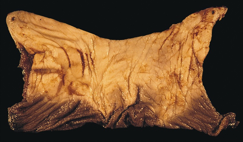
Mucosal and wrinkled portion of foreskin
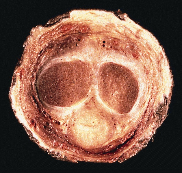
Cross section
Microscopic (histologic) description
- Skin (Am J Surg Pathol 2017;41:1542, Mills: Histology for Pathologists, 5th Edition, 2019)
- Thin skin covering
- Stratified squamous keratinized epithelium
- Loosely connected to deeper parts of the organ
- Mucosa
- Stratified squamous epithelium
- Up to 10 cell thickness
- Nonkeratinizing at glans penis, keratinized after circumcision
- Lamina propria
- Loose connective tissue
- Small vessels
- Sparse lymphocytic infiltrate
- Penile fascia
- Loose connective tissue
- Small arteries, dorsal veins and nerve bundles
- Adipose tissue
- Corpora cavernosa
- Interanastomosed vascular spaces
- Thicker muscle wall
- Corpus spongiosum
- Vascular spaces
- Straight thin muscle wall
- Abundant elastic fibers
- Urethra
- Prostatic urethra: urothelium
- Membranous urethra: stratified or ciliated pseudostratified columnar epithelium
- Penile urethra: stratified or ciliated pseudostratified columnar epithelium
- Foreskin
- Inner foreskin: continues glans squamous mucosa
- Outer foreskin: squamous keratinized epithelium with no adnexa
- Loose connective tissue with discontinuous smooth muscle bundles (dartos)
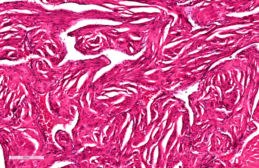
Microscopic (histologic) images
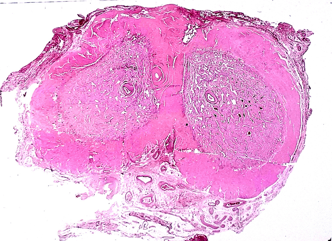
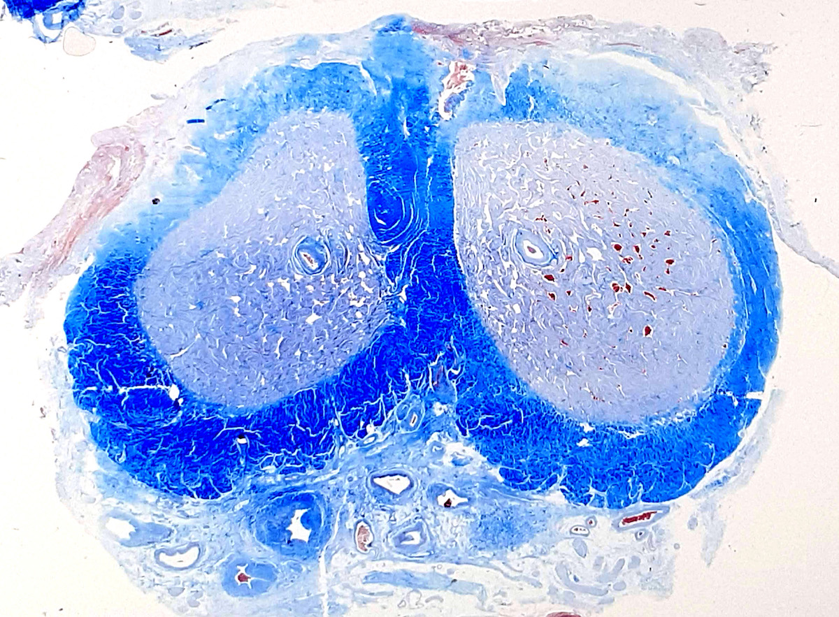
Contributed by Diego F. Sanchez, M.D. and Antonio L. Cubilla, M.D.
Penile shaft surgical margin
Surgical margin connective tissue
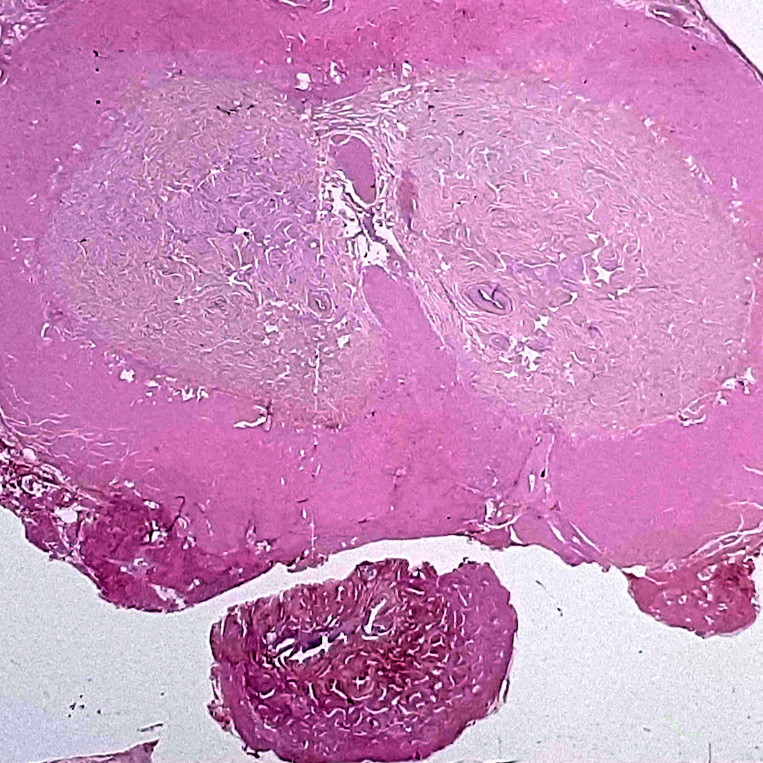
Erectile corpora
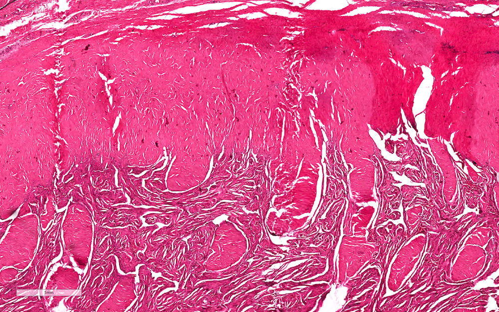
Albuginea and corpus cavernosum
Corpora cavernosa
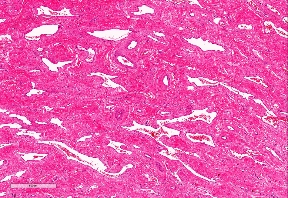
Corpus spongiosum
Glans
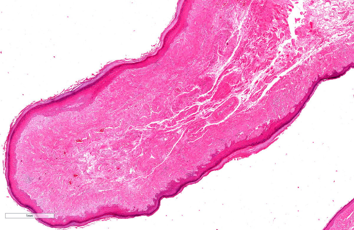
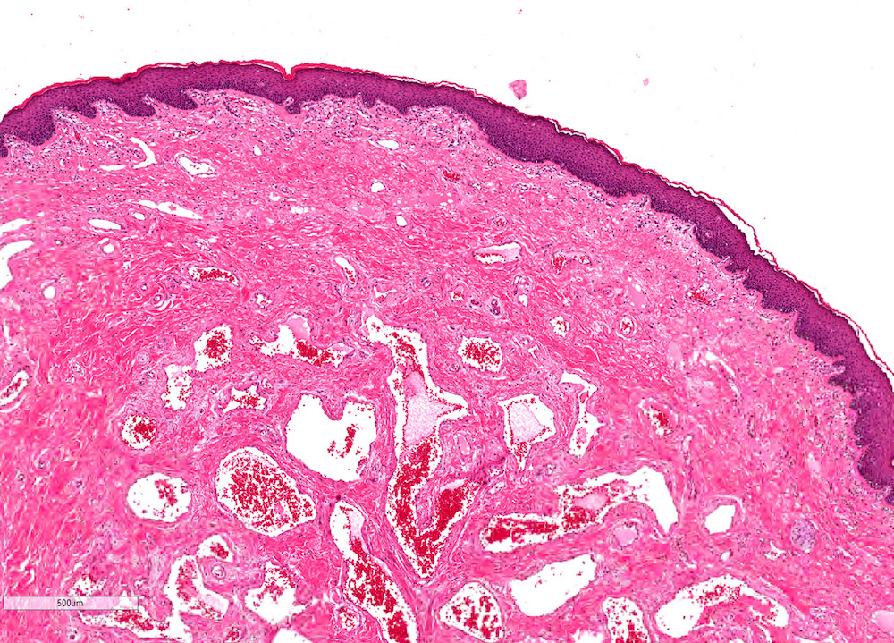
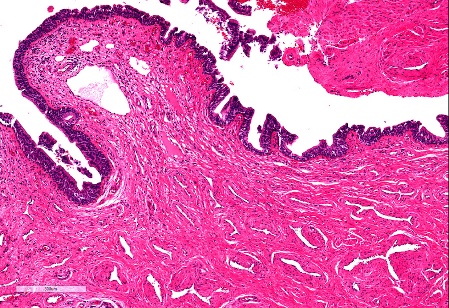
Foreskin in anatomical position
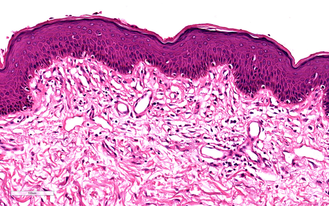
Foreskin epithelium
Foreskin dartos
Penile urethra
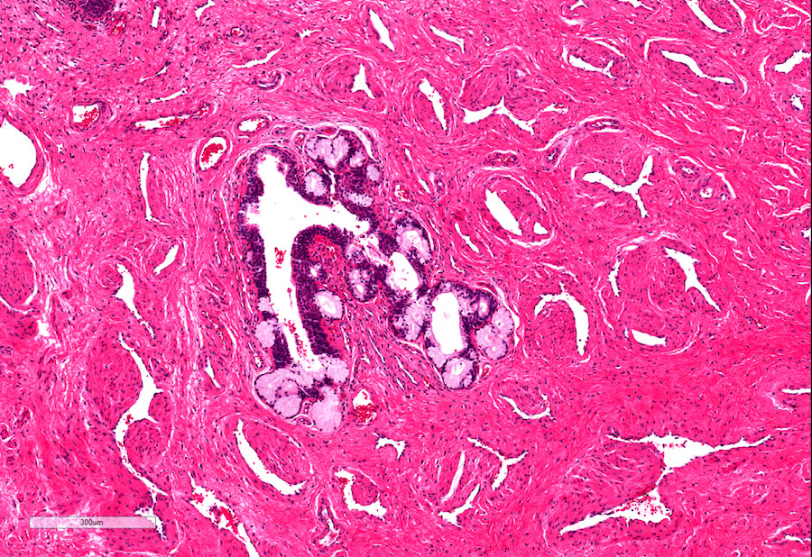
Littre glands
Virtual slides
Images hosted on other servers:

Penile shaft
Positive stains
- CD117: c-kit positive interstitial cells, similar to those in gut (J Sex Med 2007;4:66)
- CK7: urethral upper epithelial layer
- p63 and 34 beta E12: urethral basal layer is positive (Mills: Histology for Pathologists, 5th Edition, 2019)
- GATA3: both urethral layers (Hum Pathol 2013;44:2760)
- PSA: occasional urethral glands (Hum Pathol 2002;33:905)
- CK6: foreskin all layers (Differentiation 2018;103:86)
- CK10: foreskin suprabasal layers (Differentiation 2018;103:86)
Negative stains
- CD20: urethral superficial and basal lining (Hum Pathol 2013;44:2760)
- p63: urethral superficial lining
Additional references
Practice question #1
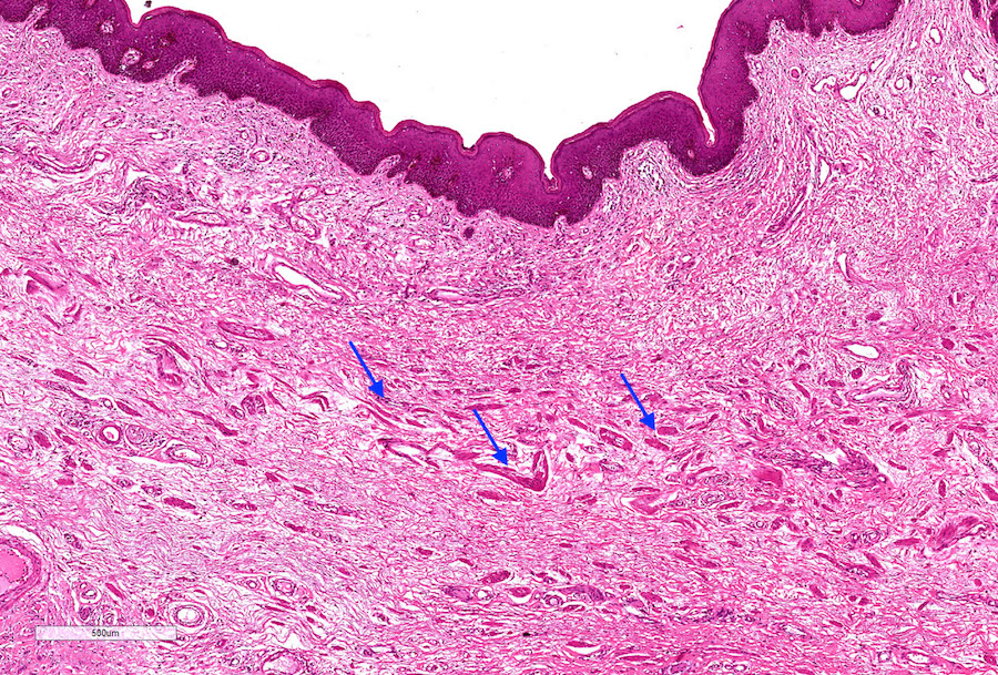
- What does the eosinophilic material (arrow) in this section of the foreskin correspond to?
- Amyloid
- Collagen fibers
- Elastic fibers
- Smooth muscle
- Vascular spaces
Practice answer #1
D. Smooth muscle (dartos) represented by discontinuous fascicles intermixed with connective tissue
Comment Here
Reference: Anatomy / histology of penis
Comment Here
Reference: Anatomy / histology of penis
Practice question #2
- What is the epithelium of the penile urethra classified as?
- Pseudostratified columnar
- Simple columnar
- Simple cuboidal
- Simple squamous
- Urothelium
Practice answer #2
