Prostate gland & seminal vesicles
Nonneoplastic
Urethral polyp
Author: Y. Albert Yeh, M.D., Ph.D.
Editorial Board Member: Bonnie Choy, M.D.
Deputy Editor-in-Chief: Maria Tretiakova, M.D., Ph.D.

Last author update: 11 August 2021
Last staff update: 25 April 2024
Copyright: 2003-2025, PathologyOutlines.com, Inc.
PubMed search: urethral polyp prostate pathology
Table of Contents
Definition / general | Essential features | Terminology | ICD coding | Epidemiology | Sites | Pathophysiology | Etiology | Clinical features | Diagnosis | Radiology description | Radiology images | Prognostic factors | Case reports | Treatment | Clinical images | Gross description | Gross images | Microscopic (histologic) description | Microscopic (histologic) images | Cytology description | Cytology images | Positive stains | Negative stains | Sample pathology report | Differential diagnosis | Additional references | Practice question #1 | Practice answer #1 | Practice question #2 | Practice answer #2Cite this page: Yeh YA. Urethral polyp. PathologyOutlines.com website. https://www.pathologyoutlines.com/topic/prostateurethralpolyp.html. Accessed August 28th, 2025.
Definition / general
- Uncommon, benign, polypoid or papillary growths protruding into the urethral lumen (J Urol 2015;193:2095)
- 2 types: prostatic type urethral polyp, fibroepithelial urethral polyp (J Urol 2015;193:2095, Am J Surg Pathol 2005;29:460)
Essential features
- Prostatic type urethral polyp is characterized by delicate papillae with true fibrovascular cores lined with prostatic epithelium with focal benign urothelial cells; prostatic acini are often present in the polyp (Am J Surg Pathol 1983;7:351)
- Fibroepithelial urethral polyp is composed of cloverleaf-like and club-like dense fibrovascular cores covered by benign urothelium (Am J Surg Pathol 2005;29:460)
Terminology
- Prostatic type urethral polyp, also known as prostatic urethral polyp and prostatic type epithelial polyp of the urethra (J Urol 2015;193:2095)
- Fibroepithelial urethral polyp, also known as fibroepithelial congenital urethral polyp (J Pediatr Surg 2014;49:835)
Epidemiology
- Prostatic type urethral polyp:
- Occurs in adult men, age range 27 - 41 years, uncommon in younger men (Am J Clin Pathol 1975;63:343)
- Fibroepithelial urethral polyp:
- Age range 17 - 70 years, mean of 47 years (Am J Surg Pathol 2005;29:460)
- Commonly occurs in pediatric males, usually 3 - 9 years of age (Can J Urol 2013;20:6974)
- Rare patients present during infancy or adulthood
- Rarely occurs in pediatric female patients
Sites
- Commonly occurs in posterior prostatic urethra adjacent to verumontanum or the bladder neck (J Urol 2015;193:2095)
- Anterior urethra in rare case reports
Pathophysiology
- Ectopic prostatic acinar epithelium presents focally in urothelial tract, commonly in posterior urethra, rarely in bladder neck and penile urethra
- Prostatic type urethral polyp likely caused by hyperplastic proliferations of the prostatic acinar epithelium results in overgrowth of the overlying urothelium (Am J Surg Pathol 1984;8:833)
Etiology
- Fibroepithelial urethral polyp may result from a congenital defect in the urethral wall or associated with a congenital anomaly (Br J Urol 1970;42:76, Urology 1977;9:423)
Clinical features
- Common symptoms and signs: hematuria, dysuria, hemospermia
- Urethrocystoscopic examination: a polypoid mass protruding from the posterior urethral wall (most common) or from anterior urethral wall (Urology 2020;143:238)
Diagnosis
- Ultrasonography reveals a protruding hyperechogenic polypoid mass protruding from the posterior (most common) urethral wall (Urology 2020;143:238)
- Filling defect on voiding cystourethrogram (Clin Imaging 2018;51:164)
- Appearance of a benign polyp on urethrocystoscopy followed by histopathological examination of the excised polyp
Radiology description
- Abdominal sonogram of the urinary bladder shows a hyperechogenic polypoid mass protruding to the bladder neck (Urol J 2020;18:86)
- Filling defect is present at the bladder neck of a distended bladder (Clin Imaging 2018;51:164)
Radiology images
Images hosted on other servers:

Polypoid mass

Filling defect
Prognostic factors
- Excellent prognosis for prostatic type urethral polyp and fibroepithelial urethral polyp
- Recurrence is unusual (J Urol 2015;193:2095)
Case reports
- Prostatic type urethral polyp:
- 35 year old man with urinary outflow obstruction (Diagn Cytopathol 2003;29:356)
- 47, 52 and 84 year old men with hematuria (Urology 2014;83:535)
- 62 year old man with hematuria (Arch Pathol Lab Med 2003;127:e351)
- Fibroepithelial polyp:
- 3 year old boy with intermittent acute urinary retention (Urology 2020;143:238)
- 3 year old boy with abdominal pain (Clin Imaging 2018;51:164)
- 20 year old man with urinary retention (Urol J 2009;6:301)
Treatment
- Cystoscopic excision and fulguration (J Urol 2015;193:2095)
Clinical images
Images hosted on other servers:

Urethrocystoscopic view of polyp
Gross description
- Prostatic type urethral polyp (J Urol 2015;193:2095)
- Pink-tan, exophytic papillary, filiform to rarely sessile structure
- Single or multiple polyps
- Usually < 1 cm
- Fibroepithelial urethral polyp (Clin Imaging 2018;51:164)
- Pink-tan, polypoid mass with narrow stalk
- Usually < 4 cm
Gross images
Images hosted on other servers:

Brown-tan knob-like polyp
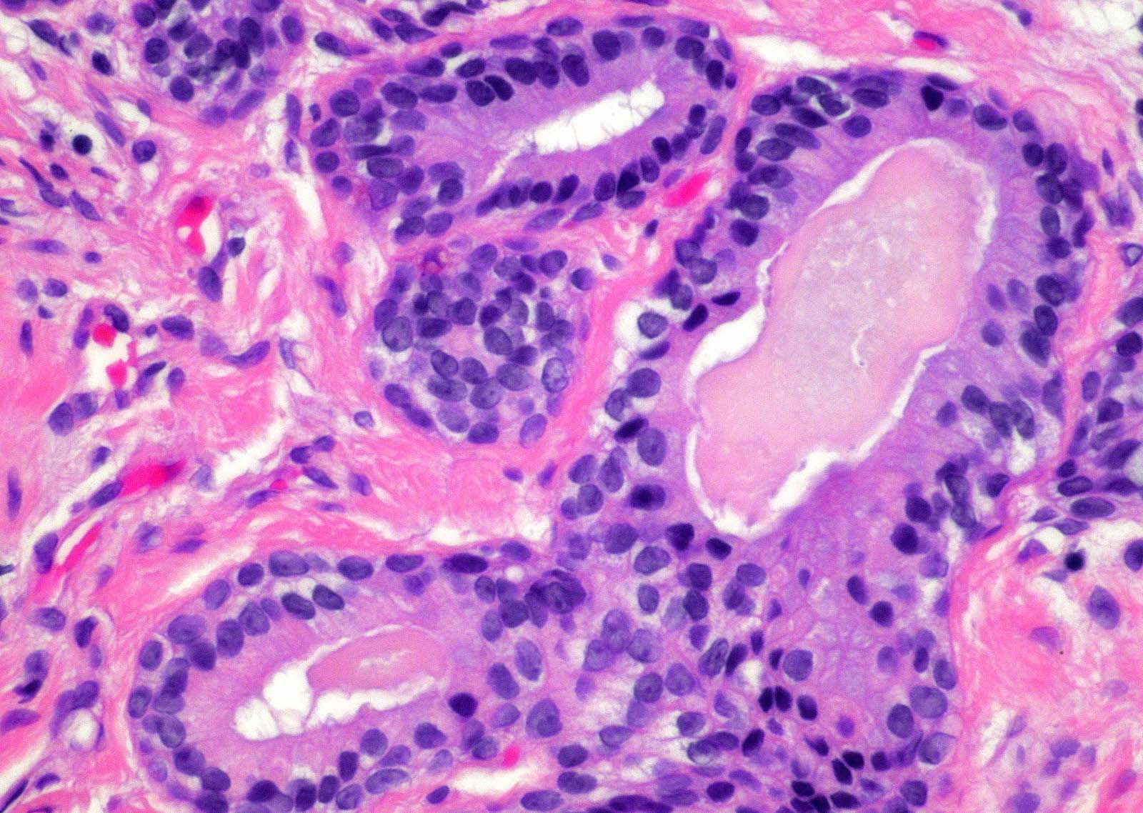
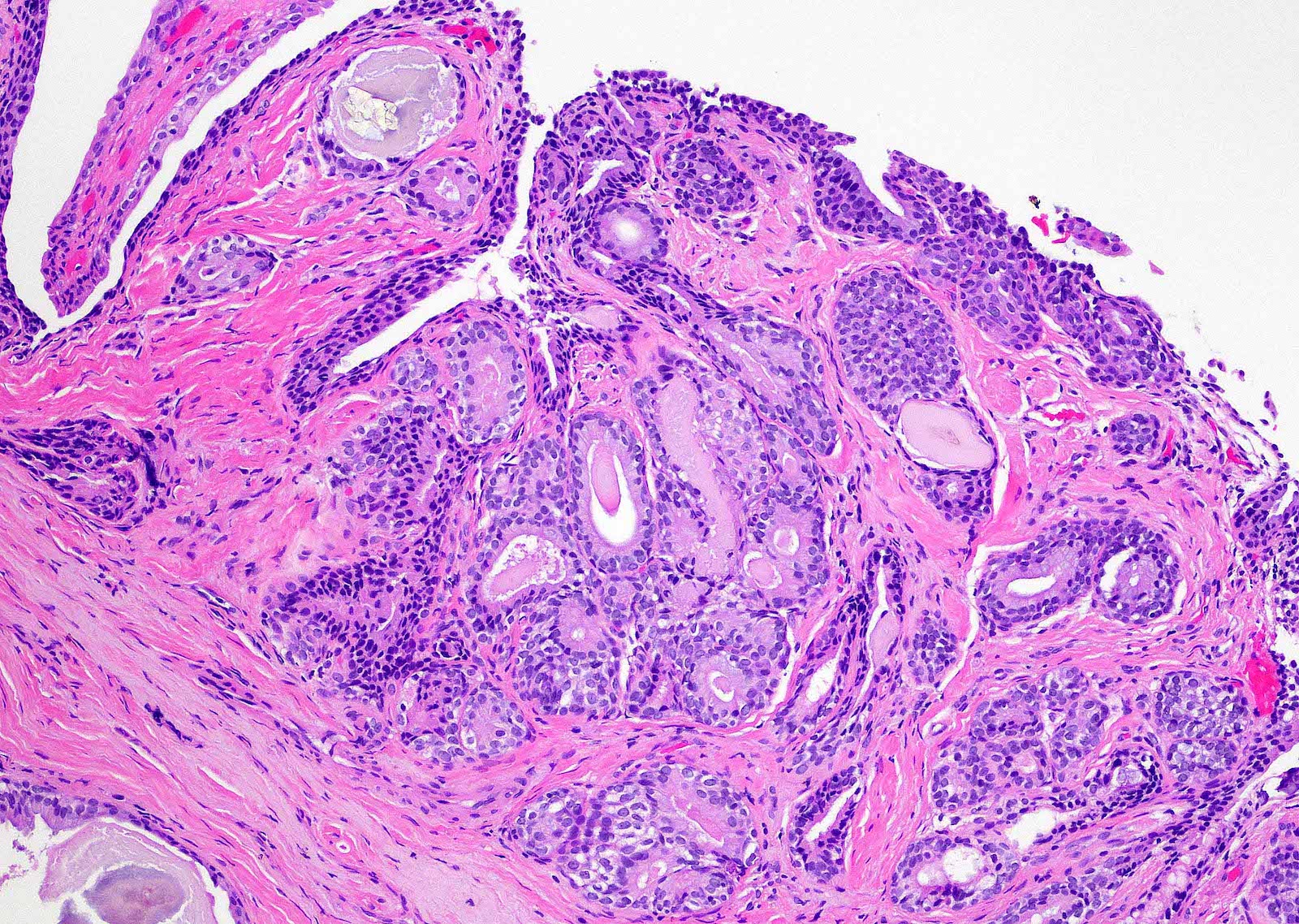
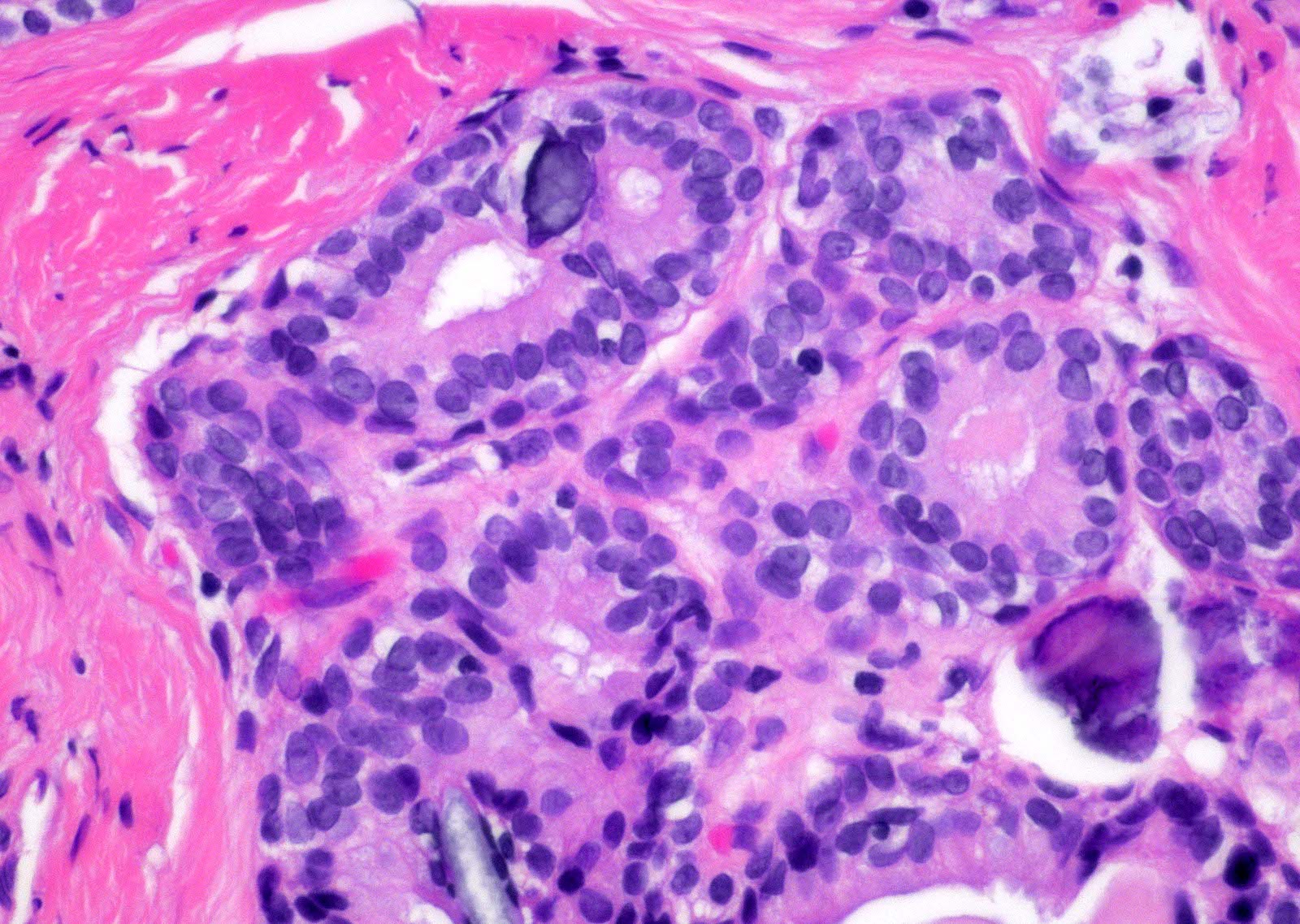
Microscopic (histologic) description
- Prostatic type urethral polyp:
- Delicate papillae with true fibrovascular cores lined by an outer layer of columnar cells and an underlying flattened to cuboidal basal cells (Am J Surg Pathol 1983;7:351)
- Focal benign urothelium is present in the benign prostatic epithelium (J Urol 2015;193:2095)
- Benign prostatic acini are present in the papillae and in adjacent fibromuscular stroma (J Urol 2015;193:2095)
- Fibroepithelial urethral polyp (Am J Surg Pathol 2005;29:460):
- Pattern 1: most common pattern includes the following features
- Broad cloverleaf-like and club-like projections covered by normal urothelium and composed of dense fibrovascular stroma with florid cytitis cystica et glandularis (most common morphologic variant)
- Back to back glands present in the stalk
- Anastomosing nests of benign urothelial cells resembling inverted papilloma
- Dilated cysts with intracystic papillary contents
- Degenerative reactive atypia of stromal cells
- Pattern 2: numerous small papillae with dense fibrous cores and areas of glandular differentiation
- Pattern 3: urothelial lined broad edematous papillae mimicking polypoid urethritis; urothelial lined broad fibrous papillae with subepithelial edema
- Pattern 1: most common pattern includes the following features
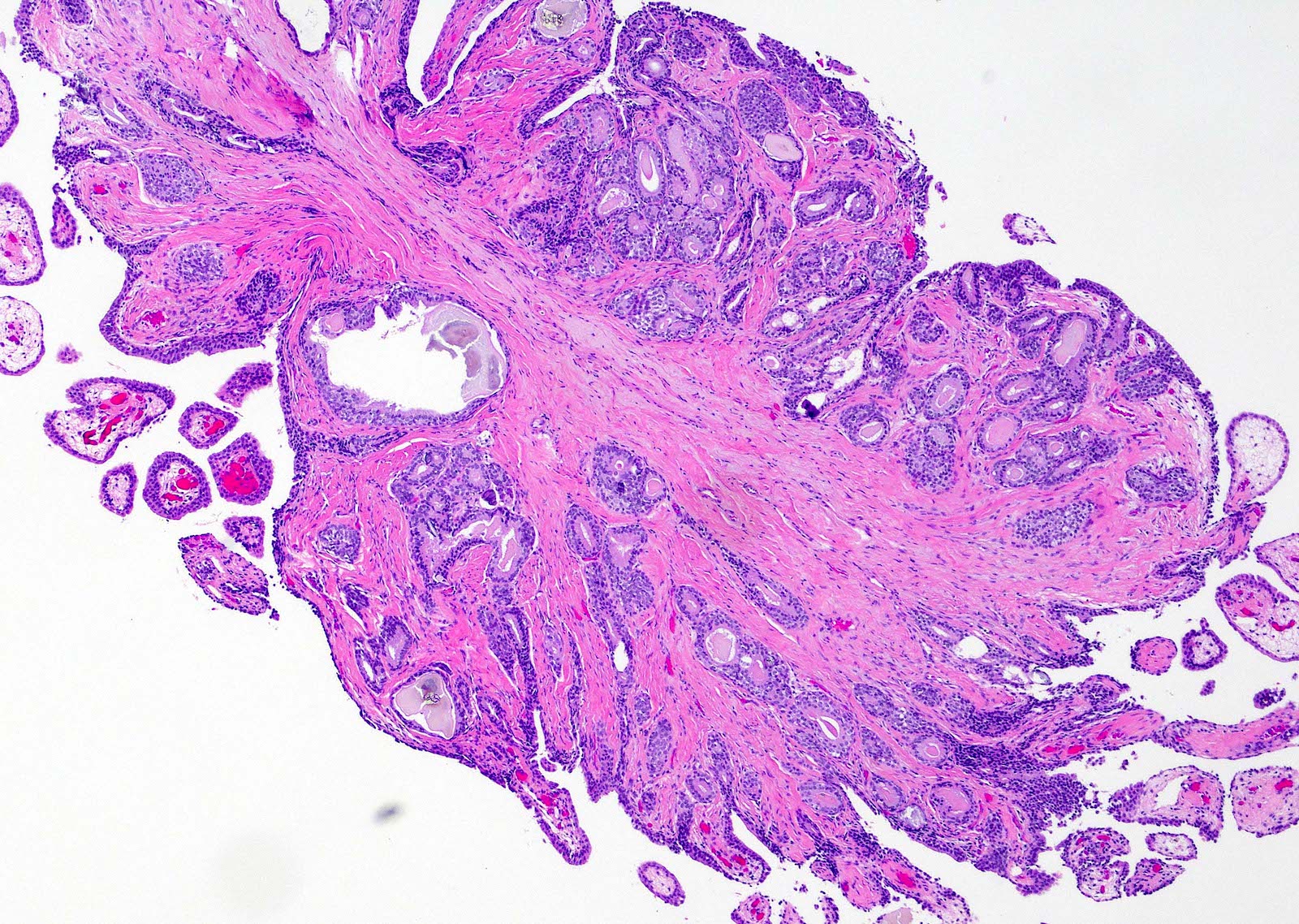
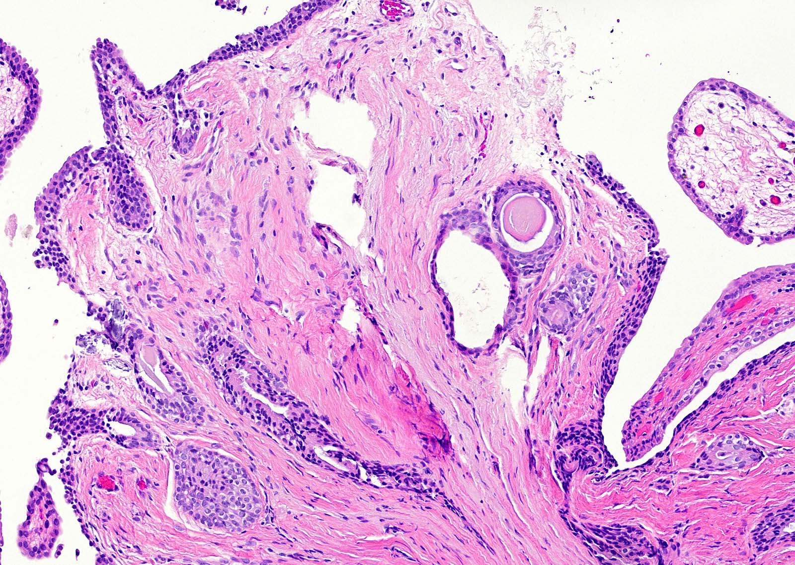
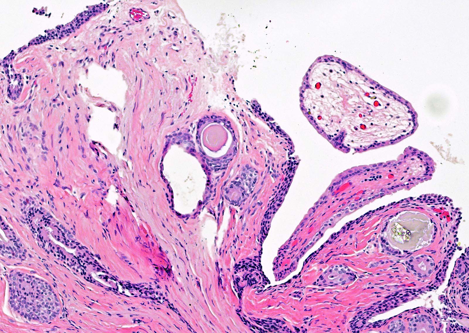
Microscopic (histologic) images
Contributed by Y. Albert Yeh, M.D., Ph.D.
Fibroepithelial polyp
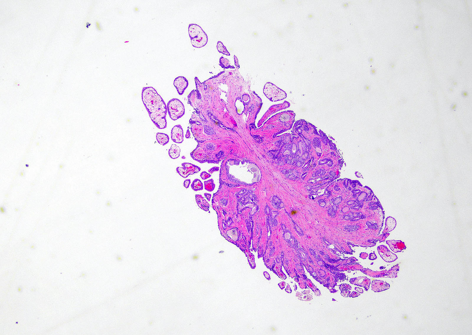
Polypoid mass
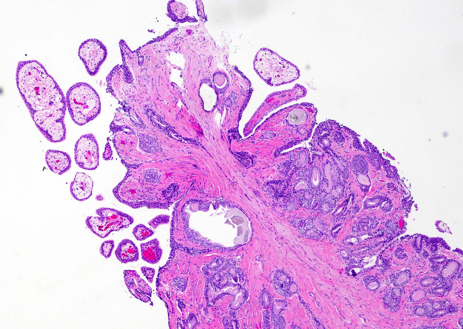
Cloverleaf-like and club-like projections
Broad based papillae
Cloverleaf-like projections
Club-like projections
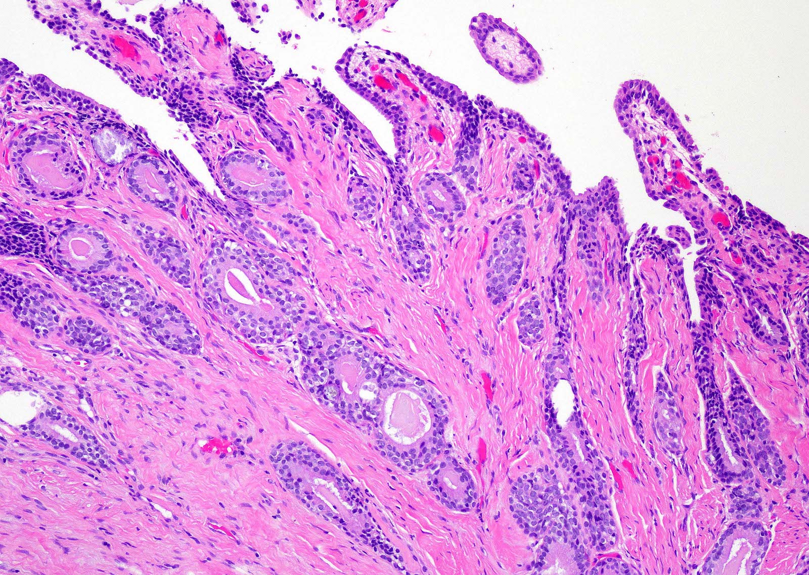
Broad based finger-like papillae
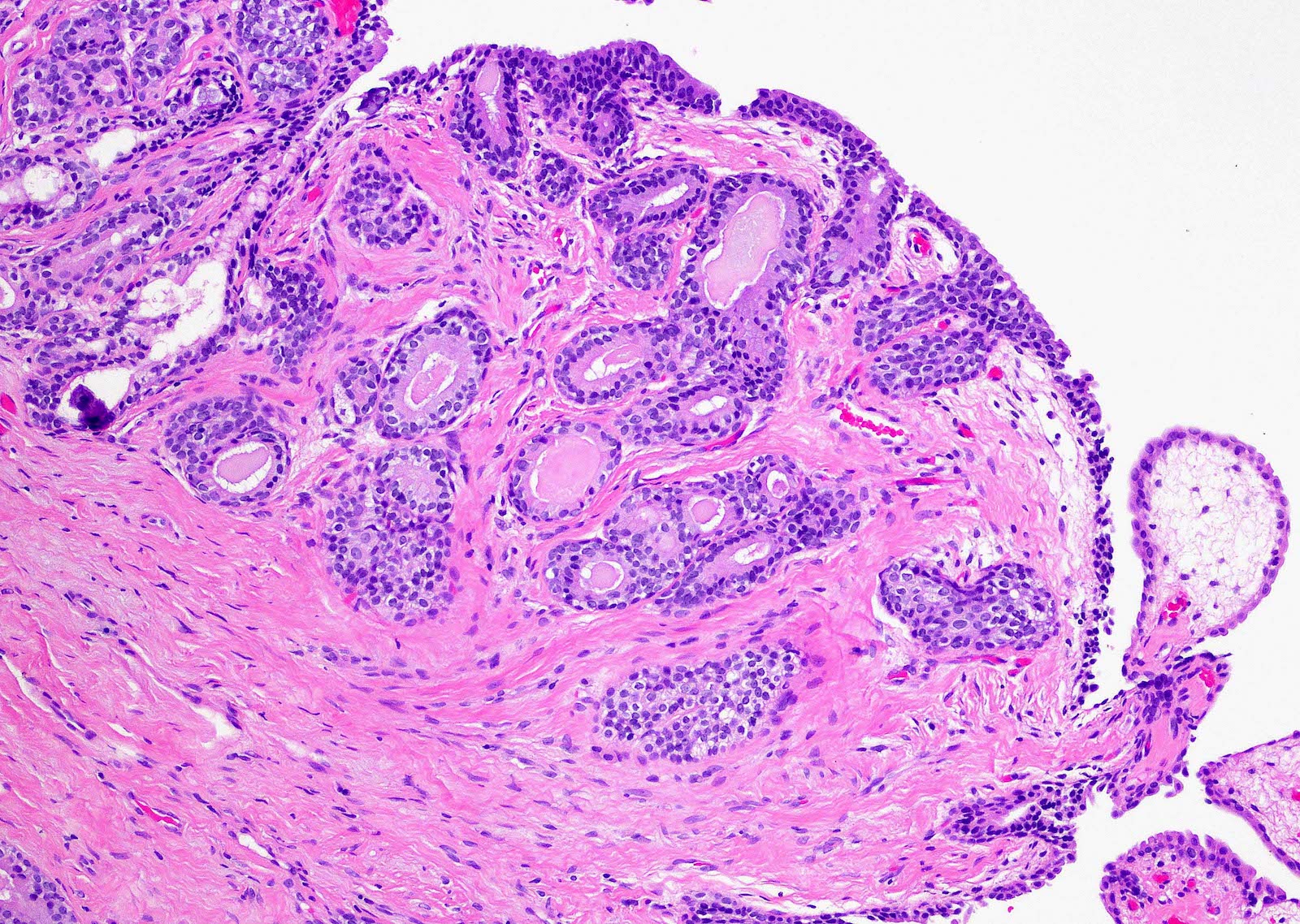
Urethritis cystica and glandularis
Polypoid and club-like projections
Focal calcifications
Cytology description
- Prostatic type urethral polyp:
- Clusters of benign columnar cells with uniform and oval nuclei (see Cytology images) (Arch Pathol Lab Med 2000;124:1047)
- Columnar cells have pale cytoplasm, fine nuclear chromatin and inconspicuous nucleoli (see Cytology images)

Positive stains
- Prostatic type urethral polyp (Am J Surg Pathol 1983;7:351):
Negative stains
- Fibroepithelial urethral polyp (Am J Surg Pathol 1983;7:351):
Sample pathology report
- Urethral lesion, prostatic, excision:
- Urethral polyp with prostatic type epithelium and changes, consistent with prostatic type urethral polyp (see comment)
- Comment: The urethral lesion shows a polypoid and papillary growth lined by prostatic type epithelium and composed of benign prostatic acini. PSA and PSAP immunohistochemical stains reveal positive cytoplasmic staining in the prostatic glandular cells. These features are consistent with prostatic urethral polyp.
- Urethral lesion, prostatic, excision:
- Cloverleaf-like and club-like projections lined by normal urothelium, consistent with fibroepithelial urethral polyp (see comment)
- Comment: The urethral lesion shows papillary projections arranged in cloverleaf-like and club-like pattern. The papillary structures are composed of broad fibrovascular cores covered by unremarkable urothelial cells. Neither urothelial proliferation nor epithelial atypia is noted. These features are consistent with fibroepithelial urethral polyp.
Differential diagnosis
- Fibroepithelial urethral polyp
- Prostatic type urethral polyp:
- Delicate papillae with true fibrovascular cores lined by benign prostatic epithelium (Am J Surg Pathol 1983;7:351)
- Urothelial papilloma:
- More slender, delicate loose fibrovascular cores lined by normal appearing urothelial cells and prominent umbrella cells with vacuolated cytoplasm (Am J Surg Pathol 2005;29:460)
- Polypoid urethritis:
- Polypoid and broad based papillary structures with marked stromal edema, acute and chronic inflammation
- No true fibrovascular cores (Am J Surg Pathol 2008;32:758)
- Prostatic type urethral polyp:
- Prostatic type urethral polyp
- Fibroepithelial polyp:
- Broad cloverleaf-like and club-like projections covered by normal urothelium
- No prostatic type epithelium (Am J Surg Pathol 2005;29:460)
- Urothelial papilloma:
- Slender fibrovascular cores lined by normal appearing urothelial cells
- No prostatic type epithelium (Am J Surg Pathol 2005;29:460)
- Polypoid urethritis:
- Polypoid structures with marked stromal edema, acute and chronic inflammation
- No true fibrovascular cores
- No prostatic type epithelium (Am J Surg Pathol 2008;32:758)
- Prostatic ductal adenocarcinoma:
- Complex prostatic glands lined with pseudostratified columnar cells with mild to severe nuclear atypia (J Urol 2015;193:2095)
- Fibroepithelial polyp:
Additional references
- Prostatic type urethral polyp: Urol Int 1993;50:114, Urology 1984;24:499, Eur Urol 1989;16:92, J Urol 1984;131:120, Histopathology 1987;11:789, Br J Urol 1993;72:981, J Urol 1989;142:1218, J Urol 1981;126:129
- Fibroepithelial polyp: Transl Pediatr 2020;9:272, J Endourol Case Rep 2016;2:90, J Surg Case Rep 2019;2019:rjz320
Practice question #1
A 50 year old man presented with dysuria and hematuria for 3 months. Cystoscopic examination showed a polypoid mass protruded from the posterior wall of the prostatic urethra. Cystoscopic excision of the polyp and histopathological examination were performed. The microphotograph of the urethral polyp is shown above. What is the diagnosis?
- Fibroepthelial urethral polyp
- Polypoid urethritis
- Prostatic type urethral polyp
- Urethral caruncle
Practice answer #1
Practice question #2
A 40 year old man presented with dysuria for 2 months. During cystoscopy, a polypoid mass protruding from the posterior urethra was detected. Surgical excision of the polyp was performed and microscopic examination showed a papillary growth lined by tall benign columnar cells with pale eosinophilic cytoplasm. In the stroma are some glands lined by benign columnar cells with underlying flattened epithelial cells. What is the diagnosis?
- Fibroepithelial polyp
- Polypoid urethritis
- Prostatic type urethral polyp
- Urethral caruncle
Practice answer #2
