

Soft tissue
Pericytic (perivascular)
Angioleiomyoma
Authors: Ohoud Aljarbou, M.D., Omar P. Sangueza, M.D.
Editorial Board Member: Jose G. Mantilla, M.D.
Editor-in-Chief: Debra L. Zynger, M.D.

Last author update: 8 November 2021
Last staff update: 8 November 2021
Copyright: 2003-2024, PathologyOutlines.com, Inc.
PubMed search: angioleiomyoma [title] vascular pathology
Table of Contents
Definition / general | Essential features | Terminology | ICD coding | Epidemiology | Sites | Pathophysiology | Etiology | Clinical features | Diagnosis | Radiology description | Prognostic factors | Case reports | Treatment | Clinical images | Gross description | Gross images | Microscopic (histologic) description | Microscopic (histologic) images | Cytology description | Positive stains | Negative stains | Molecular / cytogenetics description | Sample pathology report | Differential diagnosis | Additional references | Board review style question #1 | Board review style answer #1 | Board review style question #2 | Board review style answer #2Cite this page: Aljarbou O, Sangueza OP. Angioleiomyoma. PathologyOutlines.com website. https://www.pathologyoutlines.com/topic/softtissueangioleiomyoma.html. Accessed May 13th, 2024.
Definition / general
- Benign dermal / subcutaneous neoplasm arising in vascular smooth muscle; there is strong overlap with myopericytoma and these lesions are considered to be part of the same spectrum of disease (J Ultrasound Med 2016;35:1669)
Essential features
- Angioleiomyoma is a benign smooth muscle tumor
- Preoperative diagnosis is not certain, microscopic examination is needed for accurate diagnosis
- Complete excision is curative
Terminology
- Vascular leiomyomas or angiomyomas (J Ultrasound Med 2016;35:1669)
ICD coding
- ICD-O: 8894/0 - angiomyoma
Epidemiology
- F:M = 1.7:1.0
- Fourth to sixth decades (J Ultrasound Med 2016;35:1669)
- Accounts for 5% of benign soft tissue neoplasms (J Ultrasound Med 2019;38:1201)
Sites
- Extremity, particularly the lower leg (50 - 70%), followed by the head and trunk (J Ultrasound Med 2016;35:1669, Tumori 2014;100:148e)
- Other sites less common
Pathophysiology
- Histogenesis is uncertain (J Ultrasound Med 2016;35:1669)
Etiology
- Minor trauma, venous stasis and hormonal changes, especially estrogen (J Ultrasound Med 2016;35:1669)
Clinical features
- Painful, solitary, slow growing nodule (J Ultrasound Med 2019;38:1201)
- Well demarcated subcutaneous lesions classically found on the lower extremities but can also present on the head, trunk and upper extremities (J Cutan Pathol 2017;44:342)
- Difficult to distinguish from other smooth muscle tumors and few are diagnosed pre-operatively (J Cutan Pathol 2017;44:342)
- Presence of pain may be helpful in the diagnosis but is unreliable, as only 58% of tumors present with pain (J Cutan Pathol 2017;44:342)
- Pain can be caused by local ischemia from contraction of the tumoral vessels and may be mediated by nerve fibers (J Ultrasound Med 2019;38:1201)
- Multiple subcutaneous angioleiomyomas have been reported in a patient with AIDS (Br J Dermatol 2002;147:563)
- Clinical differential diagnosis includes glomus tumors, spiradenomas, angiolipomas or neuromas
Diagnosis
- Dermoscopic findings: symmetric pink-reddish tumor with vessels, white structures and the absence of ulceration (J Eur Acad Dermatol Venereol 2019;33:693)
Radiology description
- On ultrasonography: subcutaneous masses with an oval shape, well defined margins, a homogeneous structure, hypervascularity and a small size (< 2 cm) (J Ultrasound Med 2016;35:1669)
- On MRI: well circumscribed mass with slightly hyperintense signal on T1 weighted images, heterogeneous and hyperintense signal on T2 weighted and STIR images and heterogeneous enhancement on contrast enhanced T1 weighted images
Prognostic factors
- No known prognostic factors
Case reports
- 40 year old man of North African descent noted a nodule on the rim of the helix of his right ear (Case Rep Otolaryngol 2017;2017:8289710)
- 51 year old presented with a slowly growing mass in the right mid foot in the region of the medial arch (J Clin Diagn Res 2016;10:PD07)
- 69 year old woman presented with a painful nodule on her left posterior heel (Open Access Maced J Med Sci 2017;5:436)
- 70 year old man with a 3 year history of a slowly growing nodule on the fifth right toe (J Cutan Pathol 2019;46:301)
- 82 year old man presented with a 1 month history of lower back pain and paresthesias involving the soles of both feet (Skeletal Radiol 2018;47:293)
Treatment
- Surgical excision is considered a safe and effective method of treatment (J Cutan Pathol 2017;44:342)
Clinical images
Images hosted on other servers:

Foot mass

Palate mass

Lip mass

Hand tumor
Gross description
- Sharply demarcated, spherical, gray-white or brown tumor
- Usually measuring less than 2 cm in diameter in approximately 80% of cases
- Reference: Cancer 1984;54:126
Gross images
Images hosted on other servers:

Circumscribed lip mass

Homogenous cut surface
Microscopic (histologic) description
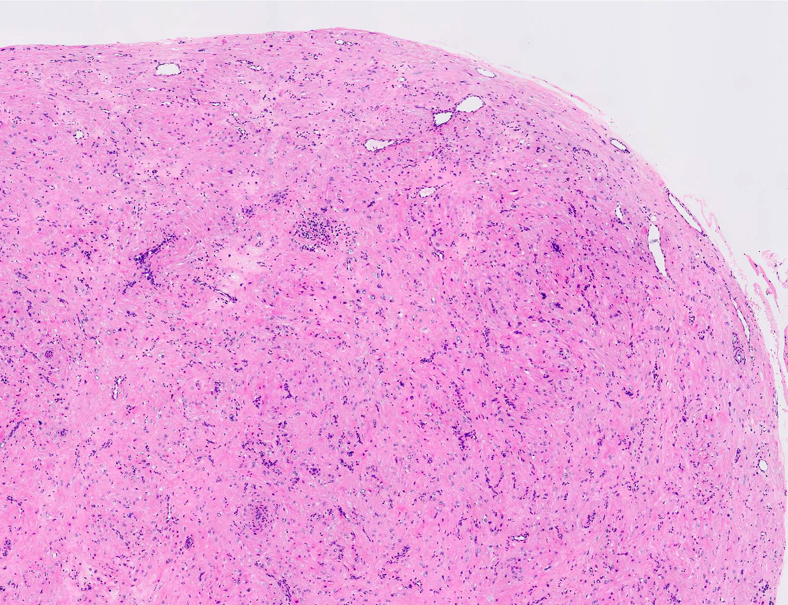
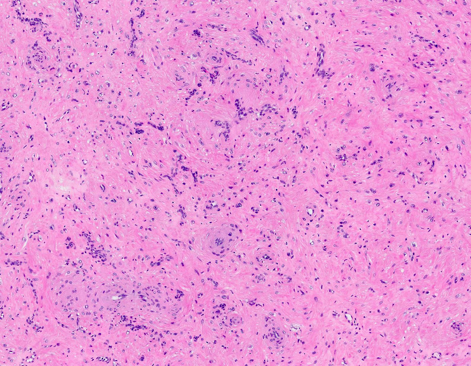
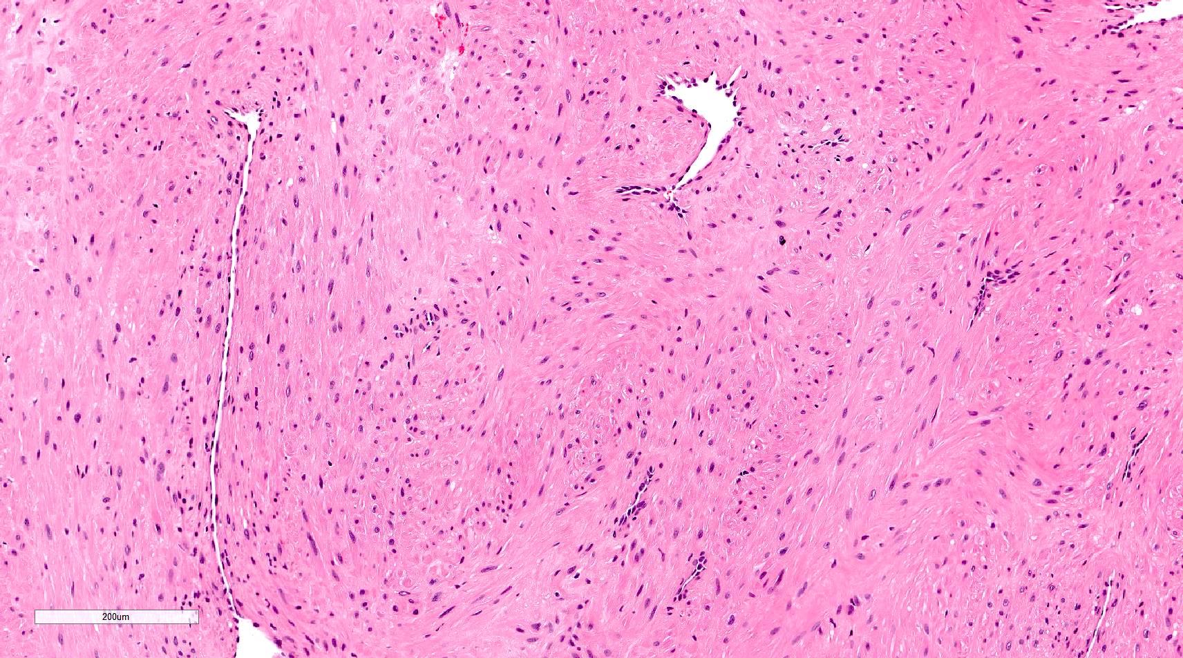
- Encapsulated proliferation of eosinophilic smooth muscle cells with minimal nuclear pleomorphism
- Rounded or slit-like veins with muscular walls present within the tumor (J Cutan Pathol 2017;44:342)
- Tumors can further be classified into 3 subcategories, first described by Morimoto et al:
- Solid type: smooth muscle bundles surround numerous small slit-like channels
- Cavernous type: dilated vascular channels, the walls of which are difficult to distinguish from the intervascular smooth muscle
- Venous type: thick walled vessels that are easily distinguished from the intervascular smooth muscle
- Epithelioid and pleomorphic variants are reported in a few cases
- Epithelioid type: composed of cells with round to oval nuclei and a moderate amount of finely granular eosinophilic cytoplasm with occasional vacuoles (Am J Dermatopathol 1998;20:213)
- Pleomorphic type: marked nuclear pleomorphism but only rare or absent mitoses (Am J Dermatopathol 2000;22:268)
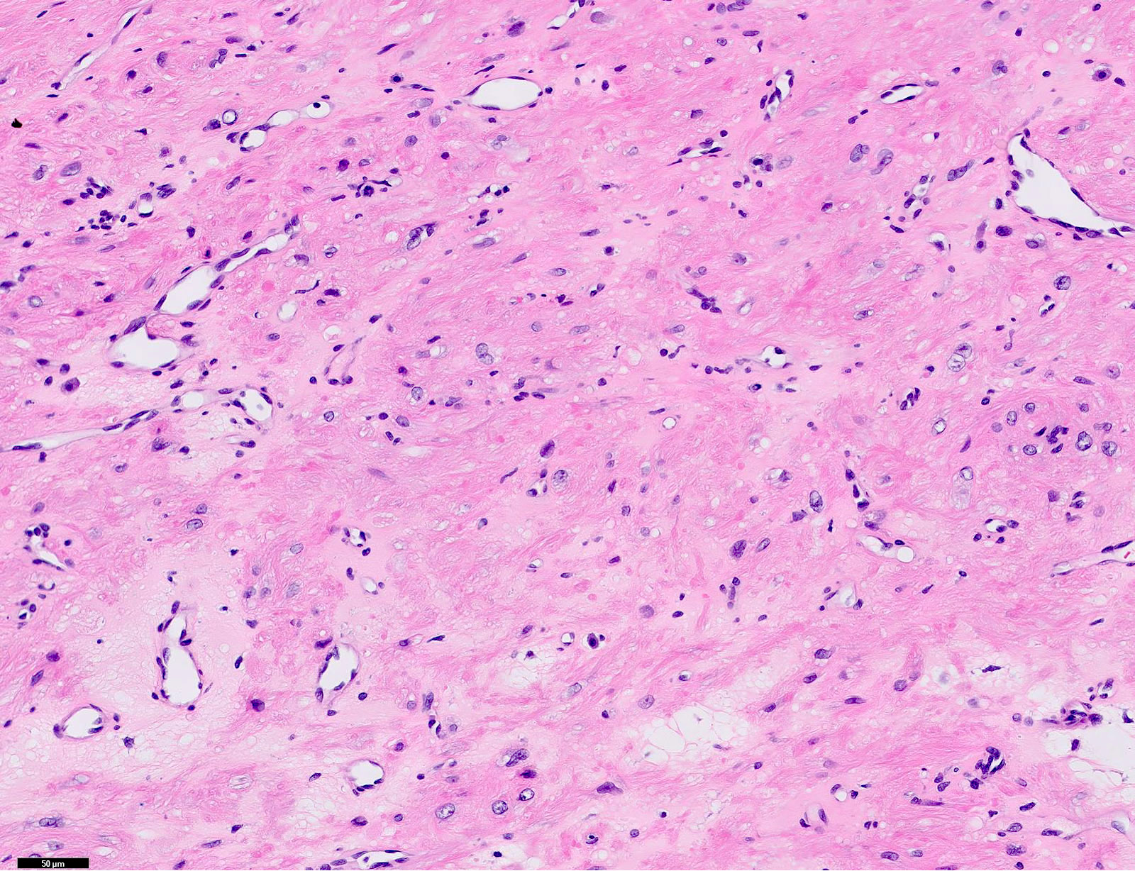
- Calcification and marked degeneration, including hyalinization and myxoid changes, may be present (J Cutan Pathol 2017;44:342, J Ultrasound Med 2019;38:1201)
- Fat can be seen in a few cases; not to be mistaken for angiomyolipoma (Cancer 1984;54:126)
Microscopic (histologic) images
Contributed by Ohoud Aljarbou, M.D., Jijgee Munkhdelger, M.D., Ph.D. and Andrey Bychkov, M.D., Ph.D.
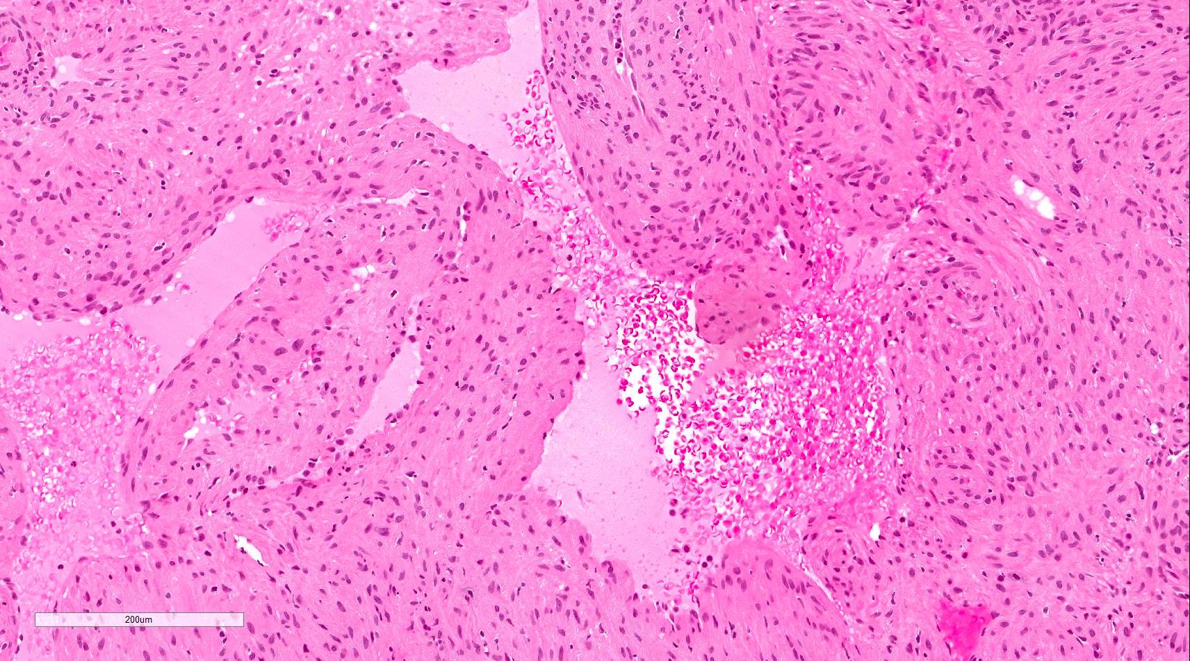
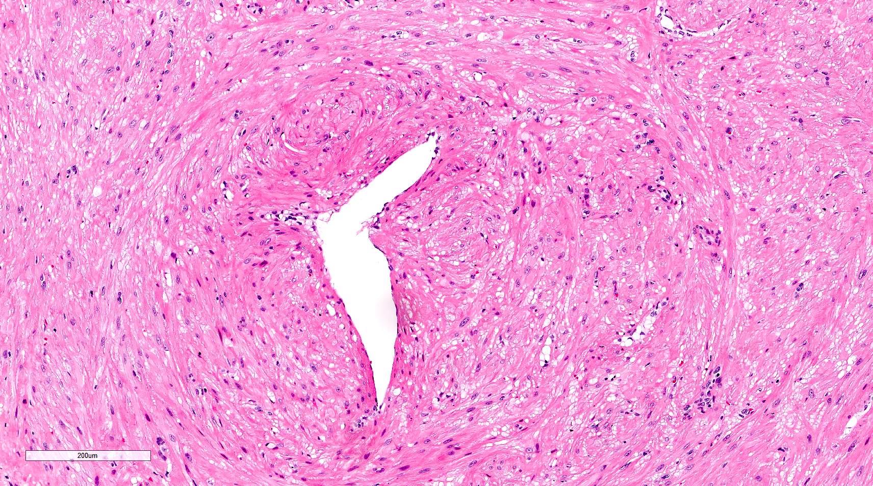
Smooth muscle fascicles and vascular channels

Lack atypia and mitosis
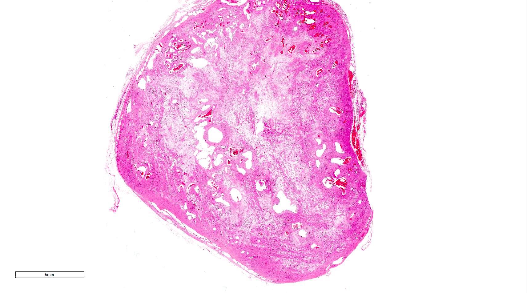
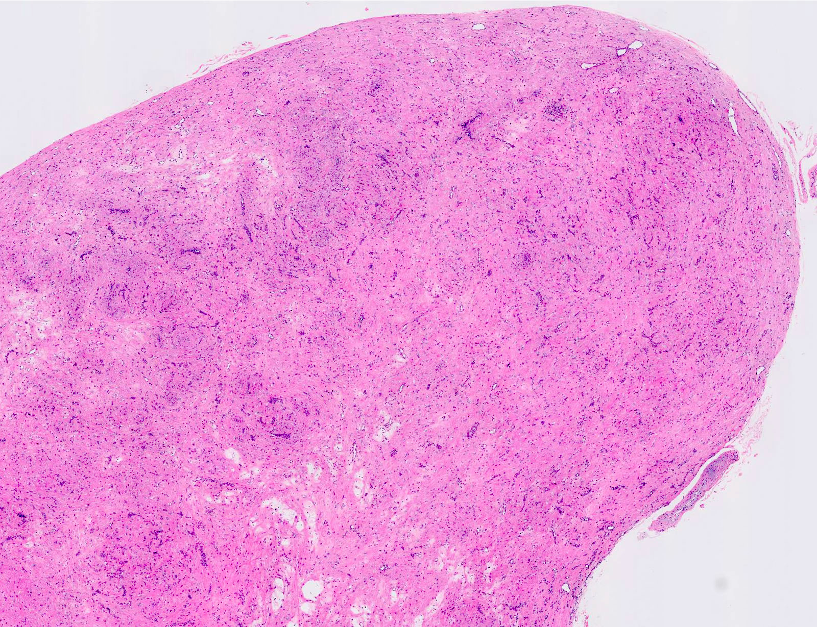
Well defined mass
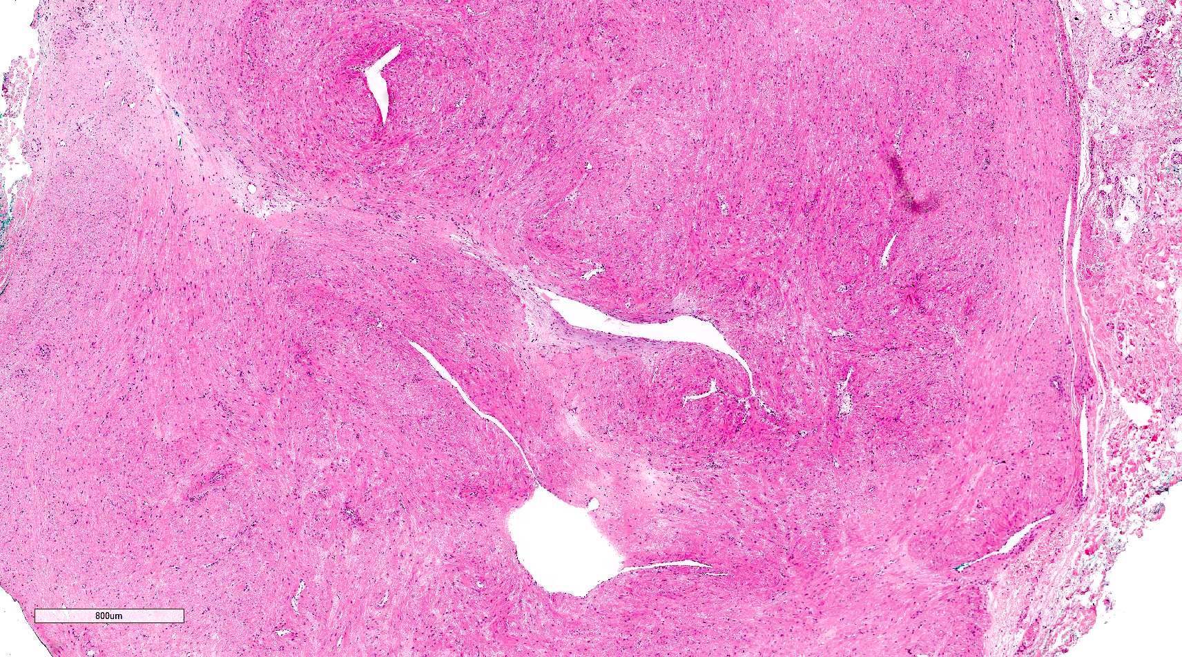
Edema and hyalinization

Small fascicles of smooth muscles
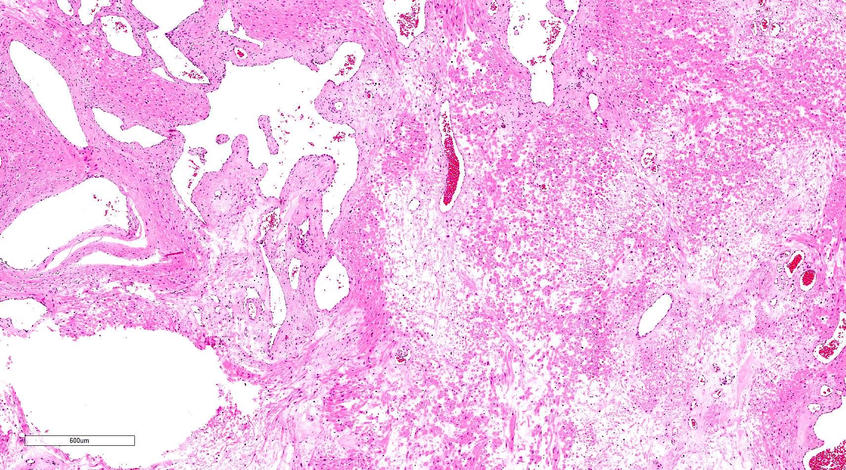
Well defined mass with vascular component
Fascicles of smooth muscles
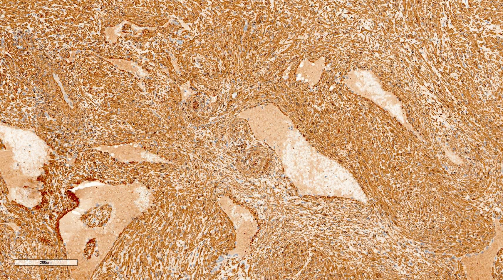
SMA

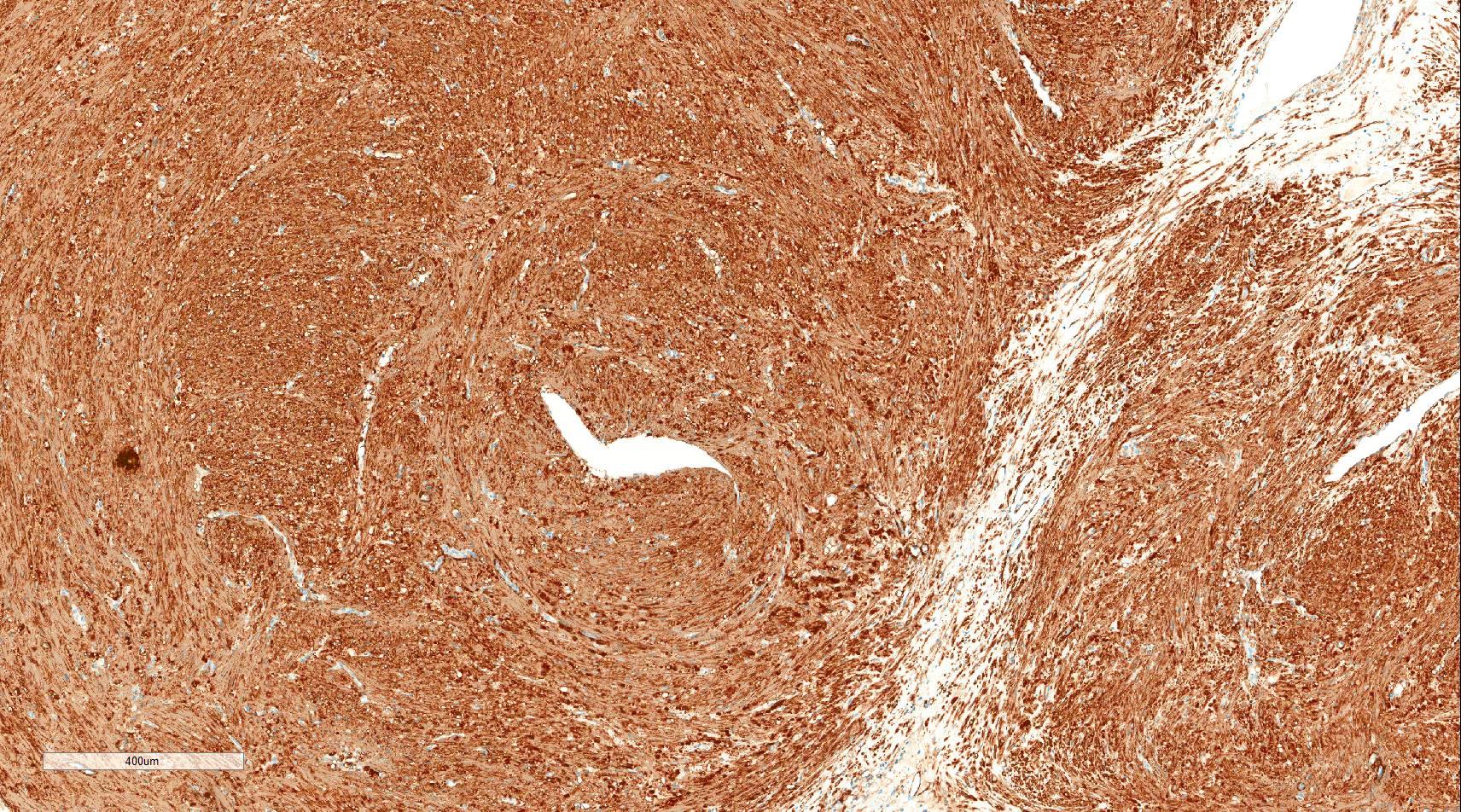
Desmin
Low power
Well defined nodule
Smooth muscle bundles, vessels

Organizing thrombus
Thin walled blood vessels

Thick walled blood vessels
Cytology description
- Variable proportions of benign smooth muscle cells and uniform spindle cells
- Dissociated or arranged in small fascicles
- Small fragments of a collagenous matrix in the background (Monogr Clin Cytol 2017;22:68)
- The limitations encountered are very small, hard or painful skin lesions that may be difficult to aspirate (Diagn Cytopathol 2002;27:161)
- Abundant connective tissue and calcification can also cause problems in obtaining adequate material (Diagn Cytopathol 2002;27:161)
Positive stains
- Actins (alpha smooth muscle actin and HHF35) and calponin (diffuse positivity) and diffusely or focally positive for h-caldesmon
- Desmin has variable positivity, observed in 75% of solid type, 50% of the venous type and only 18% of the cavernous type (Hum Pathol 2007;38:645)
Negative stains
Molecular / cytogenetics description
- Diploid or near diploid with no consistent chromosome aberrations
- Few cases show karyotypic aberrations (Cancer Genet Cytogenet 2010;199:147)
Sample pathology report
- Skin, lower leg mass, excision:
- Angioleiomyoma, completely excised (see comment)
- Comment: The grossly noted tumor is 1 cm. The resection margins are negative. There is no evidence of atypia or malignancy.
Differential diagnosis
- Myopericytoma:
- Concentric perivascular arrangement
- Most are negative for desmin
- Also express alpha smooth muscle actin, HHF35 and calponin
- Angiomyolipoma:
- Glomus tumor:
- Leiomyoma:
- No prominent rounded or slit-like veins with muscular walls present
Additional references
Board review style question #1
- The image shown above is from a 45 year old woman with a right leg mass. SMA and desmin are positive. Which of the following is the most appropriate diagnosis?
- Angioleiomyoma
- Myopericytoma
- Glomus tumor
- Angiomyolipoma
Board review style answer #1
A. Angioleiomyoma. A mixture of smooth muscle bundles arranged in small fascicles and intervening vascular channels is noted. The concentric perivascular spindle cell proliferation which is characteristic for myopericytoma is absent. There are no glomus cells seen. Adipocytes are not seen in this image, which makes angiomyolipoma less likely.
Comment Here
Reference: Angioleiomyoma
Comment Here
Reference: Angioleiomyoma
Board review style question #2
- Which of the following modalities is the gold standard for the diagnosis of angioleiomyoma?
- Ultrasonography findings
- Dermoscopic findings
- MRI findings
- Microscopic examination
Board review style answer #2
D. Microscopic examination. Angioleiomyoma has no specific findings preoperatively. Microscopic findings are the gold standard for accurate diagnosis.
Comment Here
Reference: Angioleiomyoma
Comment Here
Reference: Angioleiomyoma
