Skin nonmelanocytic tumor
Adnexal tumors
Follicular derived
Trichilemmoma
Editorial Board Member: Brandon Umphress, M.D.


Last author update: 12 June 2025
Last staff update: 12 June 2025
Copyright: 2002-2025, PathologyOutlines.com, Inc.
PubMed Search: Trichilemmoma
Table of Contents
Definition / general | Essential features | Terminology | ICD coding | Epidemiology | Sites | Pathophysiology | Etiology | Clinical features | Diagnosis | Prognostic factors | Case reports | Treatment | Clinical images | Microscopic (histologic) description | Microscopic (histologic) images | Positive stains | Negative stains | Sample pathology report | Differential diagnosis | Additional references | Practice question #1 | Practice answer #1 | Practice question #2 | Practice answer #2Cite this page: Vanderbeck K, Torres-Cabala CA. Trichilemmoma. PathologyOutlines.com website. https://www.pathologyoutlines.com/topic/skintumornonmelanocytictrichilemmoma.html. Accessed August 14th, 2025.
Definition / general
- Benign follicular neoplasm with outer root sheath differentiation (trichilemmal differentiation) (Arch Dermatol 1962;86:430, Am J Dermatopathol 2018;40:561, Curr Diagn Pathol 2007;13:273)
- Benign adnexal proliferation with clear cell features
Essential features
- Bland keratinocytic tumor with thickened eosinophilic basement membrane and endophytic / lobular architecture
- CD34 positive in most cases, often focal (Clin Exp Dermatol 2006;31:807, Am J Dermatopathol 2018;40:561)
- Frequent clear cell change due to glycogenated cytoplasm
Terminology
- Tricholemmoma
ICD coding
- ICD-10: D23 - other benign neoplasms of skin
Epidemiology
- Predominantly affects middle aged adult patients (J Cutan Pathol 1990;17:45, Am J Dermatopathol 2018;40:561, Curr Diagn Pathol 2007;13:273)
- May present earlier in patients with Cowden syndrome (typically pediatric cases, late adolescence) (J Cutan Pathol 1985;12:83, J Int Med Res 2019;47:3918)
Sites
- Head and neck are the most common locations, with most tumors appearing on the central face (Arch Dermatol 1973;107:866, Arch Dermatol 1978;114:286, J Clin Pathol 2007;60:129)
Pathophysiology
- Originates from the outer root sheath of the pilosebaceous unit, specifically the follicular infundibulum (Arch Dermatol 1962;86:430, Am J Dermatopathol 2018;40:561, Curr Diagn Pathol 2007;13:273)
Etiology
- Connection to human papillomavirus (tricholemmal verrucae) has been suggested but remains controversial (Arch Dermatol 1978;114:286, Arch Dermatol 1984;120:859, J Cutan Pathol 1991;18:193, Br J Dermatol 2014;171:1073)
- Cowden syndrome
- Autosomal dominant genodermatosis caused by a mutation in the PTEN tumor suppressor gene on chromosome 10q22-23 (Nat Genet 1996;13:114)
- Characterized by multiple mucosal papillomas, mucosal fibromas, trichilemmomas, hamartomas (Nat Genet 1996;13:114)
- Hamartomatous lesions in the gastrointestinal tract (esophagus, stomach, colon) (Cancers (Basel) 2019;11:844)
- Soft tissue tumors such as lipomas, neurofibromas, lymphatic malformations, PTEN hamartoma of soft tissue (Cancers (Basel) 2019;11:844)
- Associated with an increased risk of visceral malignancies, including thyroid, breast and gastrointestinal carcinomas (J Dermatol Surg Oncol 1979;5:12, Br J Dermatol 1979;100:667, Am J Dermatopathol 2018;40:561, Curr Diagn Pathol 2007;13:273, Cancers (Basel) 2019;11:844)
Clinical features
- Verrucous or smooth exophytic and dome shaped lesions
Diagnosis
- Histologic evaluation is required for diagnosis
- Clinically presents as nonspecific papules or nodules, often mistaken for verruca or basal cell carcinoma
Prognostic factors
- While generally benign, multiple trichilemmomas may indicate the presence of Cowden syndrome (J Dermatol Surg Oncol 1979;5:12, Br J Dermatol 1979;100:667, Am J Dermatopathol 2018;40:561, Curr Diagn Pathol 2007;13:273)
Case reports
- 28 year old woman with desmoplastic trichilemmoma arising in nevus sebaceous (Int J Clin Exp Pathol 2019;12:3953)
- 28 year old woman with multiple erythematous lesions over the left ankle (J Cutan Pathol 2017;44:93)
- 34 year old man with trichilemmoma of scalp (Cureus 2024;16:e72141)
- 36 year old man with pigmented desmoplastic trichilemmoma arising in nevus sebaceous (Skin Appendage Disord 2023;9:309)
- 38, 54 and 74 year old men with nasal vestibule tumors (Head Neck Pathol 2012;6:492)
- 55 year old man with a lesion on his right upper eyelid (J Cutan Pathol 2007;34:22)
- 58 year old woman with a lesion on the scalp (An Bras Dermatol 2017;92:836)
- 67 year old man with desmoplastic trichilemmoma of scalp (J Int Med Res 2019;47:3918)
- 78 year old man with desmoplastic histology clinically mimicking carcinoma (An Bras Dermatol 2014;89:796)
Treatment
- No treatment required in majority of cases unless cosmetic or symptomatic concerns arise
- Options for treatment are localized and include curettage, shave excision or surgical excision
- Laser may also be used
- Mohs micrographic surgery has been used (Cureus 2024;16:e61910)
- Patients should understand that an associated malignancy (including basal cell carcinoma) may be concealed; they should be offered surgery for clear margins or may opt for observation (BMJ Open Ophthalmol 2020;5:e000513)
Clinical images
Images hosted on other servers:

Pale papule of the upper lip

Central ulceration
Microscopic (histologic) description
- Lobular squamoproliferative lesion with pushing and rounded border
- Connection with the epidermis and pilosebaceous apparatus can be appreciated
- Hyperkeratosis and stromal clefting is common
- May show verrucous hyperplasia of epidermis (J Clin Pathol 2007;60:129)
- Cells with pale or clear staining cytoplasm (due to glycogenation) are characteristic but may be challenging to find, as they are particularly dependent on sampling and artifactual changes in the tissue (J Clin Pathol 2007;60:129)
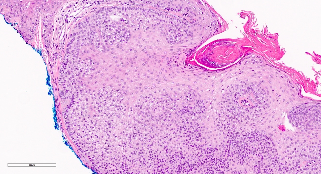
- Peripheral palisading of cuboidal or columnar basaloid cells are usually present (Cureus 2024;16:e61910, J Clin Pathol 2007;60:129)
- Reverse nuclear polarity of peripheral cells
- Distinct basement membrane resembling the hair outer root sheath zone below the level of the follicular isthmus (Arch Dermatol 1962;86:430, Am J Dermatopathol 2018;40:561, Curr Diagn Pathol 2007;13:273)
- Squamous eddies are often appreciated
- Mucin deposition is rare and typically focal
- Associated with a thickened and prominent basement membrane that surrounds nests of cells (BMJ Open Ophthalmol 2020;5:e000513)
- Desmoplastic trichilemmoma
- Characterized by a prominent desmoplastic stroma that typically appears at the center of the tumor (J Clin Pathol 2007;60:129, J Cutan Pathol 1990;17:45)
Microscopic (histologic) images
Contributed by Carlos A. Torres-Cabala, M.D., Nicholas Turnbull, M.B.Ch.B. and Richard A. Carr, M.B.Ch.B.
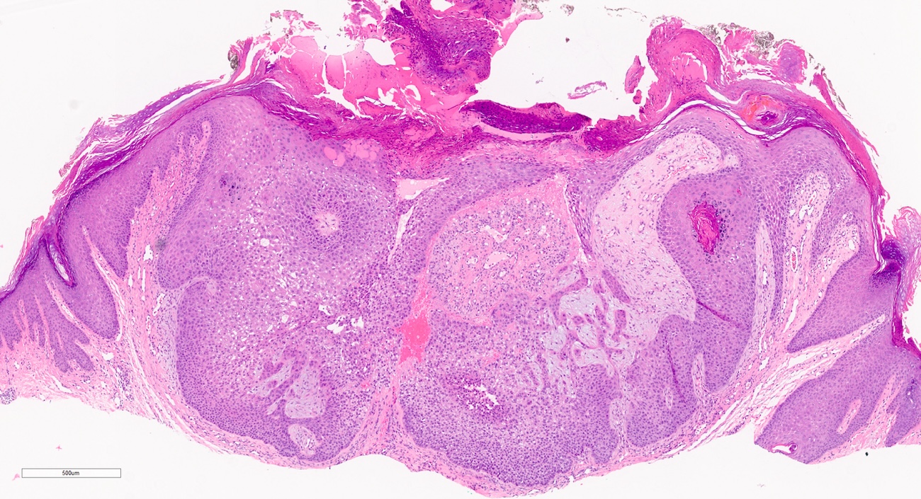
Bulbous keratinocytic predominantly basaloid proliferation
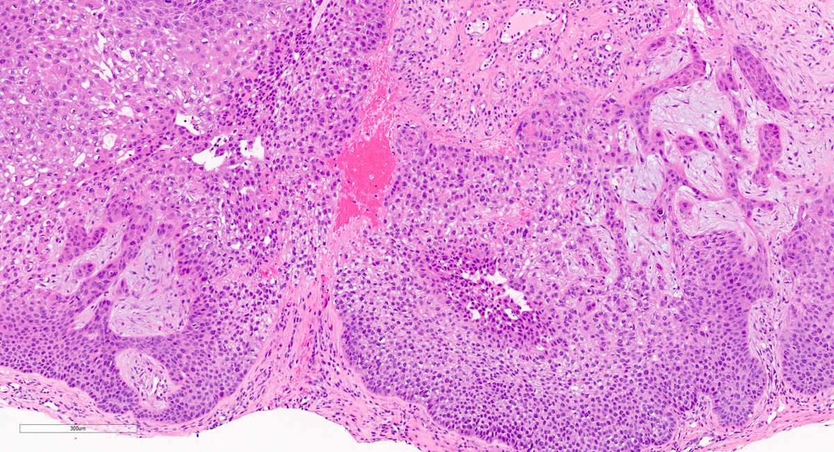
Basaloid to clear cells
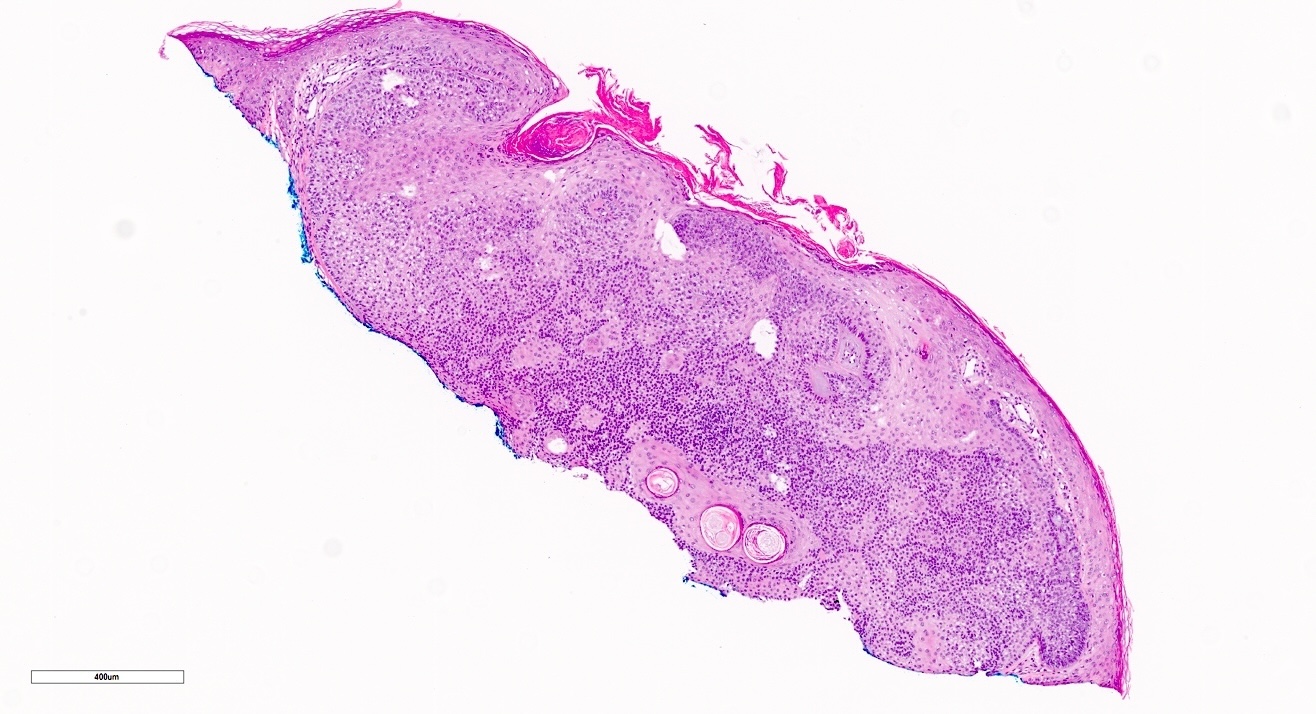
Basaloid proliferation with pseudohorn cysts
Clear cells
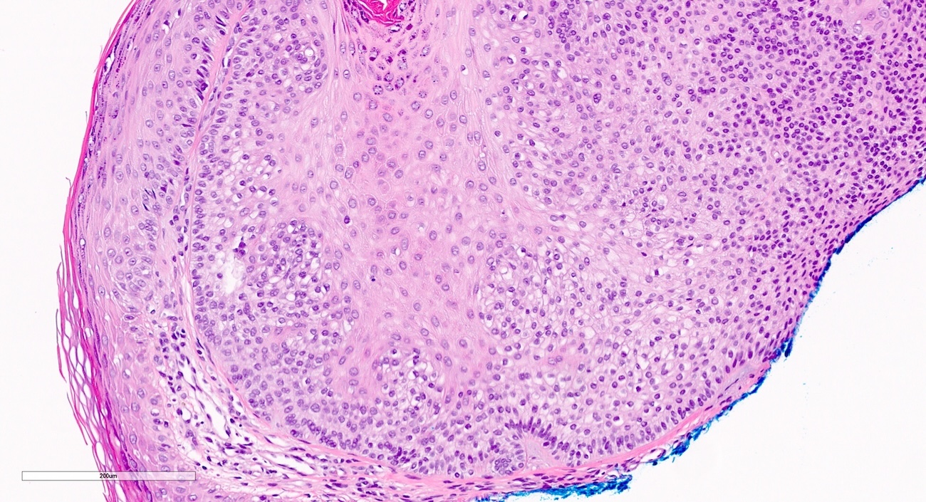
Peripheral palisading
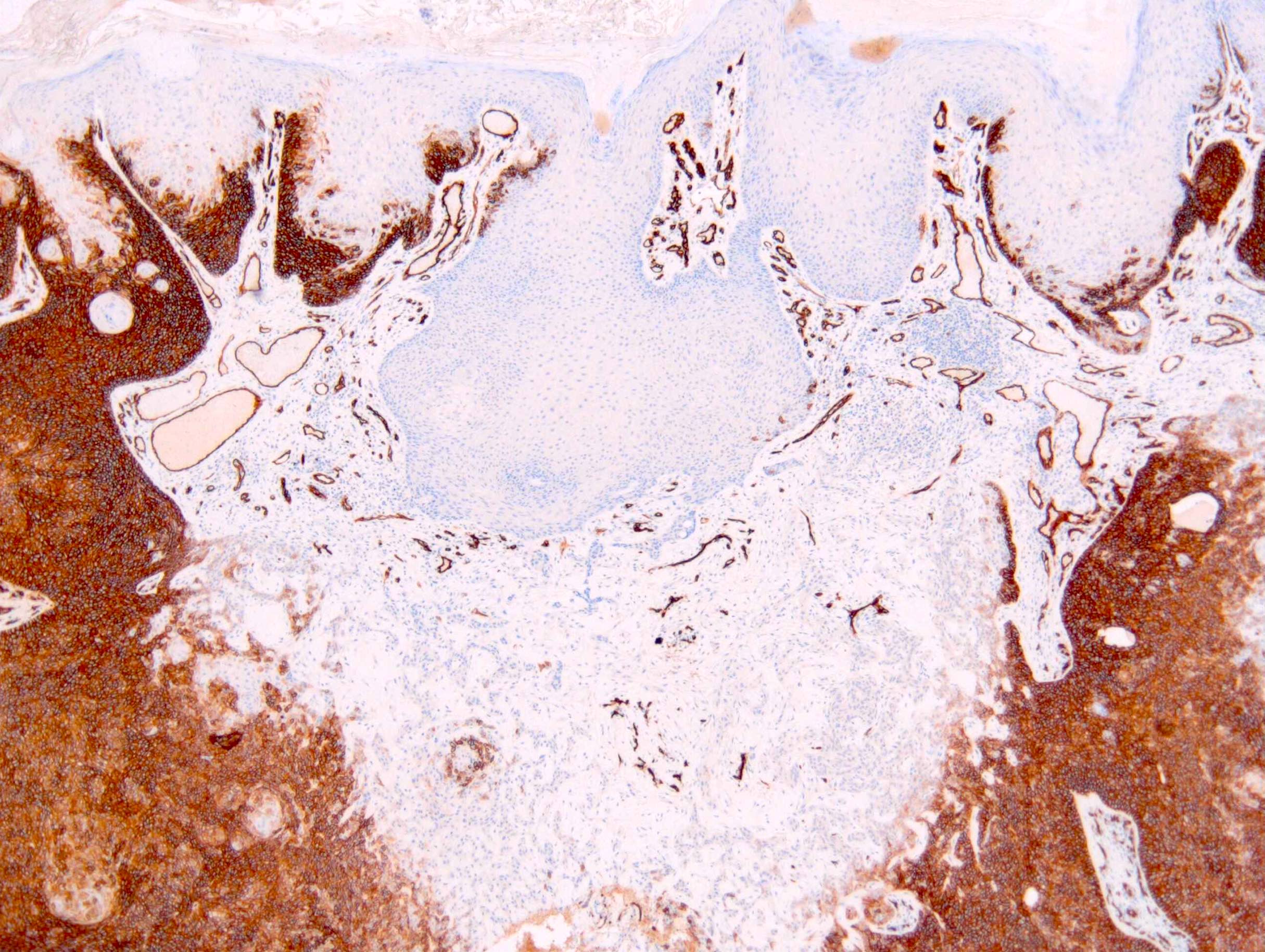
CD34 immunohistochemical study
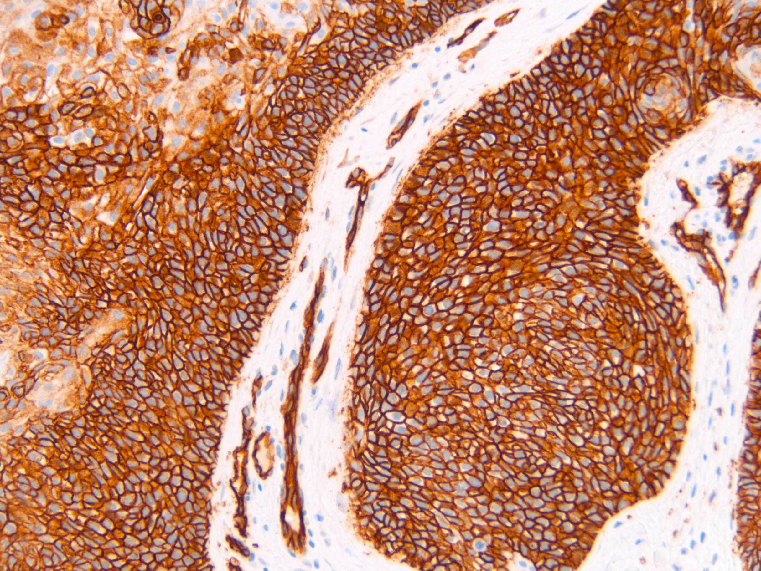
CD34 positive in trichilemmoma
Positive stains
- CD34: positive in most cases, often focal; may require deeper sections (Am J Dermatopathol 2018;40:561, J Cutan Pathol 2016;43:535, Head Neck Pathol 2012;6:492)
- CK7, EMA: typically expressed in a patchy pattern (Ophthalmic Plast Reconstr Surg 2016;32:e9)
- PAS (periodic acid-Schiff): highlights glycogenated cytoplasm (Head Neck Pathol 2012;6:492)
Negative stains
- BerEP4 (Am J Dermatopathol 2018;40:561, J Cutan Pathol 2016;43:535)
- CK17: occasionally positive at the rim periphery of tumor (Am J Dermatopathol 2022;44:886)
Sample pathology report
- Skin, right side of nose, punch biopsy:
- Trichilemmoma with desmoplastic features (see comment)
- Comment: A bland appearing folliculocentric and lobular tumor is appreciated. Cytologically, there are 2 main populations of tumor cells, which include centrally located cells with pale to clear cytoplasm as well as basaloid cells at the periphery. Palisading is appreciated. A thickened basement membrane is also noted throughout. The overlying epidermis demonstrates hyperkeratosis. Dysplasia is not identified. The findings are in keeping with a trichilemmoma. Multiple trichilemmomas may be associated with Cowden syndrome. Clinical correlation recommended.
Differential diagnosis
- Clear cell acanthoma:
- Characterized by epidermal acanthosis with prominent clear cell changes in the epidermal cells (Dermatopathology (Basel) 2020;7:26)
- Overlying parakeratosis and attenuation of the granular layer are commonly seen
- Sharp demarcation adjacent to normal epidermis
- Lacks lobular growth pattern and prominent basement membrane
- Inverted follicular keratosis:
- Keratinocytic neoplasm with inverted / endophytic growth pattern showing small basaloid cells and tightly organized squamous eddies (Indian Dermatol Online J 2016;7:177)
- Clear cells are not typically seen
- Viral wart (verruca):
- Hyperkeratotic and papillomatous lesions with typically prominent granular layer and inward turning of the rete ridges
- Marked hyperkeratosis, with columns of parakeratosis and associated hemorrhage
- Basal cell carcinoma:
- May resemble trichilemmoma, particularly the basaloid variant
- Clefting between tumor cells and stroma with mucin is seen, not the clefting between stroma commonly seen in follicular tumors including trichilemmoma
- Cytologic atypia and mitotic activity is usually evident
- BerEP4 is usually strong and diffuse (except in superficial or eroded cases)
- CD34 is negative
- CK17 expected to be positive (strong and diffuse) (Am J Dermatopathol 2022;44:886)
- May occur with trichilemmoma (Am J Dermatopathol 1996;18:43, Cureus 2024;16:e61910)
- Squamous cell carcinoma:
- Particularly if squamous cell carcinoma exhibits follicular differentiation
- Cellular pleomorphism, brisk mitotic activity and atypical mitotic figures
- Invasive border
- CD34 is negative
- Trichilemmal carcinoma:
- Malignant (J Clin Aesthet Dermatol 2021;14:25, Front Oncol 2023;12:1078272)
- Significant cytologic atypia and brisk and abnormal mitotic activity (Front Oncol 2023;12:1078272, J Clin Aesthet Dermatol 2021;14:25)
- Desmoplasia may be seen at the periphery rather than centrally in desmoplastic trichilemmoma
- Lacks circumscription or symmetry
- CD34 is typically negative
- Tumor of follicular infundibulum:
- Exhibits superficial, horizontal plate-like growth pattern that grows parallel to the epidermis
- Lobular growth pattern not appreciated
- Multiple anastomosing fronds of tumor cells (An Bras Dermatol 2014;89:964)
Additional references
Practice question #1
The tumor shown above was taken from the cheek of a 20 year old man. What is the expected staining pattern?
- BerEP4 positive
- Colloidal iron special stain highlights mucin
- Focal CD34 may be expected but requires adequate sectioning to find
- PAS special stain fails to highlight cytoplasmic glycogen
Practice answer #1
C. Focal CD34 may be expected but requires adequate sectioning to find. Trichilemmomas often exhibit focal CD34 reactivity (adequate sectioning may be required). Answer A is incorrect because BerEP4 is negative in trichilemmomas, while positive in keratinocytic carcinomas (namely basal cell carcinoma). Answer D is incorrect because PAS often highlights glycogenated cytoplasm within trichilemmomas. Answer B is incorrect because mucin is not a hallmark feature of trichilemmoma.
Comment Here
Reference: Trichilemmoma
Comment Here
Reference: Trichilemmoma
Practice question #2
The tumor shown above was taken from the forehead of a 25 year old woman. There are multiple tumors. What syndrome is likely present and what gene is involved?
- Brooke-Spiegler syndrome, CYLD
- Cowden syndrome, CYLD
- Cowden syndrome, PTEN
- Peutz-Jeghers syndrome, LKB1::STK11
Practice answer #2
C. Cowden syndrome, PTEN. Cowden syndrome is associated with multiple trichilemommas, hamartomas and visceral tumors. Answer B is incorrect because Cowden syndrome is associated with mutations in PTEN on 10q23.31. Answer D is incorrect because Peutz-Jeghers syndrome is an autosomal dominant polyposis syndrome with hamartomatous gastrointestinal polyps and mucosal pigmentation. Answer A is incorrect because Brooke-Spiegler syndrome is associated with spiradenoma, cylindroma, trichoepithelioma, milia and even basal cell adenoma of salivary gland.
Comment Here
Reference: Trichilemmoma
Comment Here
Reference: Trichilemmoma
