

Soft tissue
General
Histology-brown and white adipose
Author: Norbert Sule, M.D., Ph.D.
Resident / Fellow Advisory Board: Erna Forgó, M.D.
Editorial Board Member: Borislav A. Alexiev, M.D.

Last author update: 13 July 2021
Last staff update: 13 July 2021
Copyright: 2002-2024, PathologyOutlines.com, Inc.
PubMed Search: Brown adipose[TI] white adipose[TI]
Table of Contents
Definition / general | Essential features | Embryogenesis | Terminology | Sites | Physiology | Pathophysiology | Clinical features | Radiology images | Gross description | Gross images | Microscopic (histologic) description | Microscopic (histologic) images | Positive stains | Negative stains | Electron microscopy description | Additional references | Board review style question #1 | Board review style answer #1 | Board review style question #2 | Board review style answer #2Cite this page: Sule N. Histology-brown and white adipose. PathologyOutlines.com website. https://www.pathologyoutlines.com/topic/softtissueadiposewhitefat.html. Accessed May 12th, 2024.
Definition / general
- Brown adipose tissue (BAT)
- Specialized adipose tissue
- Primary function is body temperature regulation in newborns and hibernating animals
- Minimal residual brown fat is present in human adults
- White adipose tissue (WAT)
- Most tissues contain fat cells (adipocytes) in isolation, in clumps or may constitute a prominent component or main cell type (in adipose tissue)
- Compromises 20% of body weight in males, 25% in females
- Primary function is fat / energy storage
- Additional role in thermal insulation and mechanical cushion
- Beige / brite adipose tissue (Endocrinology 2013;154:2992)
- Cells interspersed within white adipose tissue and capable of transformation into brown-like adipocyte after cold exposure or adrenergic stress
- Multilocular droplets
- High mitochondrial density
Essential features
- 3 types of adipose tissue (white, brown and beige)
- Primary role of white adipose tissue is energy storage
- Primary role of brown adipose tissue is heat production
Embryogenesis
- Brown adipose tissue
- Brown but not white adipocytes are derived from myogenic factor 5 (Myf5) expressing muscle precursor cells under the control of the PRDM16 switch (J Mol Cell Biol 2010;2:23)
- Protein tyrosine phosphatase 1B is a modulator of brown fat adipogenesis (PLoS One 2011;6:e16446)
- White adipose tissue
- Adipocytes are derived from mesenchymal cells
Terminology
- Brown adipose tissue
- Also called normal brown fat
- White adipose tissue
- Also called normal white fat
- Cells are called adipocytes
Sites
- Brown adipose tissue
- More conspicuous in infants (5% of body weight) and children; replaced with white fat over time
- Persists in adults in interscapular region, neck, mediastinum, axilla, retroperitoneum (perirenal)
- Substantial amounts of metabolically active brown fat are present in healthy adults (N Engl J Med 2009;360:1518)
- White adipose tissue
- Subcutaneous, mediastinum, abdomen, retroperitoneum, perirenal
Physiology
- Brown adipose tissue
- Main function is heat production (nonshivering thermogenesis)
- Cold exposure leads to sympathetic stimulation of brown fat via norepinephrine binding to beta adrenergic receptors, then oxidation of fatty acids and heat production
- Brown fat mitochondria express uncoupling protein (UCP1), which uncouples electron transport from the phosphorylation ADP to generate ATP, deriving from the oxidation of lipids
- Generated energy is released as heat and conducted to the rest of the body through a dense vascular network of tissue (Physiol Rev 2004;84:277)
- Also important in animals coming out of hibernation, allowing them to rewarm quickly
- White adipose tissue
- Triglycerides circulate in blood as chylomicrons; the enzyme lipoprotein lipase, produced by adipocytes and present on luminal surface of endothelium, converts triglycerides to free fatty acids; free fatty acids are taken up by adipocytes and converted to triacylglycerol, which is stored within cytoplasmic lipid droplet of adipocytes
- Main function is energy storage as lipid from 3 sources:
- Dietary fat
- Triglycerides synthetized by liver
- Triglycerides synthetized from glucose by adipocytes
- Additional functions:
- Endocrine (adipocyte derived peptide)
Pathophysiology
- Brown adipose tissue
- Moderate intermittent stress can stimulate brown adipose tissue growth and thermogenic activity
- Increased presence is associated with alcohol abuse, malnourishment and cachexia (may maintain body temperature in those with diminished subcutaneous fat), cardiovascular disease and Duchenne muscular dystrophy (Arch Pathol Lab Med 1992;116:1152, Arch Pathol Lab Med 1988;112:550)
- White adipose tissue
- Stored lipid is mobilized to other tissues depending on metabolic needs, influenced by hormones and sympathetic nervous system
- Adipocytes also convert androstenedione to estrone, the main source of estrogen in men and postmenopausal women
- White adipose tissue is heterogeneous, different classes of adipocytes have differing metabolism and ability to communicate with other tissues by secretion of peptides, lipids and miRNAs, which affect systemic metabolism differently (J Clin Invest 2019;129:3990)
Clinical features
- Brown adipose tissue
- Brown fat thermogenesis is visible with a thermal (infrared) camera in infants over neck and interscapular area
- White adipose tissue
- Obesity: increased white adipose tissue mass
- Depot specific differences of white adipose tissue are due to the different gene expression profile:
Subcutaneous white adipose tissue Visceral white adipose tissue Primary role: energy storage Primary role: energy storage Insulin sensitive Insulin resistant Low lipolysis High lipolysis
- Increased body fat is a major contributing factor in insulin resistance and the pathogenesis of type 2 diabetes (J Clin Invest 2019;129:3990)
- Lipodystrophy (complete or partial loss of body fat)
Radiology images
Images hosted on other servers:

Sites of brown fat
Gross description
- Brown adipose tissue
- Red-brown (less lipid) to tan (more lipid) due to vascularity and numerous mitochondria
- Has glandular and lobular appearance
- White adipose tissue
- Homogeneous, yellow, greasy surface separated by thin fibrous septa
- References: Young: Wheater's Functional Histology, 6th Edition, 2013, Mills: Histology for Pathologists, 3rd Edition, 2007
Gross images
Contributed by Norbert Sule, M.D., Ph.D.
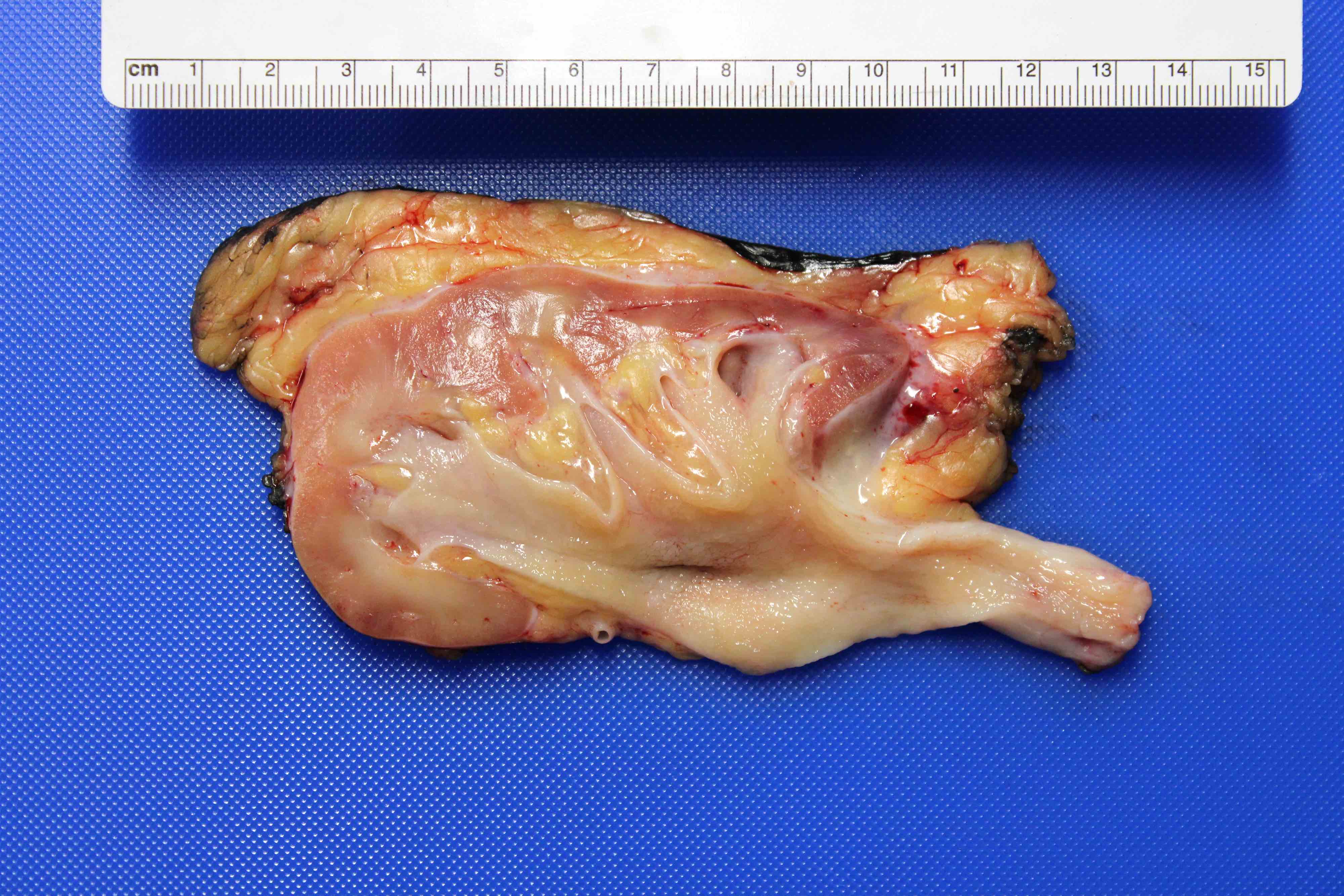
Perirenal and sinusoidal adipose tissue

Subcutaneous fibroadipose tissue
Microscopic (histologic) description
- Brown adipose tissue
- Lobules of adipocytes separated by fibrous septa
- Adipocytes are smaller (25 - 40 microns) than in white fat and are polygonal, with eccentrically located nucleus displaying fine indentations
- Multivacuolated cytoplasm results from numerous small lipid droplets
- Cytoplasm is acidophilic and granular due to large number of mitochondria, multivacuolated and granular cytoplasm and central spherical nucleus
- Fibrous septae contain capillaries and nerves
- White adipose tissue
- Uniform large spherical cells up to 120 microns
- Small lipid droplets fuse and form a single large intracytoplasmic droplet
- Nucleus is pushed and compressed to periphery of the cell by the lipid droplet, resulting in a thin crescent shape
- Adipocytes are clear on a routine H&E stained section because cytoplasmic fat dissolves during tissue processing
- Nucleus may have central vacuole or Lochkern (derived from German - loch: hole; kern: nucleus) (J Oral Maxillofac Pathol 2016;20:339)
- Very thin membranes are present between cells
Microscopic (histologic) images
Contributed by Norbert Sule, M.D., Ph.D.
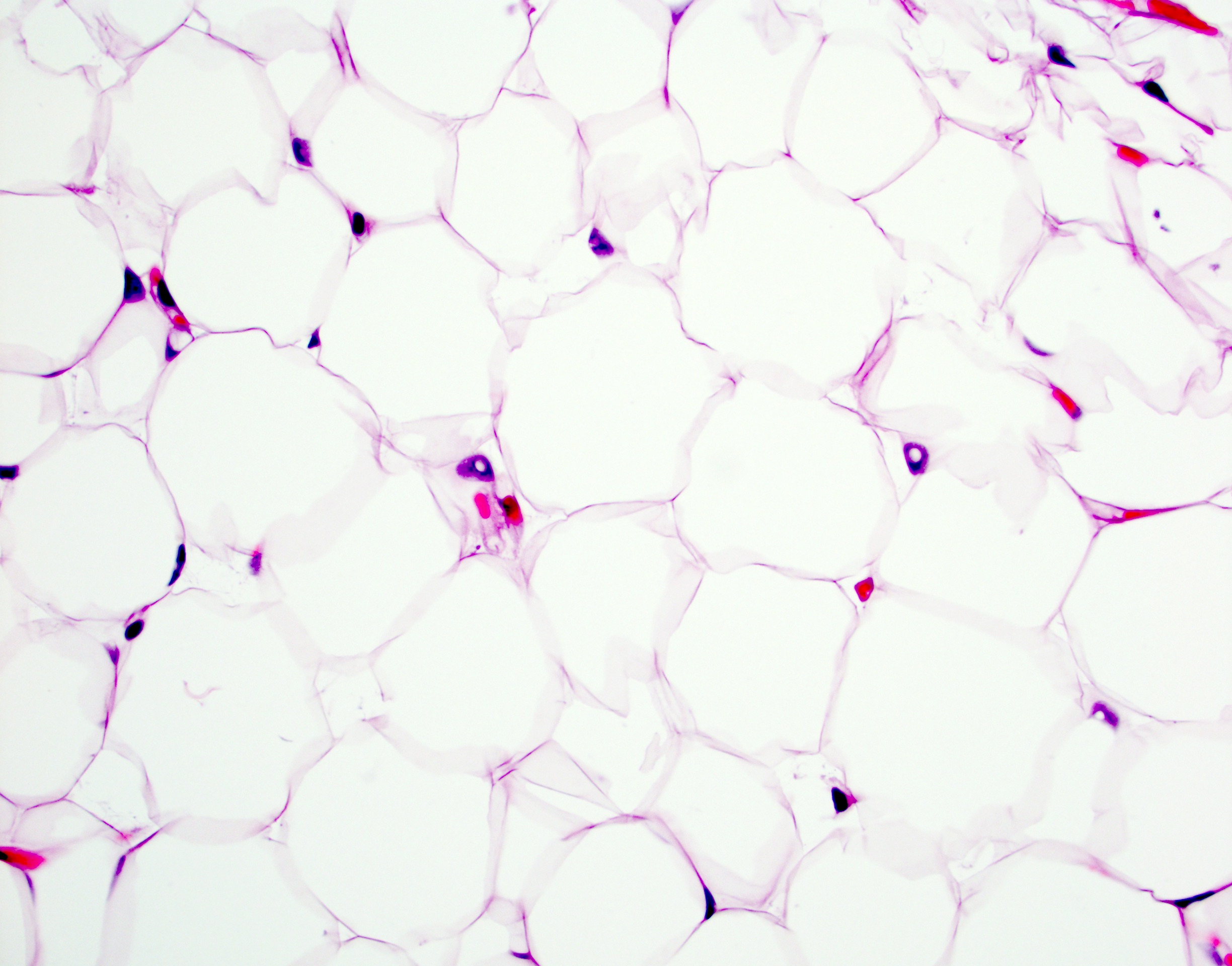
White adipose: Lochkern in nuceli
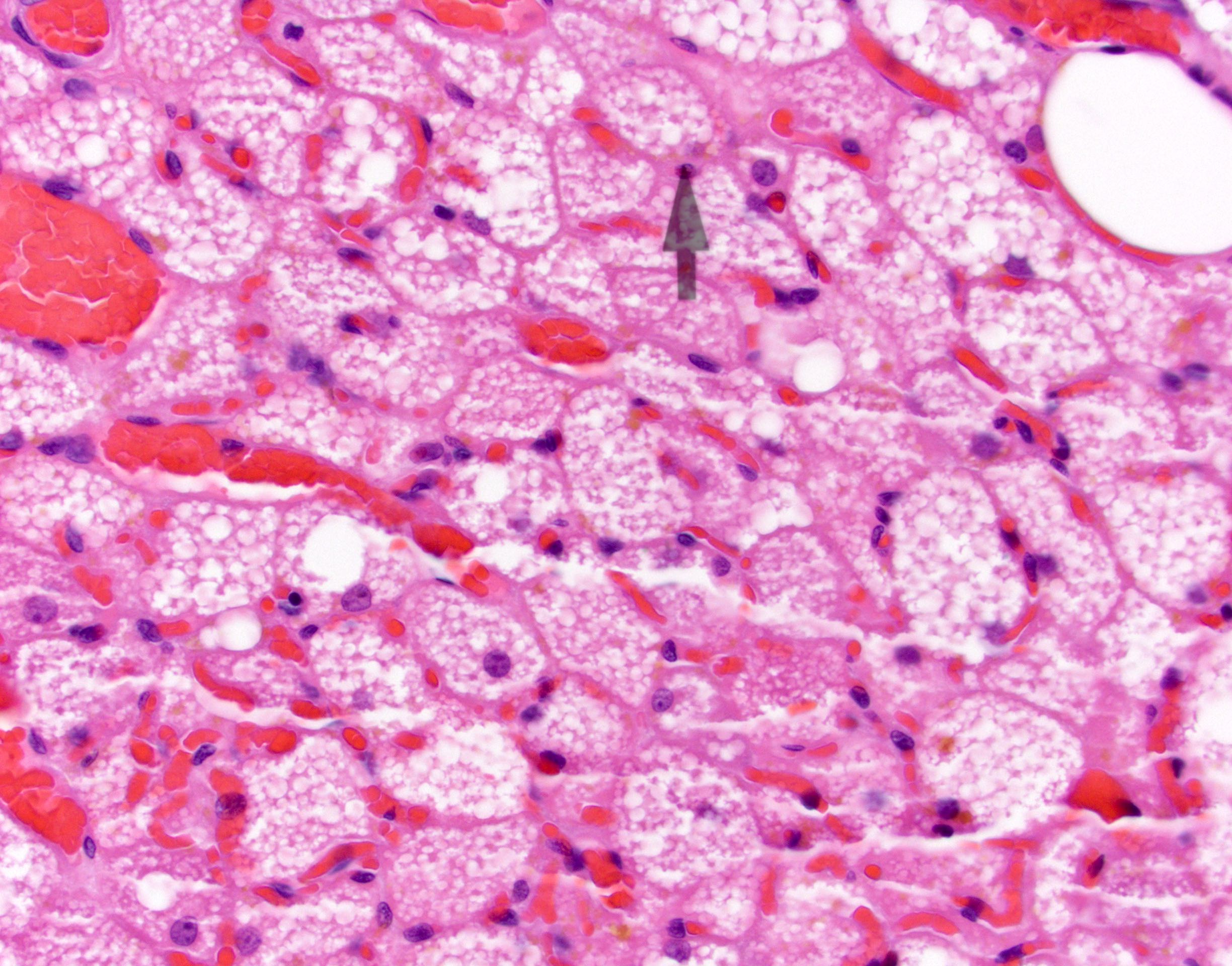
Brown fat
Positive stains
- Brown adipose tissue
- S100
- CD31 (Arch Pathol Lab Med 2006;130:480
- Also mitochondrial membrane uncoupling protein (J Histochem Cytochem 1993;41:759)
- White adipose tissue
- Vimentin (nonspecific), S100 (primarily for mature adipocytes), calretinin (also stains lipoma and liposarcoma) (Hum Pathol 2006;37:312)
- Oil red O and Sudan black stain neutral fat (must use frozen tissue because tissue processing removes fat)
- Oil red O also stains most carcinomas
- Nile blue sulfate stains neutral fat pink-red and stains fatty acids / phospholipids blue
Negative stains
- Brown adipose tissue
- White adipose tissue
- References: Young: Wheater's Functional Histology, 6th Edition, 2013, Mills: Histology for Pathologists, 3rd Edition, 2007
Electron microscopy description
- Brown adipose tissue
- Numerous mitochondria
- Numerous smaller lipid droplets
- White adipose tissue
- Preadipocytes: spindle cells with abundant endoplasmic reticulum and small spherical mitochondria
- Mature adipocyte:
- Large irregular central lipid droplet with multiple minute additional droplets at the periphery, representing ongoing fusion with the main fat droplet
- No membrane is present around the droplets
- Thin rim of residual cytoplasm contains cellular organelles, such as mitochondria and endoplasmic reticulum
- Nucleus is flattened against cytoplasmic membrane
- References: Young: Wheater's Functional Histology, 6th Edition, 2013, Mills: Histology for Pathologists, 3rd Edition, 2007
Additional references
Board review style question #1
What is the approximate size of a white fat cell?
- 0.1 micrometers
- 1 micrometer
- 10 micrometers
- 100 micrometers
Board review style answer #1
D. 100 micrometers. White adipose tissue cells are one of the largest cells in the body, measuring approximately 100 micrometers.
Comment Here
Reference: Histology-brown and white adipose
Comment Here
Reference: Histology-brown and white adipose
Board review style question #2
Which of the following is a characteristic feature of brown fat tissue?
- Largest fat depot in the body
- Multilocular vacuolization in the cytoplasm
- Nucleus is pushed and compressed against the cytoplasmic membrane
- Primary role is energy storage
Board review style answer #2
B. Multilocular vacuolization in the cytoplasm. Brown fat represents only 2 - 3% of body fat. The stored fat is present in multiple small droplets within the cytoplasm. The nucleus maintains its round shape and is not compressed against the cytoplasmic membrane. It has a primary role of thermoregulation.
Comment Here
Reference: Histology-brown and white adipose
Comment Here
Reference: Histology-brown and white adipose
