
Stomach
Carcinoma
Hereditary diffuse gastric cancer
Deputy Editor-in-Chief: Raul S. Gonzalez, M.D.

Last author update: 16 April 2020
Last staff update: 26 May 2021
Copyright: 2020, PathologyOutlines.com, Inc.
PubMed Search: Hereditary diffuse gastric cancer[TI] stomach pathology

Table of Contents
Definition / general | Essential features | Terminology | Epidemiology | Sites | Pathophysiology | Etiology | Clinical features | Radiology description | Case reports | Treatment | Gross description | Gross images | Microscopic (histologic) description | Microscopic (histologic) images | Positive stains | Negative stains | Molecular / cytogenetics description | Sample pathology report | Differential diagnosis | Additional references | Board review style question #1 | Board review style answer #1 | Board review style question #2 | Board review style answer #2Cite this page: Nowak K, Chetty R. Hereditary diffuse gastric cancer. PathologyOutlines.com website. https://www.pathologyoutlines.com/topic/stomachhdgc.html. Accessed May 14th, 2024.
Definition / general
- Autosomal dominant associated cancer syndrome
- Associated with diffuse gastric and invasive lobular breast cancers
- Caused by inactivating germline mutations in CDH1 (E-cadherin)
Essential features
- Autosomal dominant associated cancer syndrome
- Caused by inactivating germline mutations in CDH1 (E-cadherin)
- Associated with poorly cohesive carcinoma (signet ring carcinoma)
- Precursor lesions include signet ring carcinoma in situ and signet ring cells with pagetoid spread (pTis)
- Loss of membranous E-cadherin immunohistochemical staining
- Associated risk of developing lobular breast cancer
Terminology
- Older terms include: E-cadherin associated hereditary gastric cancer, familial diffuse gastric cancer, hereditary diffuse gastric adenocarcinoma
Epidemiology
- 10% of all gastric cancers demonstrate familial clustering (Int J Cancer 2000;87:133)
- 1 - 3% of gastric cancers that display familial clustering are hereditary (Best Pract Res Clin Gastroenterol 2009;23:147)
- 40% of hereditary gastric cancers are HDGC (Best Pract Res Clin Gastroenterol 2009;23:147)
Sites
- No predilection for specific area of stomach
Pathophysiology
- Inactivating mutation of second CDH1 allele
- CDH1 promoter methylation = most common cause of second CDH1 allele inactivation (Gastroenterology 2009;136:2137, Cancer Res 2009;69:2050, J Clin Oncol 2013;31:868)
- Loss of heterozygosity (LOH) = associated with lymph node metastasis (Gastroenterology 2009;136:2137, Cancer Res 2009;69:2050, J Clin Oncol 2013;31:868)
Etiology
- CDH1 germline mutation
Clinical features
- Variable presentation
- 40% lifetime risk of developing lobular breast cancer (JAMA Oncol 2015;1:23)
- Risk of developing gastric cancer by the age of 80 years: ~ 70% in men and ~ 56% in women (JAMA Oncol 2015;1:23)
Radiology description
- Range of findings (Cancer Imaging 2013;13:212):
- None
- Thickened gastric wall
Case reports
- 32 and 36 year old women with CDH1 mutations (Oncol Lett 2017;14:1671)
- 58 year old man with history of gastric signet ring adenocarcinoma (World J Clin Cases 2018;6:1)
- 62 year old woman with synchronous multiple primary gastrointestinal cancers with CDH1 mutations (World J Clin Cases 2019;7:1703)
Treatment
- Prophylactic total gastrectomy
Gross description
- Range:
- No gross lesions identified
- Linitis plastica
- Multiple polyps
- Summary of grossing a total gastrectomy specimen:
- Open the specimen along the greater curvature
- Ink margins
- Record all measurements
- Pin and allow to fix overnight
- Record any lesions and distance to margins
- Current recommendations include submitting the prophylactic gastrectomy specimen in toto (J Med Genet 2015;52:361)
- Take pictures in order to map out the specimen as to where sections came and where lesions occur
Gross images
Images hosted on other servers:

Total gastrectomy specimen
Microscopic (histologic) description
- Signet ring cell carcinoma in situ (pTis)
- Signet ring cells within basal membrane
- Signet ring cells with pagetoid spread (pTis)
- Second row of signet rings cells beneath normal epithelium in a gastric gland within the basal membrane
- Intramucosal signet ring carcinoma (pT1a)
- Signet ring cells restricted to the lamina propria
- Advanced HGDC (> pT1a)
- Poorly cohesive carcinoma with signet ring cells
- NB
- If no foci of signet ring cell carcinoma or in situ component is identified in a prophylactic total gastrectomy specimen , it should not be reported as negative for carcinoma, but as 'no carcinoma found in xx% of mucosa examined' (J Med Genet 2015;52:361)
Microscopic (histologic) images
Contributed by Runjan Chetty, M.B.B.Ch., Ph.D. and Altaf Taher, M.B.B.S., M.D.
Signet ring carcinoma in situ
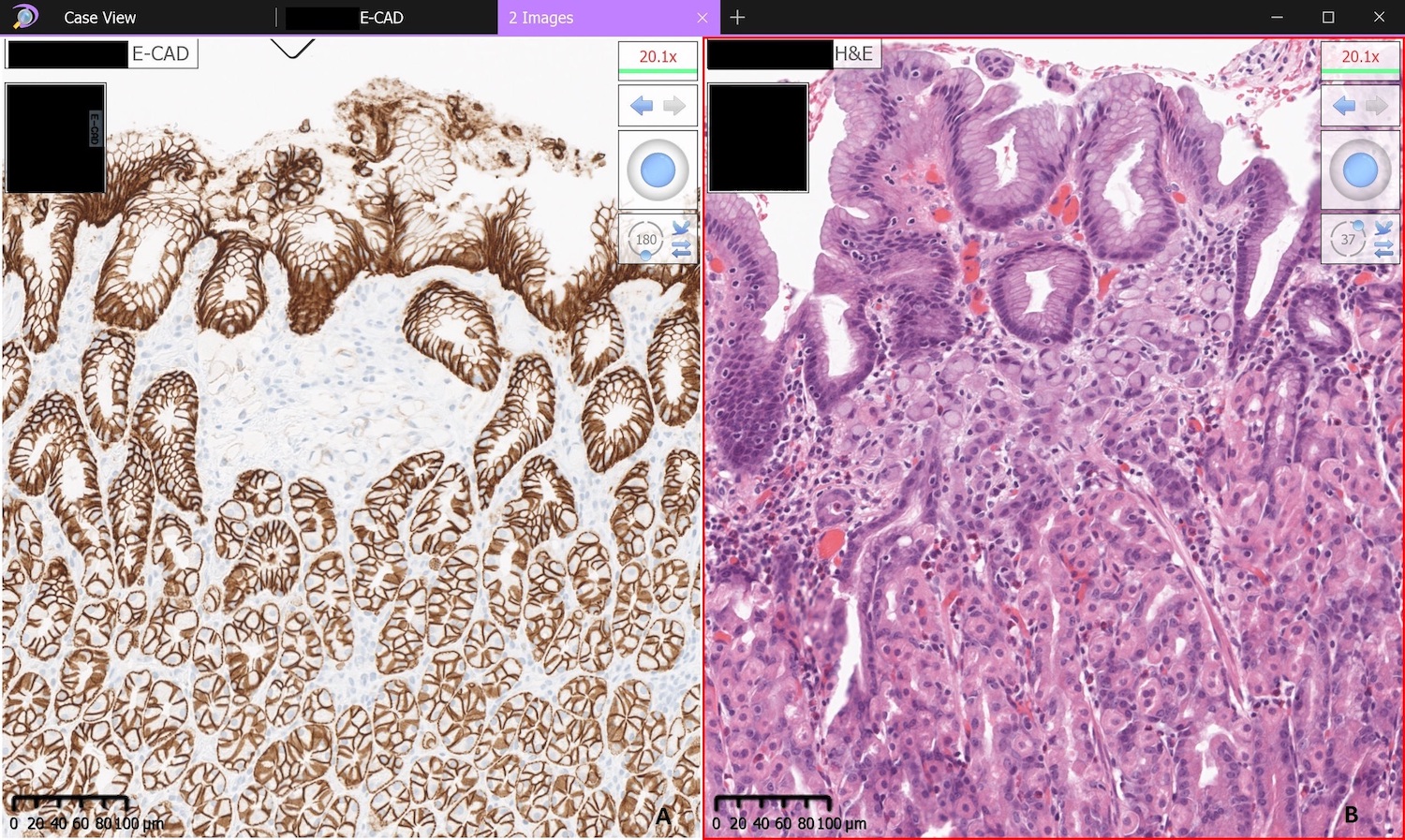
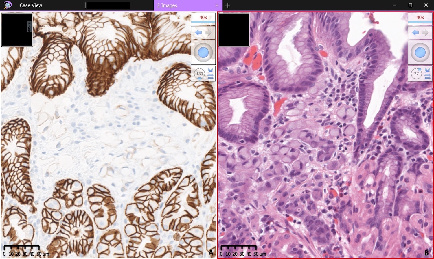
Loss of membranous E-cadherin staining
Contributed by Raul S. Gonzalez, M.D.
Signet ring carcinoma in situ
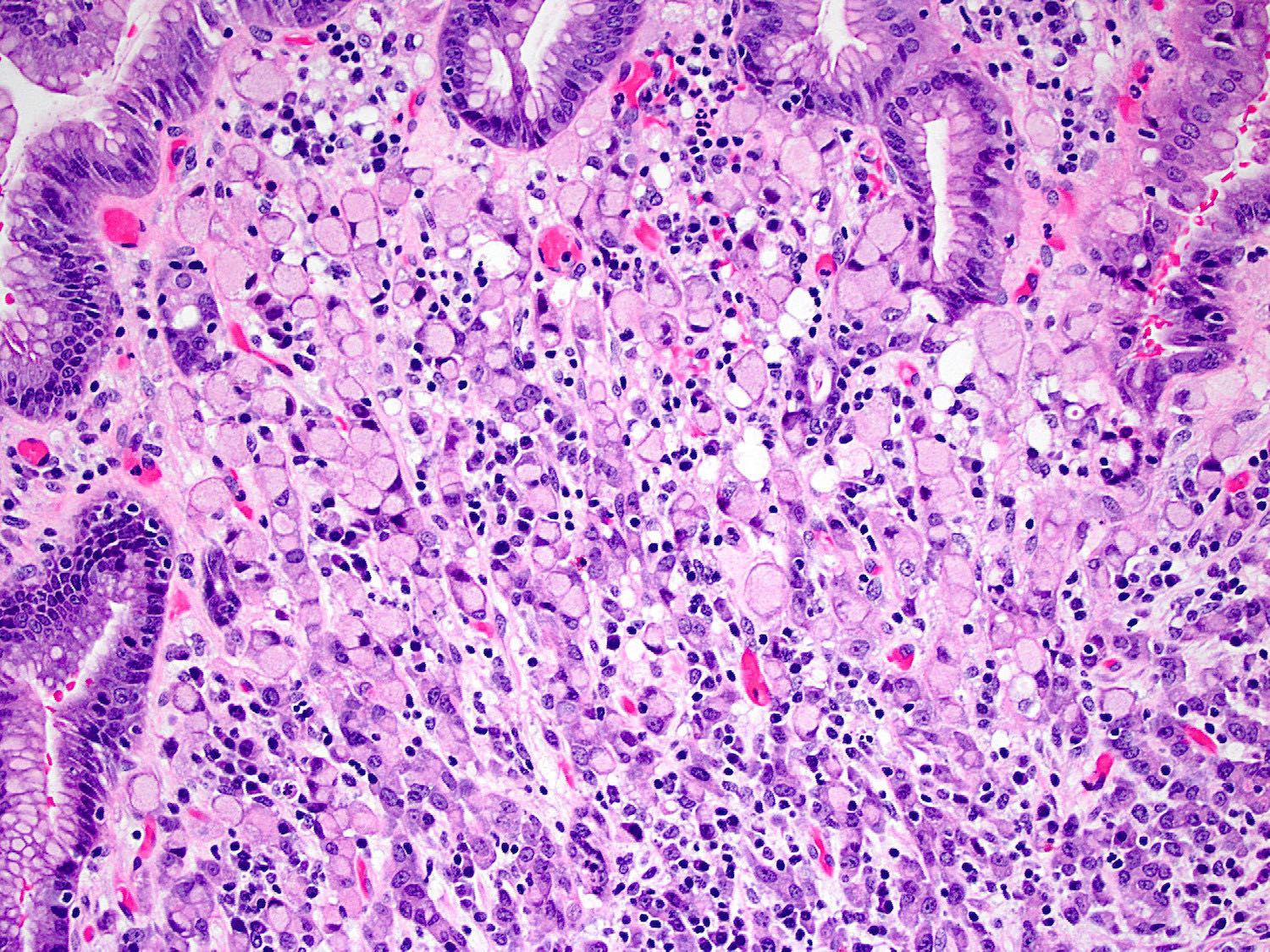
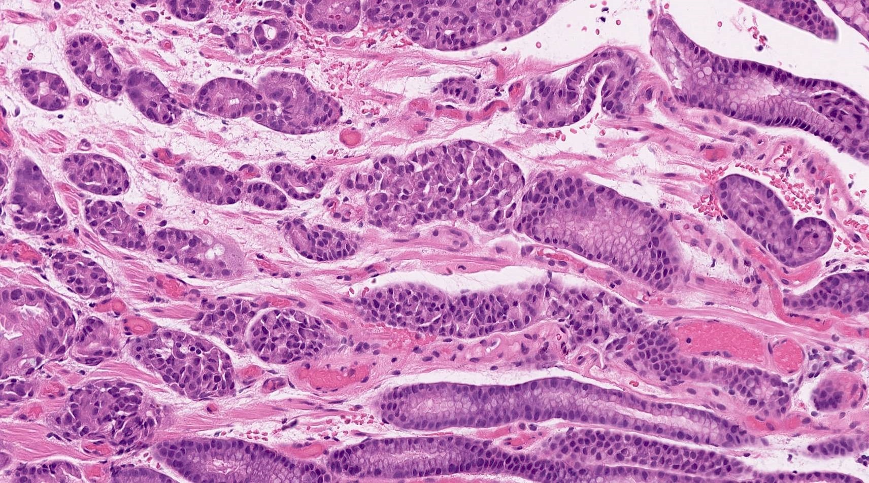
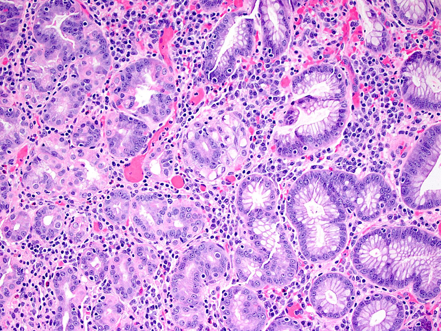
Obvious poorly cohesive carcinoma
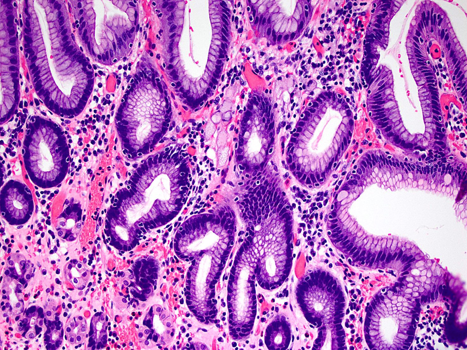
Subtle poorly cohesive carcinoma
Positive stains
- Pancytokeratin (e.g., AE1 / AE3)
- Mucicarmine
- Ki67 overexpression = more aggressive phenotype (Adv Exp Med Biol 2016;908:371, Cancer Res 2007;67:2480)
- Aberrant p53 expression = more aggressive phenotype (Adv Exp Med Biol 2016;908:371)
- Aberrant p16 expression = more aggressive phenotype (Hum Pathol 2018;74:64)
Negative stains
- Loss or reduced membranous staining of E-cadherin (J Pathol 2008;216:295, Gastroenterology 2009;136:2137)
Molecular / cytogenetics description
- CDH1 autosomal dominant mutation (Gastroenterology 2009;136:2137, Cancer Res 2009;69:2050, J Clin Oncol 2013;31:868)
- Other germline mutations reported in HDGC:
- CTNNA1 has been reported in a minority of cases (JAMA Oncol 2015;1:23, J Pathol 2013;229:621, Lancet Gastroenterol Hepatol 2018;3:489)
- No evidence for any other common genes has been found by exome sequencing (Eur J Hum Genet 2017;25:1246, Lancet Gastroenterol Hepatol 2018;3:489)
- Patients that harbor germline mutations in PALB2, MSH2, RECQL5, ATM and BRCA2 have also been described to meet the clinical criteria for HDGC (Cancer Res 2009;69:2050, Gastroenterology 2009;136:2137, J Clin Oncol 2013;31:868)
Sample pathology report
- Stomach, total gastrectomy:
- Poorly cohesive carcinoma with signet ring features (see comment and synoptic report)
- Comment: A poorly cohesive carcinoma with signet ring features is seen arising in a background of signet ring cell carcinoma in situ. The tumor cells are diffusely positive for pankeratin and demonstrate a loss of membranous E-cadherin staining. In the appropriate clinical context, genetic counseling is recommended to exclude hereditary diffuse gastric cancer, which is associated with a germline mutation in CDH1.
Differential diagnosis
- Metastatic lobular carcinoma of the breast:
- ER, PR and GCDFP are specific for metastatic breast carcinomas (Arch Pathol Lab Med 2005;129:338)
- Proton pump inhibitor induced clear cell change:
- No loss / reduced E-cadherin staining
- Look for other features of proton inhibitor induced changes (i.e. cystically dilated glands)
- No atypia (i.e., no increased nuclear to cytoplasmic ratio)
- Pseudo signet ring cells:
- Reactive change
- No loss / reduced E-cadherin staining
- No atypia (ie. no increased nuclear to cytoplasmic ratio)
- Gastric xanthoma:
- Negative for pankeratin and positive for histiocytic markers (e.g., CD68)
Additional references
Board review style question #1
Which mutation is most commonly associated with hereditary diffuse gastric cancer?
- BRCA2
- CDH1
- NF1
- SMARCA4
Board review style answer #1
Board review style question #2
The following morphology is associated with which answer?
- Loss of cytoplasmic keratin staining
- Loss of membranous E-cadherin staining
- Overexpression of CD68
- Overexpression of DOG1
Board review style answer #2
