Breast
Fibrocystic changes
Apocrine metaplasia
Editor-in-Chief: Debra L. Zynger, M.D.


Last author update: 28 May 2020
Last staff update: 15 January 2025
Copyright: 2002-2025, PathologyOutlines.com, Inc.
PubMed Search: Apocrine metaplasia breast
Table of Contents
Definition / general | Essential features | Epidemiology | Sites | Etiology | Clinical features | Diagnosis | Radiology description | Prognostic factors | Case reports | Treatment | Gross description | Microscopic (histologic) description | Microscopic (histologic) images | Cytology description | Positive stains | Negative stains | Electron microscopy description | Molecular / cytogenetics description | Sample pathology report | Differential diagnosis | Practice question #1 | Practice answer #1 | Practice question #2 | Practice answer #2Cite this page: Diez-Freire CC, Masood S. Apocrine metaplasia. PathologyOutlines.com website. https://www.pathologyoutlines.com/topic/breastapocrinemetaplasia.html. Accessed August 21st, 2025.
Definition / general
- Is considered one of the most common metaplastic changes of the breast, characterized by enlarged epithelial cells with abundant granular eosinophilic cytoplasm that can show apical snouting (Schnitt: Biopsy Interpretation of the Breast, 2nd Edition, 2012)
Essential features
- Cells with abundant eosinophilic cytoplasm and apical snouts
- Nuclei are round with variable size, vesicular chromatin with prominent nucleoli
- Supranuclear vacuoles or eosinophilic granules may be present
Epidemiology
- Common in women > 25 years of age (J Clin Pathol 2007;60:1313)
- Occasionally can be found in younger woman
Sites
- Breast parenchyma ducts and lobules
Etiology
- Unknown; the relation to hormonal imbalances is not well established
- Appears to be part of the fibrocystic complex manifested in cysts or part of ordinary duct hyperplasia (Hoda: Rosen's Breast Pathology, 4th Edition, 2014)
Clinical features
- Grossly palpable cysts can be lined by apocrine epithelium but there are no specific clinical features per se due to the apocrine metaplasia itself
- Can be concurrent with both benign and malignant lesions (J Clin Pathol 2007;60:1313)
- Apocrine cysts and hyperplasia with apocrine metaplasia were more common in the breasts of American women in New York than in Japanese women in Tokyo (Hoda: Rosen's Breast Pathology, 4th Edition, 2014)
Diagnosis
- Histologic examination of tissue removed
Radiology description
- Mammographic features are nonspecific, although an enlarging lobular or micro lobulated mass may be noted
- Heterogeneous microcalcification clusters may sometimes represent apocrine metaplasia
- These findings can lead to needle core biopsy for definitive diagnosis (AJR Am J Roentgenol 2003;180:795)
Prognostic factors
- No relationship or minimal impact on the development of mammary carcinoma (Hoda: Rosen's Breast Pathology, 4th Edition, 2014)
- Florida apocrine metaplasia with atypia may coexist with apocrine carcinoma, although most patients with short term follow up remained well
Case reports
- 48 year old woman presented with a growing palpable mass in her right breast of 3 months' duration (J Korean Soc Radiol 2018;78:103)
Treatment
- Specific treatment is not indicated for proliferative lesions with apocrine metaplasia
- Surgical excision of apocrine cysts is not indicated unless there is bloody fluid, the cysts reform or there is atypia in the cytologic specimen
- Atypical apocrine metaplasia warrants a clinical evaluation comparable to that of women with other atypical proliferative lesions, yet the precancerous significance of this abnormality remains uncertain
Gross description
- No specific gross findings are noted
- Apocrine areas may show a brown color when unfixed (Hoda: Rosen's Breast Pathology, 4th Edition, 2014)
Microscopic (histologic) description
- Cells are enlarged with abundant eosinophilic cytoplasm and apical snouts
- Supranuclear vacuoles or eosinophilic granules may be present
- Nuclei are round with variable size, vesicular chromatin with prominent nucleoli
- Can be a single or multiple layer epithelium; even papillary configurations can be seen (papillary apocrine change)
- Apocrine cells can show a degree of nuclear variability, even in simple epithelium; this is partly a product of cross cutting of a large nucleus and partly an intrinsic property of apocrine epithelium
- Moderate degrees of nuclear variation and atypia are ignored unless there is necrosis or significant mitotic activity (J Clin Pathol 2007;60:1313)
- Cytologic atypia tends to be more severe in sclerosing adenosis
- Highly complex architecture with cytologic atypia should be categorized as atypical ductal hyperplasia or ductal carcinoma in situ
Microscopic (histologic) images
Contributed by Carlos C. Diez Freire, M.D.
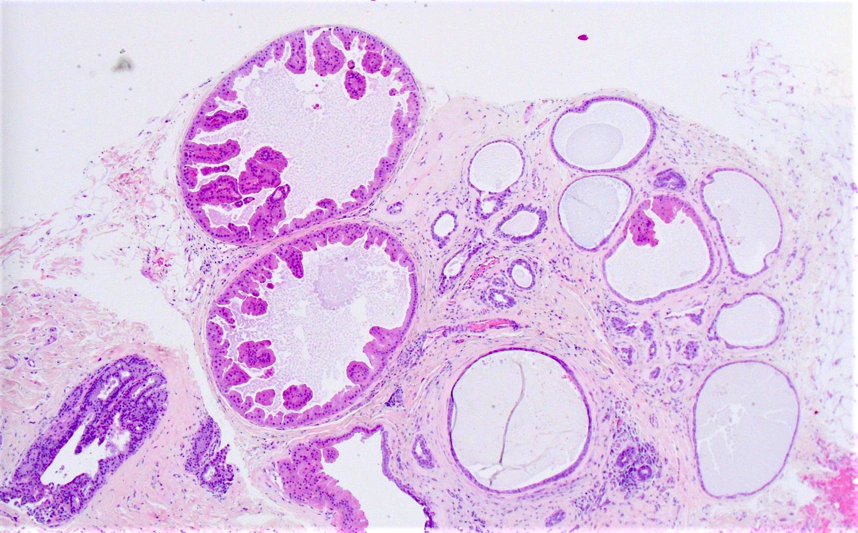
Dilated eosinophilic glands
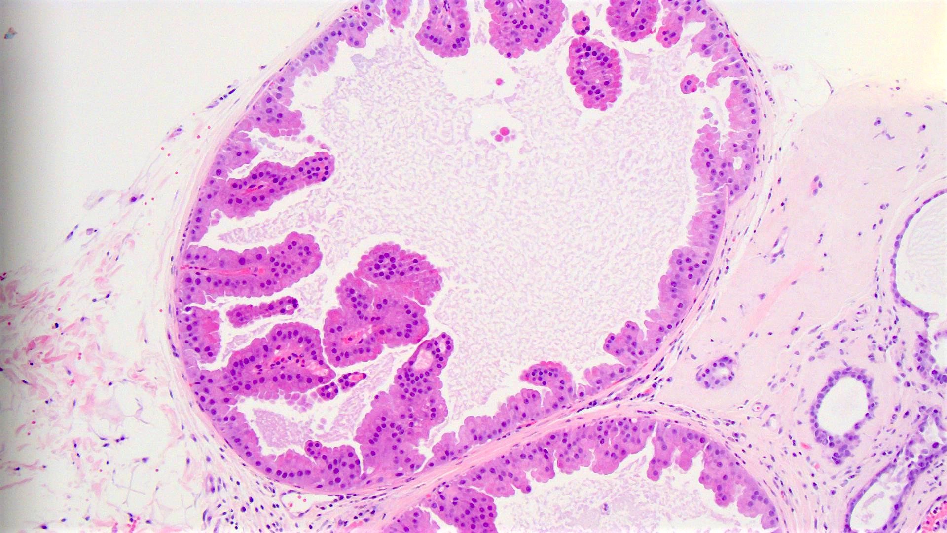
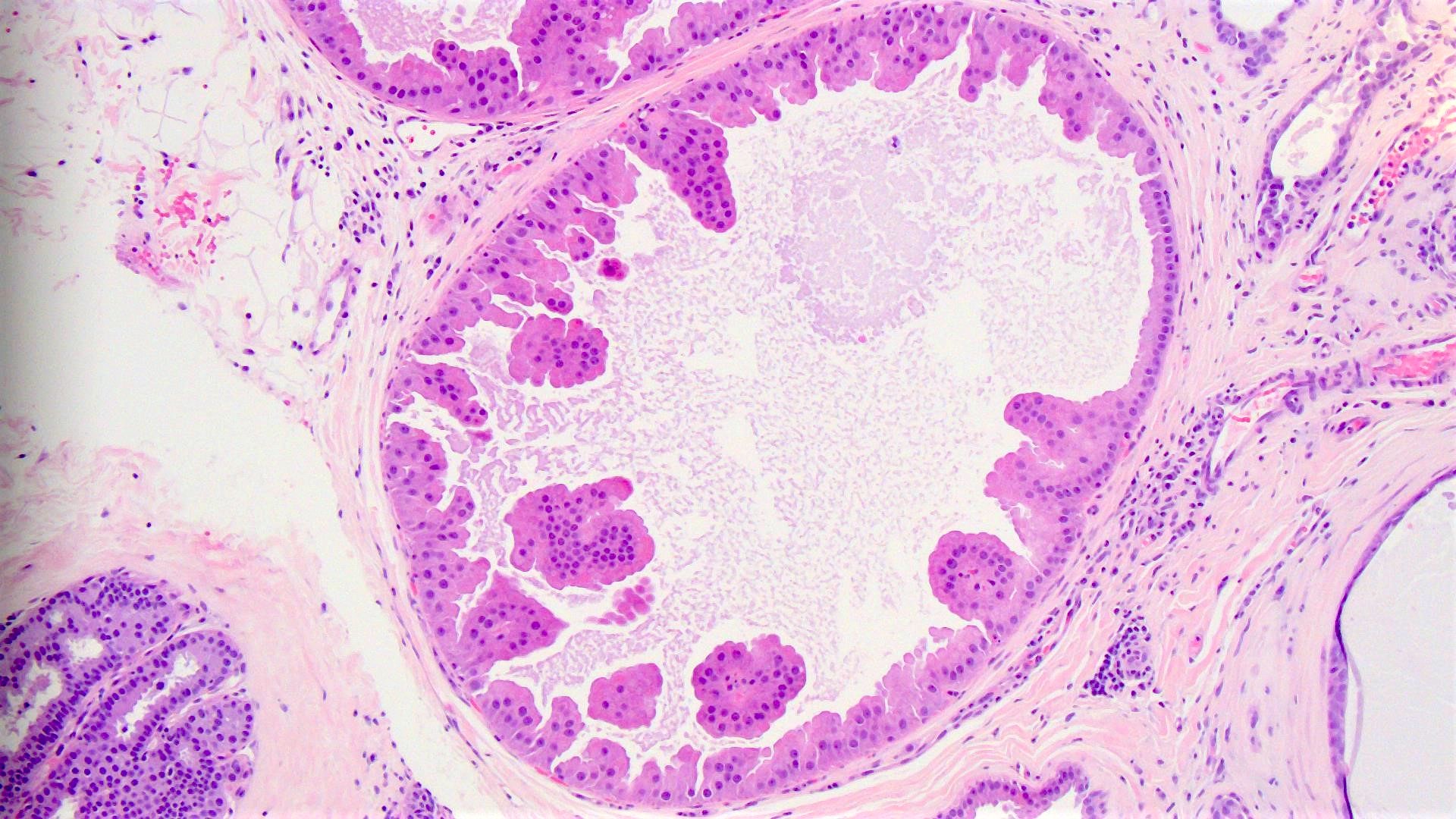
Papillary projections
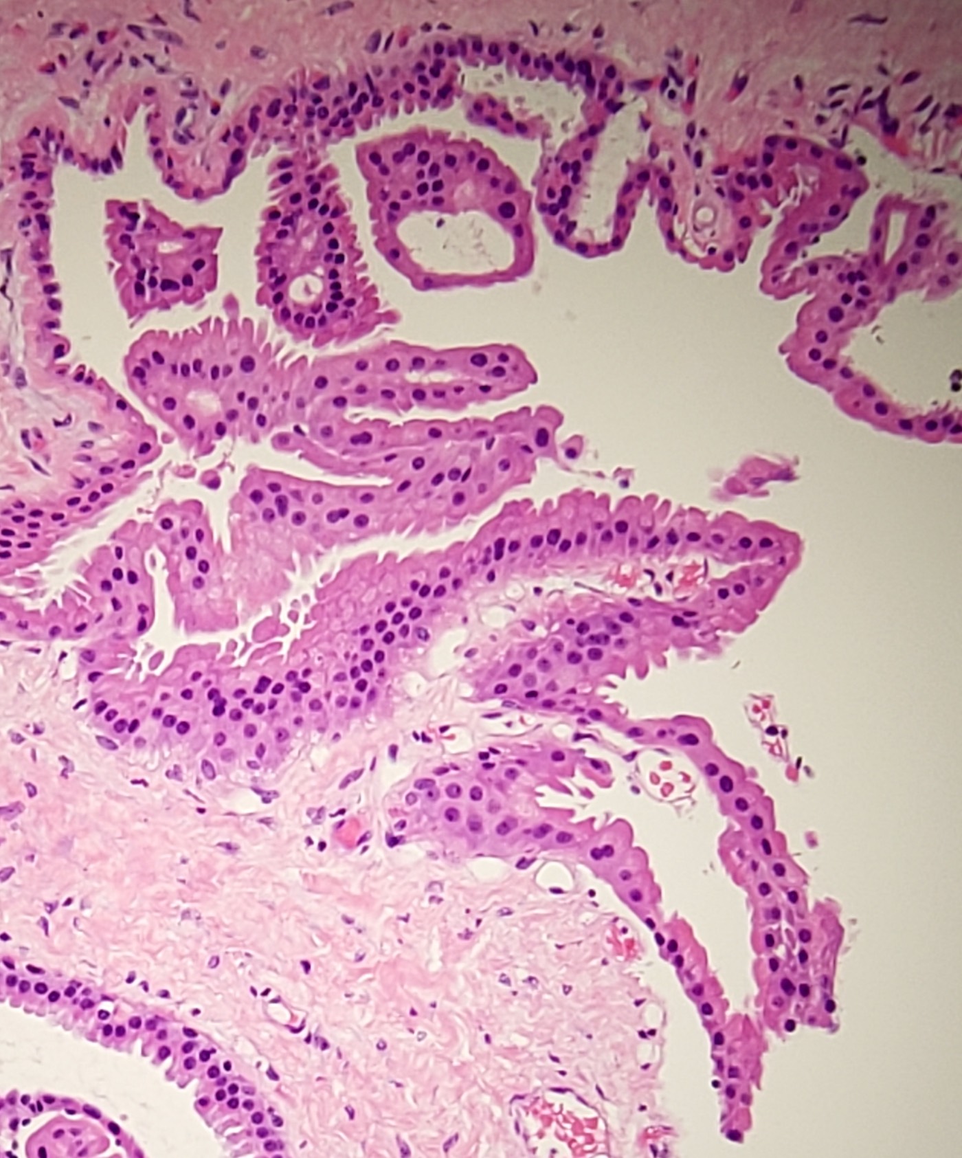
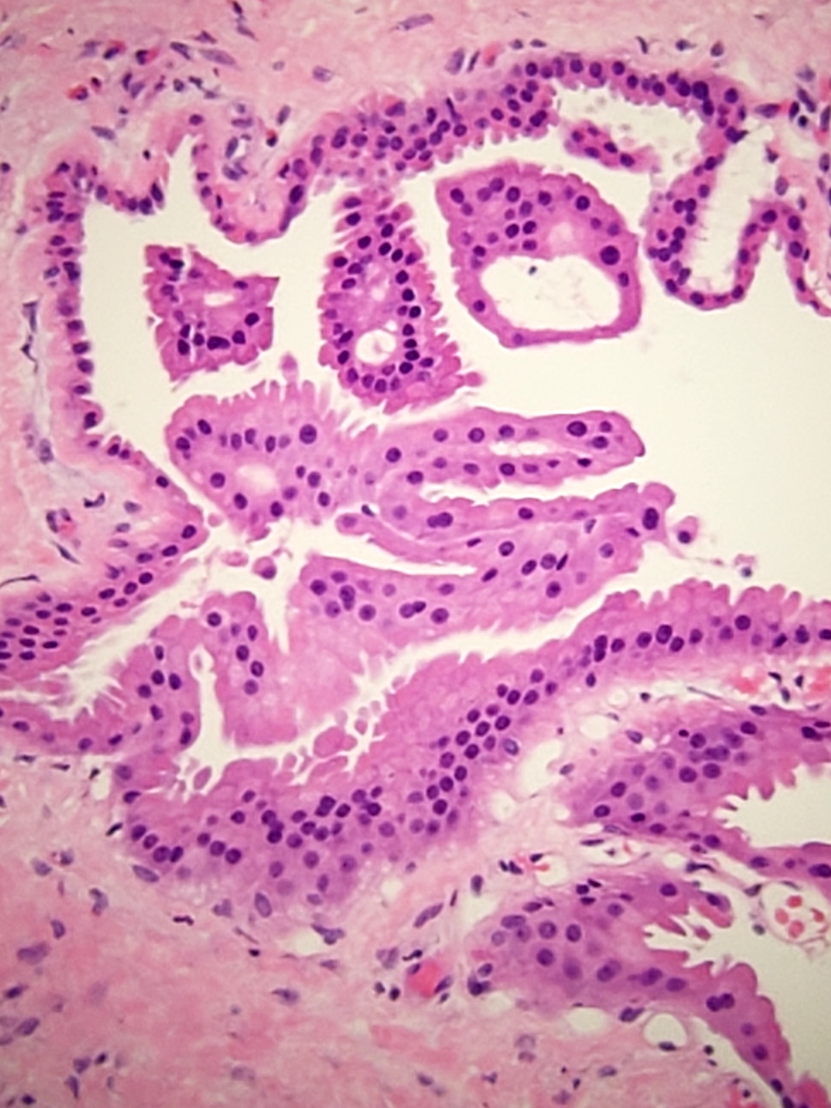
Abundant cytoplasm
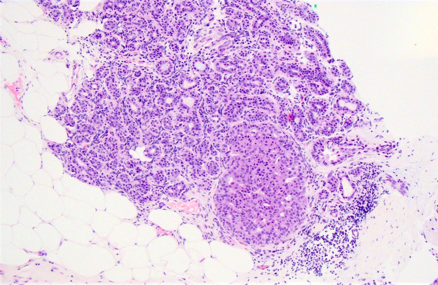
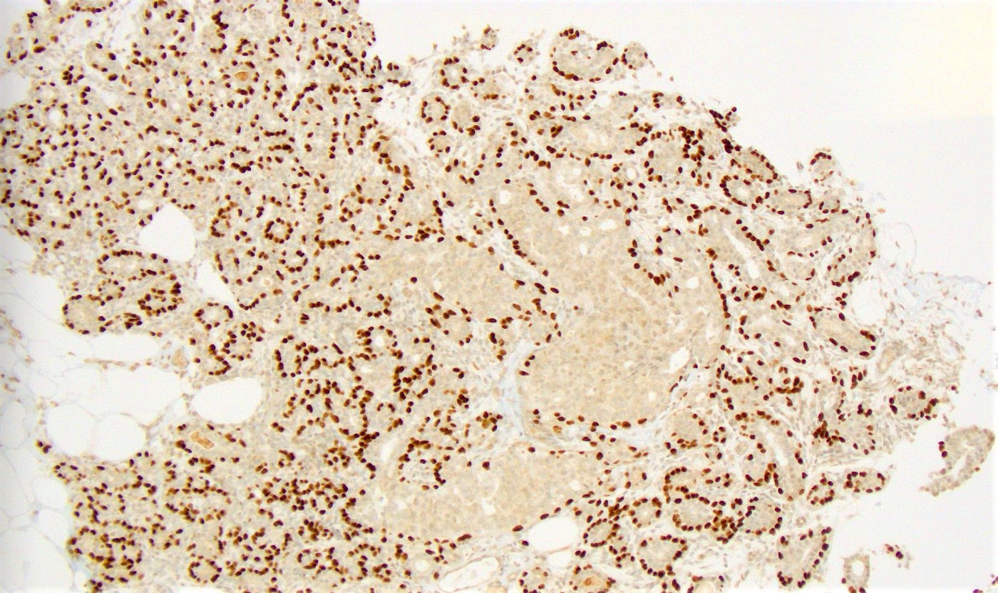
Early apocrine metaplasia and p63
Cytology description
- Nuclei of benign apocrine cells are eccentrically located with slight variation in size; the nuclear membrane is smooth with fine granular dispersed chromatin and a single prominent nucleolus
- Cytoplasm is abundant and dense with distinct cell borders with diffuse finely granular cytoplasm that stains pink / blue in PAP stains and purple / gray in Romanowsky stains
- Atypical apocrine cells can include nuclear enlargement, pleomorphism, prominent nucleoli and cellular dyshesion
- Features more concerning for carcinoma include crowded cell clusters, more pleomorphism, irregular nuclear borders, dark clumpy chromatin and increased nuclear to cytoplasmic ratio and necrosis
- Be cautious in a diagnosis of malignancy when atypical apocrine cells occur together with benign cells or if the lesion is clinically or mammographically benign (Demay: The Art & Science of Cytopathology, 2nd Edition, 2011)
Positive stains
- PAS (coarse glycolipid granules), GCDFP-15, androgen receptor (J Clin Pathol 2007;60:1313)
- EMA
- MYC nuclear staining but no amplification by FISH (Breast 2002;11:466)
Negative stains
- ER, PR
- BCL2 and rare staining for p53
- Low Ki67 but can be slightly higher in complex papillary cases with higher values associated with atypical apocrine cells (Mod Pathol 2000;13:13, Breast J 2009;15:475)
- p63 may show reduction or complete loss of myoepithelial cell layer (Am J Surg Pathol 2011;35:202)
Electron microscopy description
- Abundant endoplasmic reticulum, numerous mitochondria, intermediate filaments and secretory vesicles in the cytoplasm
Molecular / cytogenetics description
- Loss of heterozygosity in 53% (Am J Pathol 2000;157:323)
- Most common losses were at 1p, 17q and 22q and gains at 2q and 13q (Am J Pathol 2001;158:207)
Sample pathology report
- Left breast, 2 o’clock, 5 cm from nipple, ultrasound guided core biopsy:
- Papillary apocrine metaplasia and duct dilation
Differential diagnosis
- Atypical apocrine adenosis:
- Cytologic atypia with more than threefold variation in nuclear size with prominent multiple nucleoli (J Clin Pathol 2007;60:1313)
- Apocrine DCIS:
- Extensive proliferation, marked nuclear pleomorphism (J Clin Pathol 2007;60:1313)
- Architectural complexity such as well formed Roman bridges or rigid cribriform structures
Practice question #1
Apocrine metaplasia of the breast is characterized by
- Dilation of terminal ducts with periductal fibrosis and chronic inflammation
- Epithelial cells with abundant granular eosinophilic cytoplasm and prominent nucleoli
- Monotonous population of histiocytes with foamy cytoplasm distending the ducts
- Severely atypical and pleomorphic cells of mesenchymal origin
Practice answer #1
B. Apocrine metaplasia of the breast is characterized by enlarged epithelial cells with abundant granular eosinophilic cytoplasm that can show apical snouting, eccentric nuclei and prominent nucleoli.
Comment Here
Reference: Apocrine metaplasia of breast
Comment Here
Reference: Apocrine metaplasia of breast
Practice question #2
The histologic findings in the above breast images are an example of
- Apocrine metaplasia
- Intraductal papilloma
- Mammary duct ectasia
- Papillary ductal carcinoma in situ
Practice answer #2
A. Apocrine metaplasia. Cells are enlarged with abundant eosinophilic cytoplasm and apical snouts. Supranuclear vacuoles or eosinophilic granules may be present. Nuclei are round with variable size, vesicular chromatin with prominent nucleoli. Can be a single or multiple layer epithelium, even papillary configurations can be seen (papillary apocrine change).
Comment Here
Reference: Apocrine metaplasia of breast
Comment Here
Reference: Apocrine metaplasia of breast
