
Breast
Other benign tumors
Leiomyoma
Author: Reena Tomar, M.B.B.S., M.D.
Editorial Board Member: Julie M. Jorns, M.D.
Deputy Editor-in-Chief: Gary Tozbikian, M.D.

Last author update: 18 January 2024
Last staff update: 18 January 2024
Copyright: 2002-2024, PathologyOutlines.com, Inc.
PubMed Search: Breast leiomyoma

Table of Contents
Definition / general | Essential features | Epidemiology | Sites | Pathophysiology | Etiology | Clinical features | Diagnosis | Radiology description | Radiology images | Case reports | Treatment | Clinical images | Gross description | Gross images | Frozen section description | Frozen section images | Microscopic (histologic) description | Microscopic (histologic) images | Positive stains | Negative stains | Sample pathology report | Differential diagnosis | Board review style question #1 | Board review style answer #1 | Board review style question #2 | Board review style answer #2Cite this page: Tomar R. Leiomyoma. PathologyOutlines.com website. https://www.pathologyoutlines.com/topic/breastleiomyoma.html. Accessed May 6th, 2024.
Definition / general
- Benign mesenchymal tumor of smooth muscle, rare in breast (BMC Womens Health 2022;22:119, J Cutan Pathol 2019;46:343)
Essential features
- Benign smooth muscle tumor
- Spindle shaped cells with cigar shaped nuclei arranged in whorls and fascicles
- SMA, desmin, caldesmon positive
Epidemiology
- Middle aged women (BMC Womens Health 2022;22:119)
- F:M = 3:1 (Case Rep Surg 2013;2013:475215)
Sites
- Rare in breast (1% of breast neoplasms) (Eur J Breast Health 2017;13:156)
- Superficial (nipple areola complex) or deep (breast parenchyma)
- Subareolar area (Eur J Breast Health 2017;13:156)
- Nipple (Case Rep Surg 2013;2013:475215)
- More common in other sites such as uterus, gastrointestinal tract, skin and lower extremities (J Oral Maxillofac Pathol 2013;17:281)
Pathophysiology
- Arises from smooth muscles (J Cutan Pathol 2019;46:343)
- Hormonal association (J Oral Maxillofac Pathol 2013;17:281)
Etiology
- Trauma (J Med Case Rep 2013;7:49)
- Tamoxifen and antiobesity drug use associated with enlargement of breast leiomyoma (AJR Am J Roentgenol 2005;185:1595)
Clinical features
- Typically a firm, mobile lump but can be nonmobile (Breast J 2020;26:529)
- Intermittent pain (Case Rep Surg 2013;2013:475215, Breast J 2020;26:529)
- No nipple discharge (Case Rep Surg 2013;2013:475215)
Diagnosis
- Can be detected on mammography or ultrasound (AJR Am J Roentgenol 2005;185:1595, Korean J Radiol 2020;21:955)
- Excisional biopsy is diagnostic as well as therapeutic due to risk of local recurrence (Int J Sci Stud 2015;3:210, J Med Case Rep 2013;7:49)
Radiology description
- Ultrasound shows an oval, circumscribed hypoechoic mass with internal vascularity (Korean J Radiol 2020;21:955)
- MRI scan reveals an intense, homogeneous enhancement (Korean J Radiol 2020;21:955)
- Well defined lesion, hypoechoic on mammography (AJR Am J Roentgenol 2005;185:1595)
- No calcifications (Eur J Breast Health 2017;13:156)
Radiology images
Images hosted on other servers:

Well defined, circumscribed, hypoechoic mass
Case reports
- 31 year old woman with enlarging hard lesion in bilateral nipple (Case Rep Surg 2013;2013:475215)
- 40 year old woman with mass in right nipple (Breast J 2020;26:529)
- 43 year old woman with bilateral breast lump with atypical features and 46 year old woman with a 3 cm lesion in left breast (BMC Womens Health 2022;22:119)
- 44 year old woman with painless lump in right breast (Eur J Breast Health 2017;13:156)
- 47 year old woman with a 2 cm mass in sonography of left breast (AJR Am J Roentgenol 2005;185:1595)
- 59 year old woman for routine mammography (Radiol Bras 2016;49:343)
Treatment
- Excisional biopsy is therapeutic (J Med Case Rep 2013;7:49)
- Calcium channel blocker, alpha adrenergic blocker may be used to reduce pain (Case Rep Surg 2013;2013:475215)
Clinical images
Images hosted on other servers:

1 cm nodule (black arrow) on left areola

Left nipple (white arrow), nodule (black arrow)

Swollen left nipple compared with the right nipple
Gross description
- Circumscribed (Breast J 2020;26:529)
- 0.8 - 2.7 cm in size (Breast J 2020;26:529)
- Fleshy, pale, whitish (Eur J Breast Health 2017;13:156)
- Homogenous white cut surface (Breast J 2020;26:529)
Gross images
Images hosted on other servers:

Well circumscribed skin covered mass
Frozen section description
- Frozen section has been described to evaluate margins (Case Rep Surg 2013;2013:475215)
Frozen section images
Contributed by Reena Tomar, M.B.B.S., M.D.
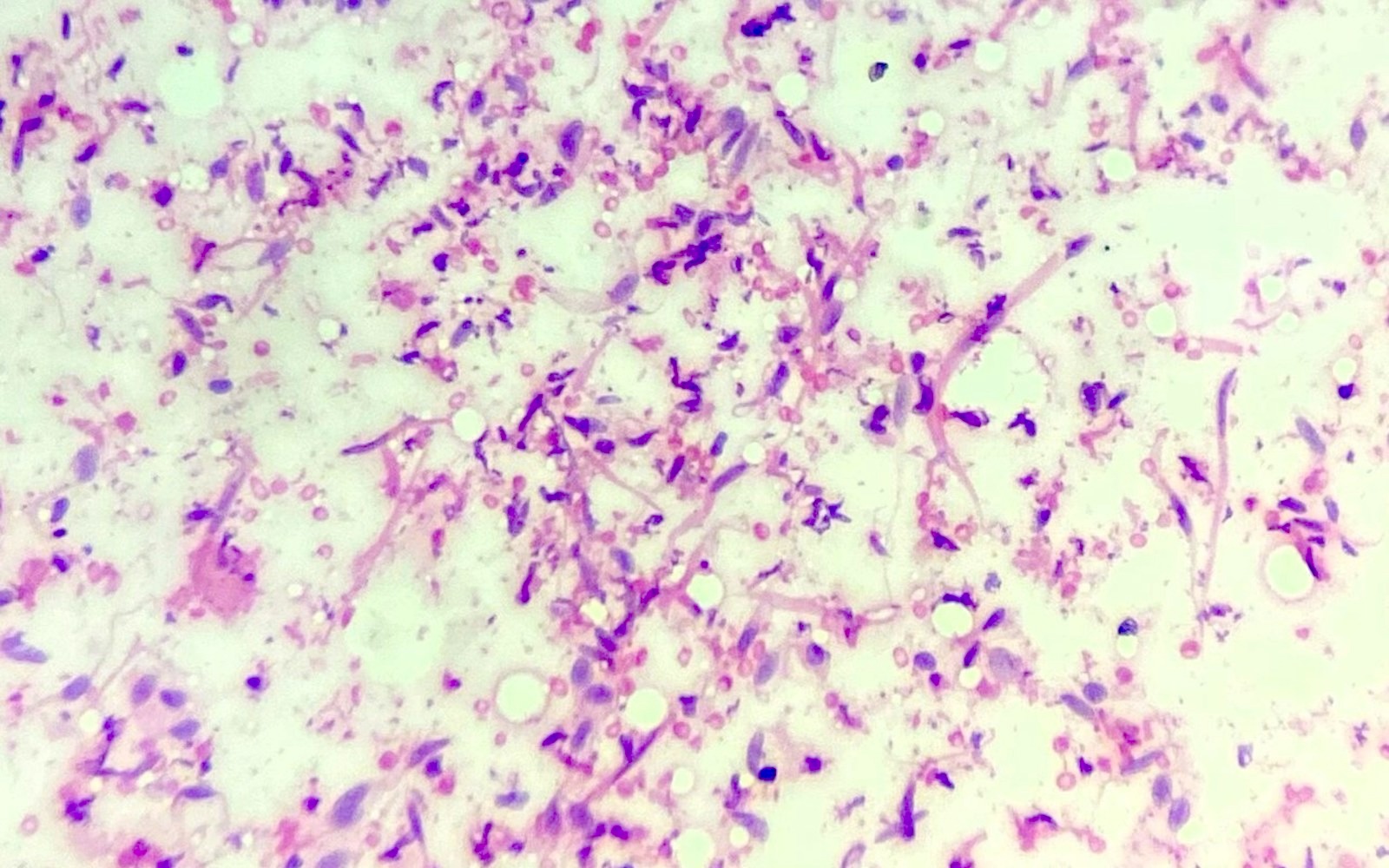
Scrapes
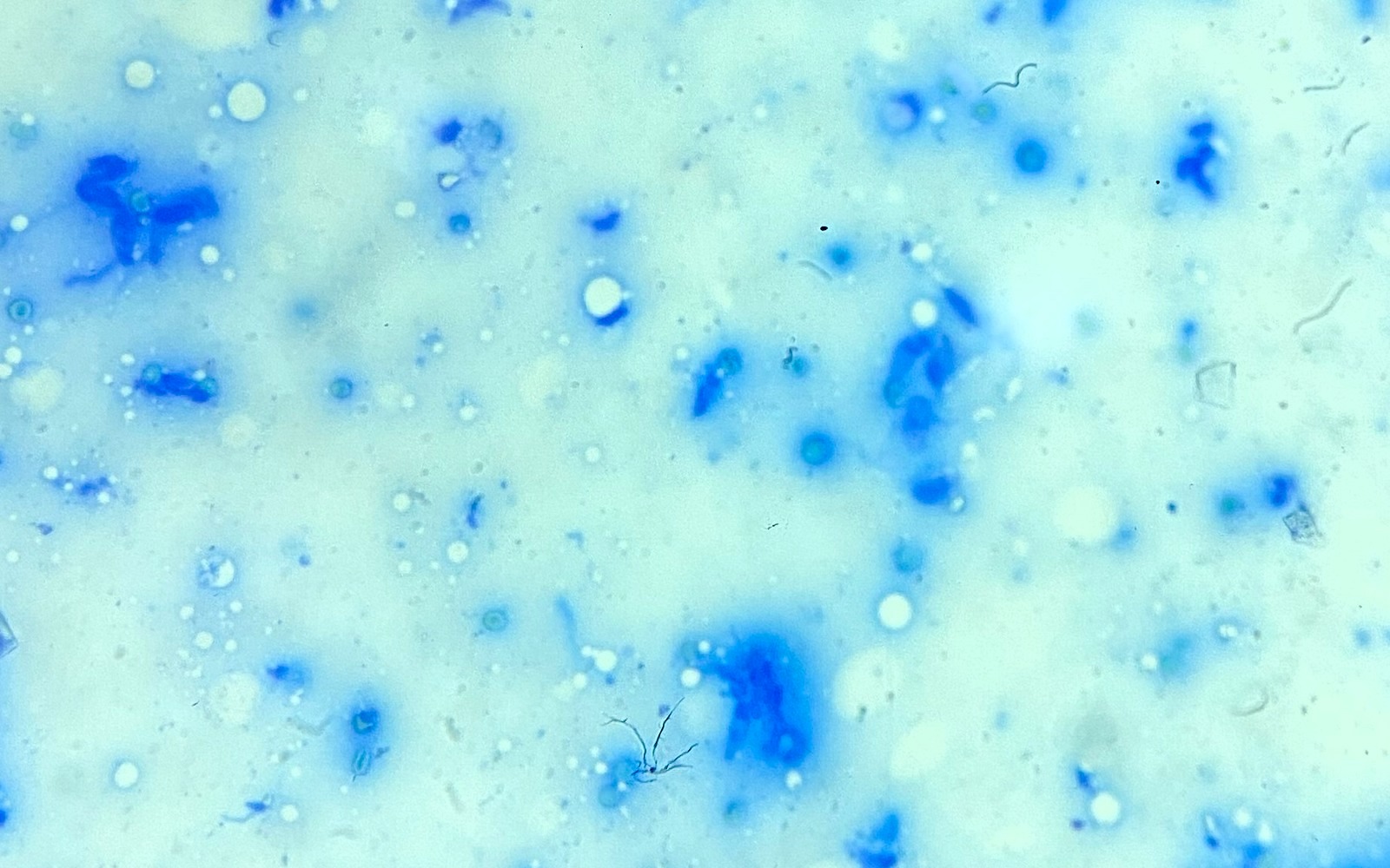
Scrapes (toluidine blue)
Microscopic (histologic) description
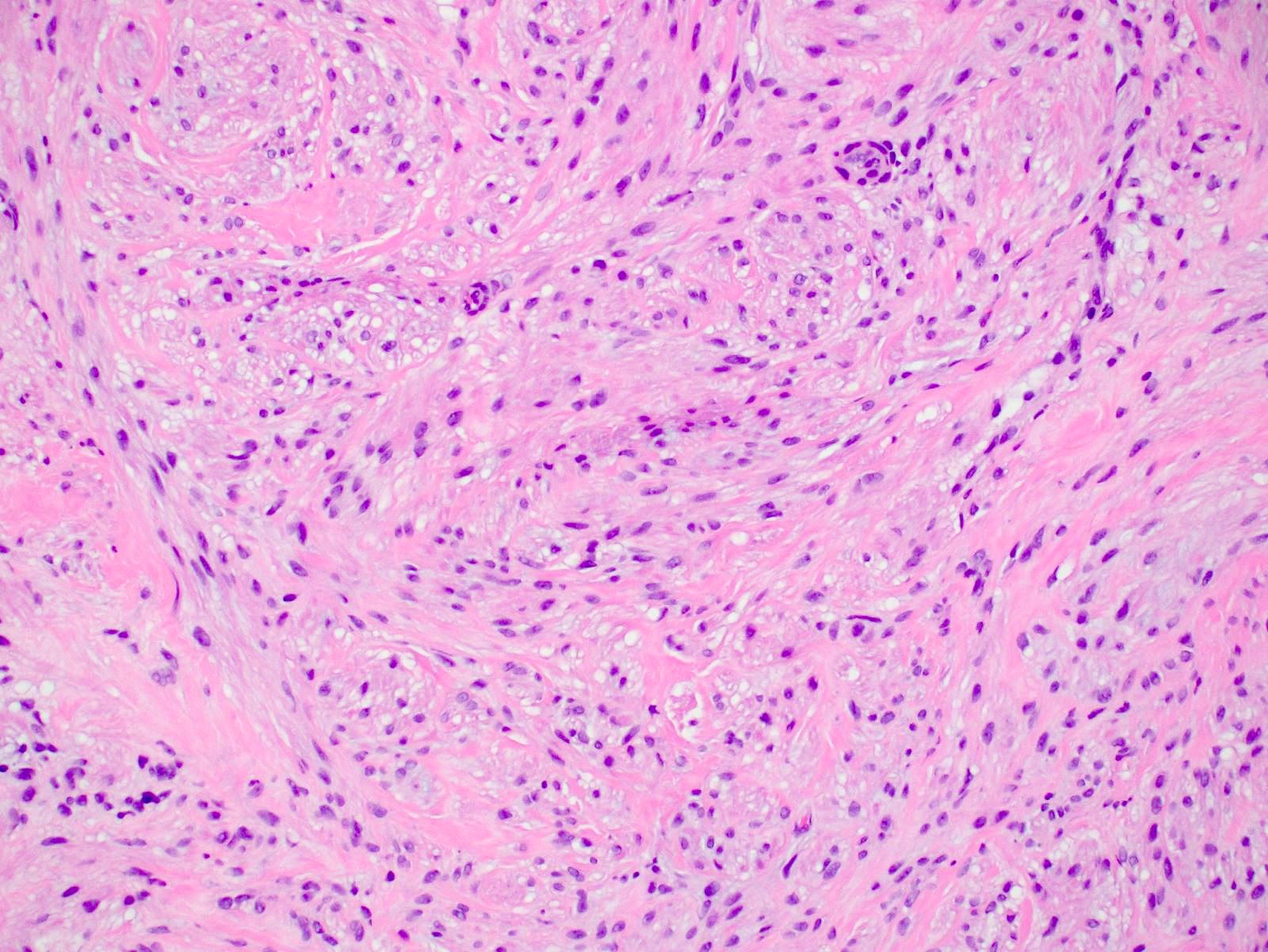
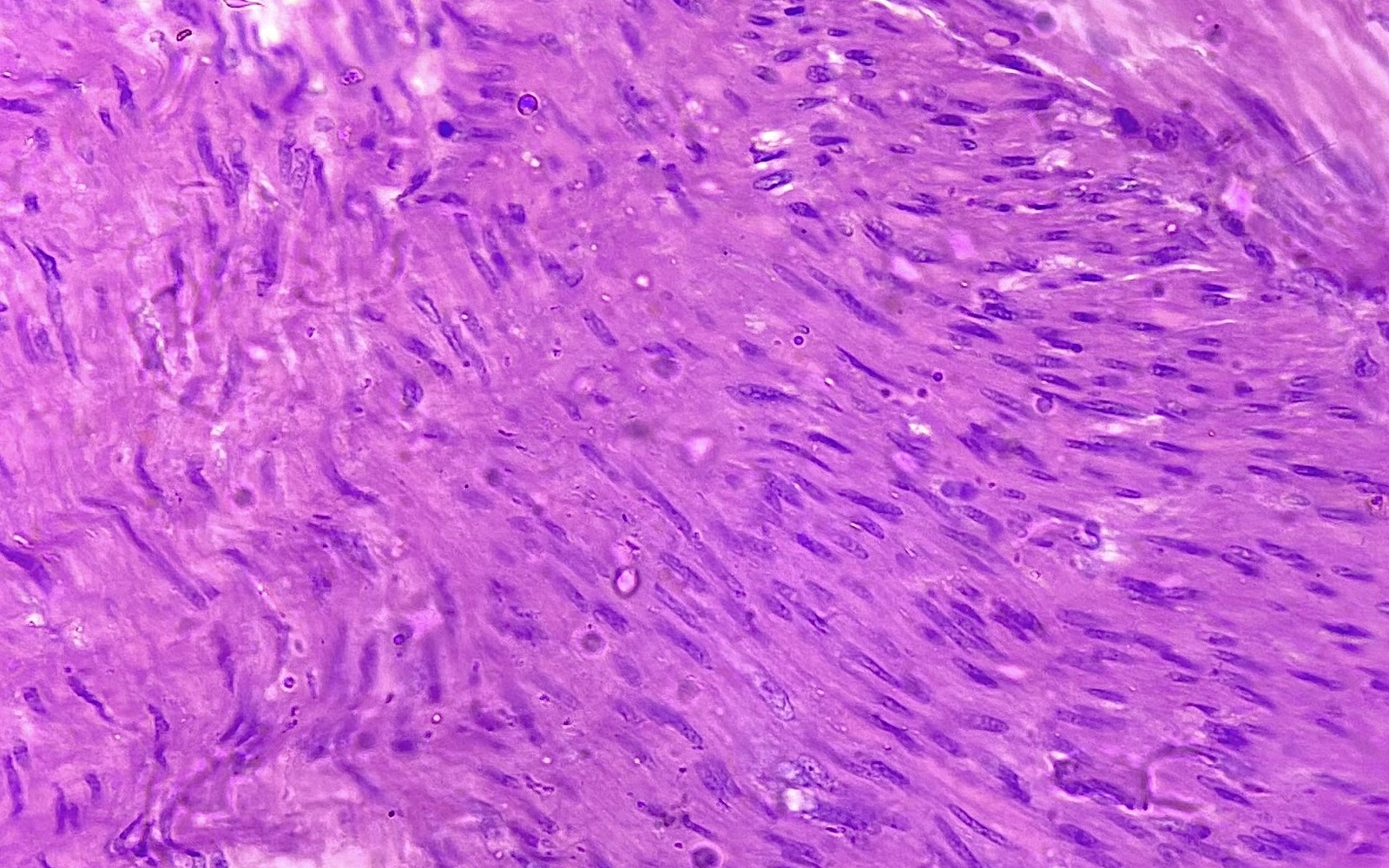
- Proliferation of monotonous spindle cells (BMC Womens Health 2022;22:119)
- Spindle cells are relatively uniform in size and shape with cigar shaped nuclei (Breast J 2020;26:529)
- Interlacing bundles of smooth muscle cells separated by a small amount of well vascularized connective tissue (J Med Case Rep 2013;7:49)
- Circumscribed, overlying epidermis is unremarkable (Breast J 2020;26:529)
- Nipple leiomyoma shows dermal lesion, glandular components are absent
- No mitosis, no necrosis, no atypia (BMC Womens Health 2022;22:119)
- Atypical leiomyoma has features of nuclear atypia and up to 3 mitosis/10 high power fields (BMC Womens Health 2022;22:119)
Microscopic (histologic) images
Contributed by Reena Tomar, M.B.B.S., M.D., Mark R. Wick, M.D. and Julie M. Jorns, M.D.

Dermal circumscribed leiomyoma
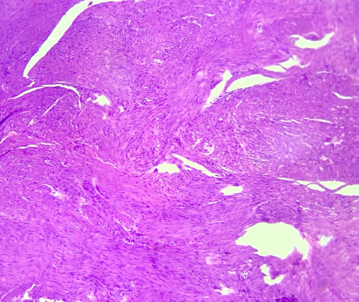
Spindle shaped cells
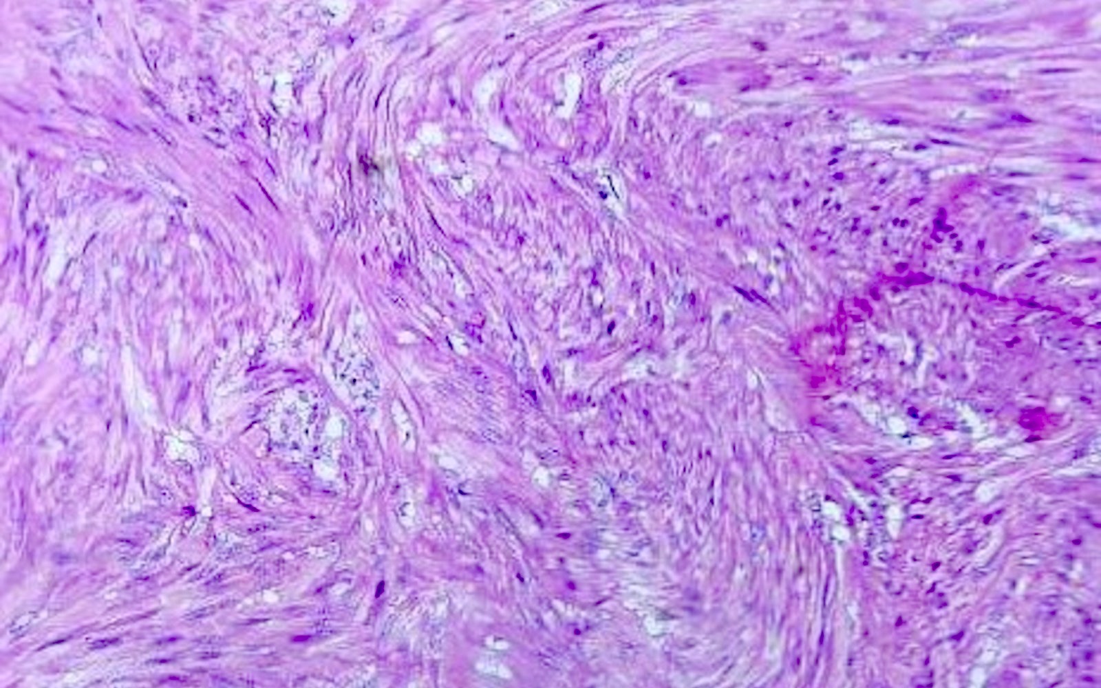
Fascicles
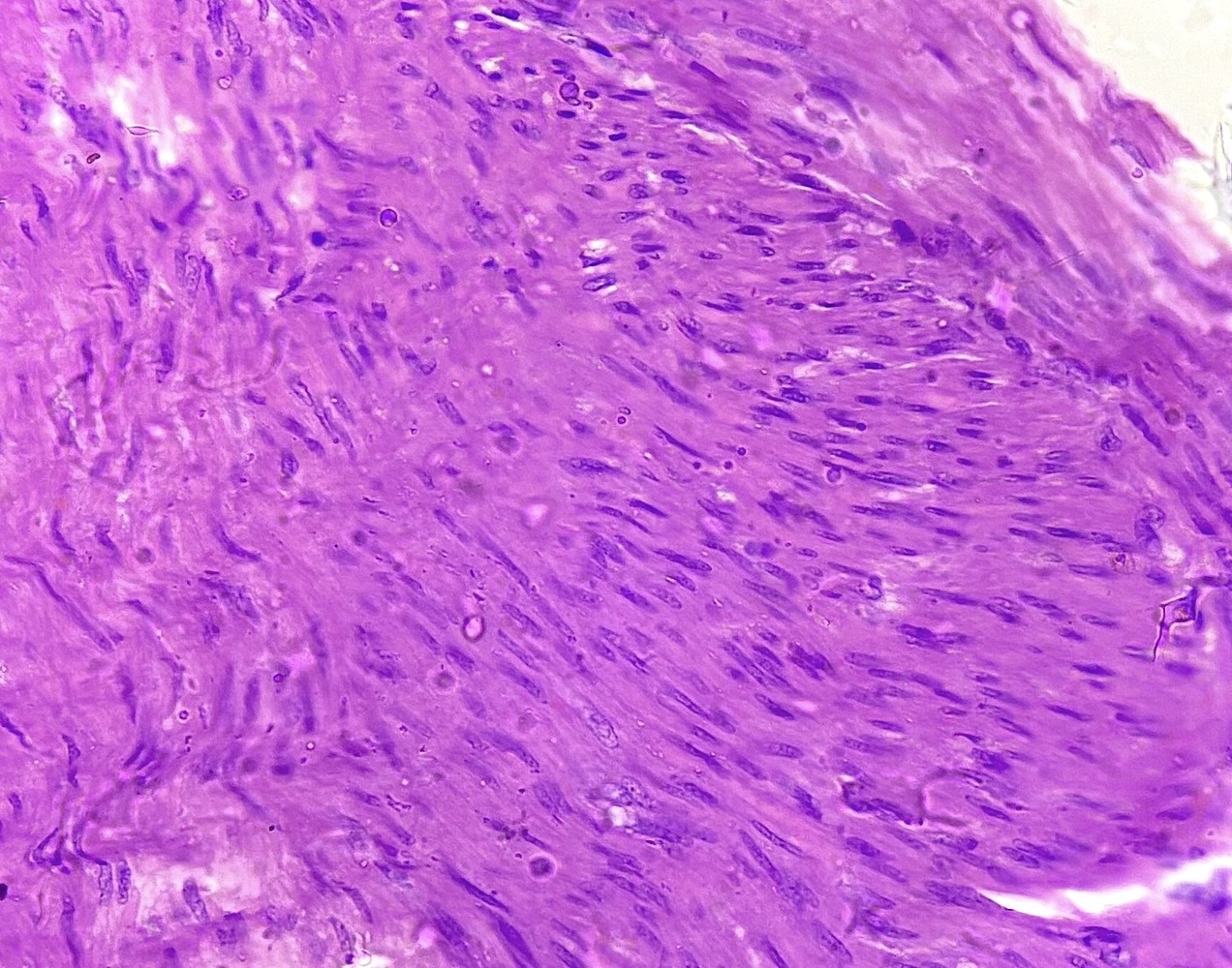
Whorls and fascicles
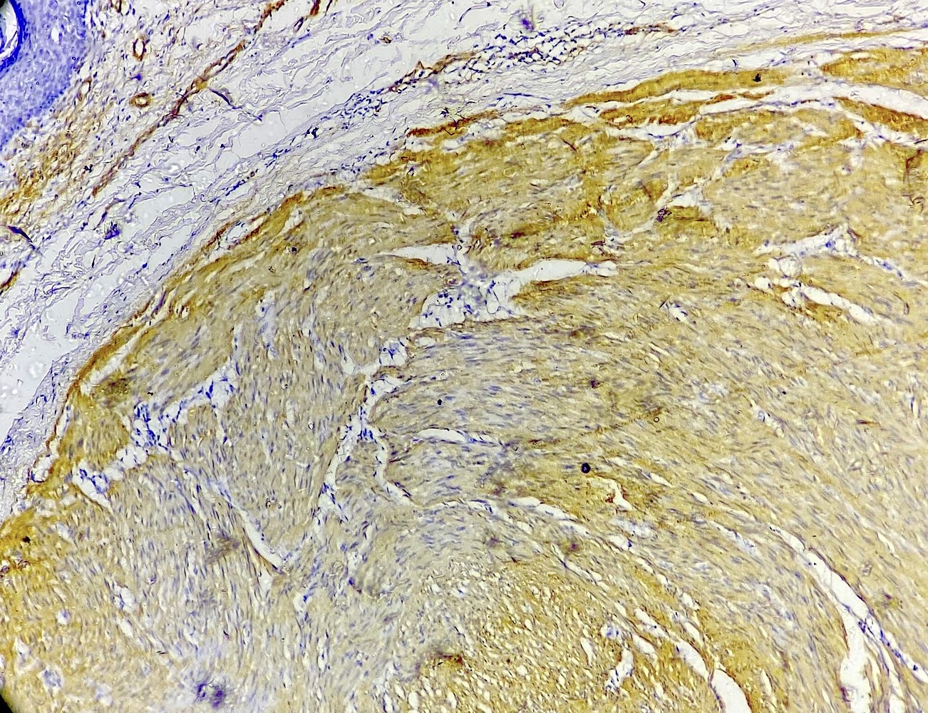
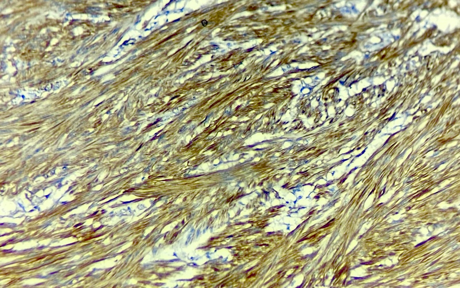
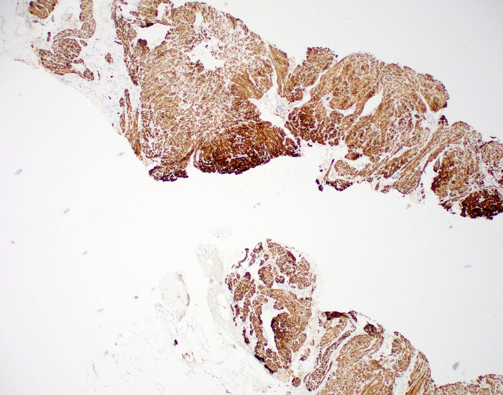
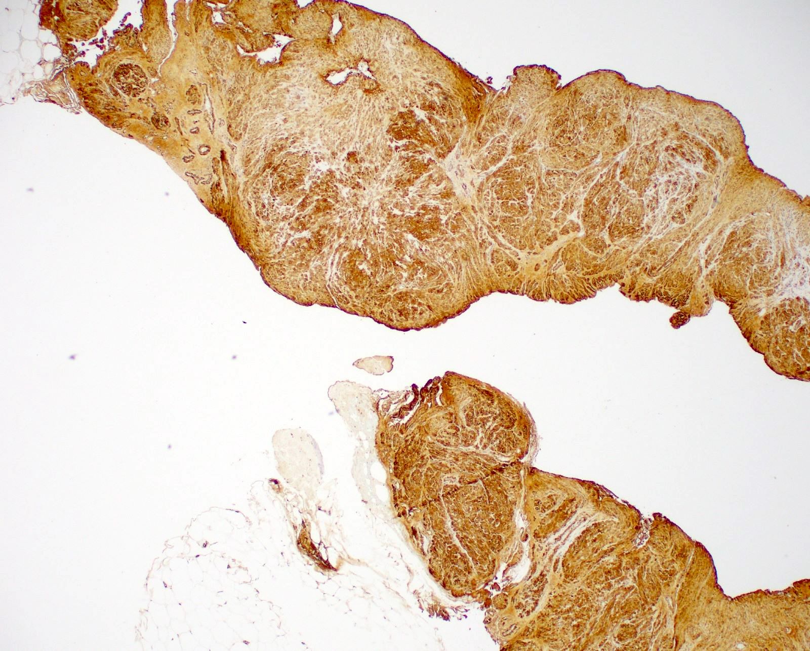
SMA
Caldesmon
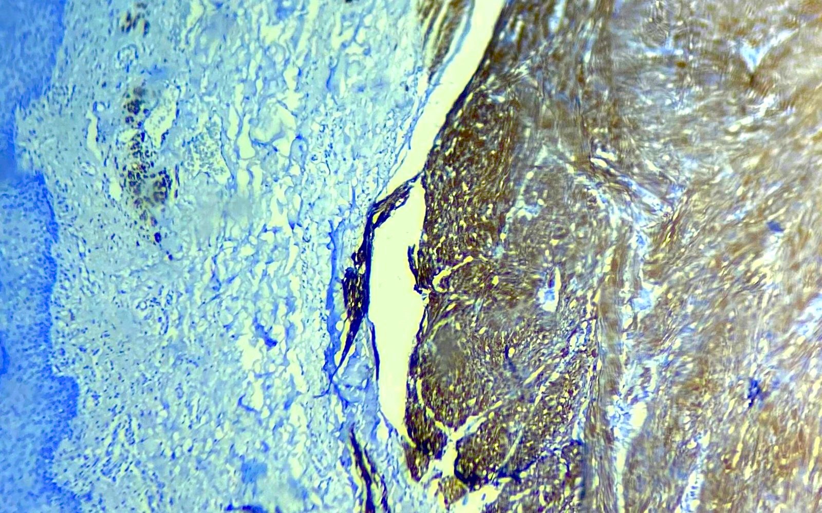
Desmin
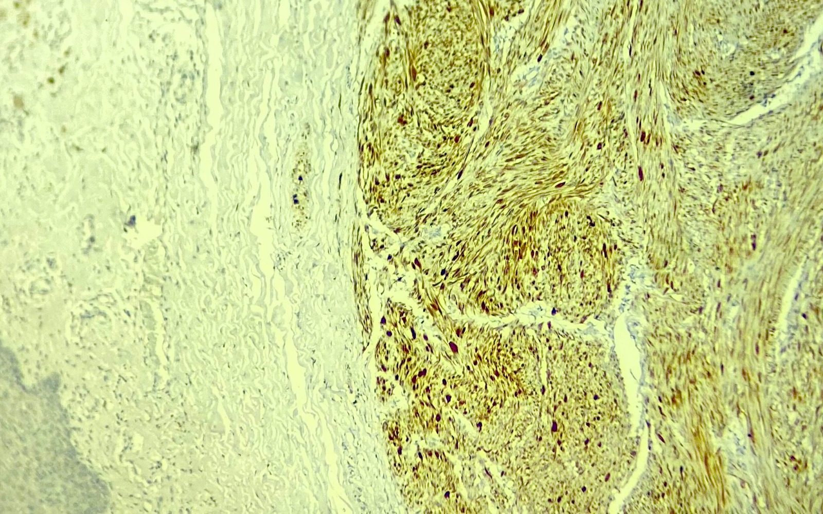
PR
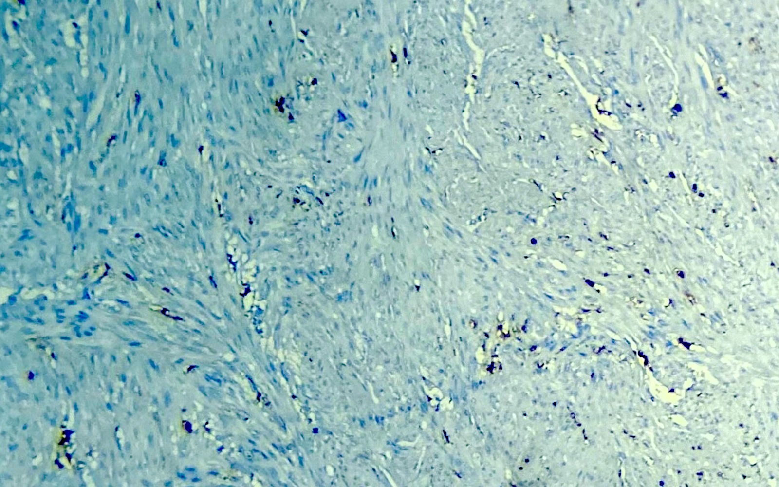
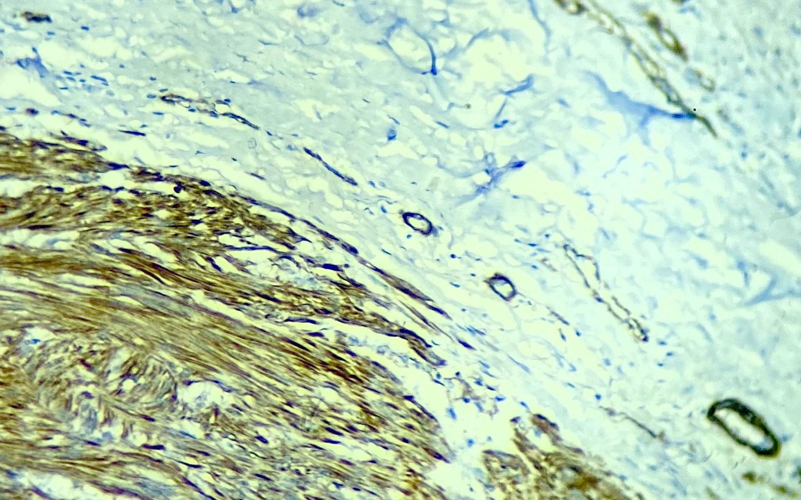
S100

Fascicles
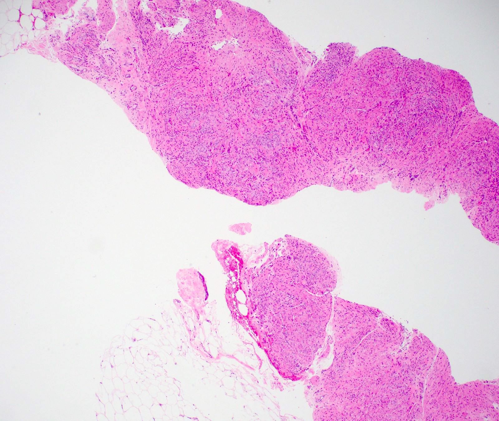
Breast core biopsy
Breast core biopsy
Positive stains
- SMA (Breast J 2020;26:529)
- Desmin (Breast J 2020;26:529)
- HHF35 (Breast J 2020;26:529)
- Caldesmon (Breast J 2020;26:529)
- Ki67 5% in atypical leiomyoma (BMC Womens Health 2022;22:119)
- ER, PR (EJMED 2023;5:26)
- Vimentin (Radiol Bras 2016;49:343)
Negative stains
Sample pathology report
- Right breast (nipple), punch biopsy:
- Nipple leiomyoma (see comment)
- Comment: There is a circumscribed dermal proliferation of monotonous spindle cells. On immunohistochemistry, spindle cells are positive for SMA and desmin and negative for S100 and CK, supporting the diagnosis.
Differential diagnosis
- Leiomyosarcoma (EJMED 2023;5:26, BMC Womens Health 2022;22:119):
- Atypical features, pleomorphism, mitosis and necrosis are seen
- Phyllodes tumor (Eur J Breast Health 2017;13:156):
- Biphasic epithelial and stromal neoplasm
- Stromal cellularity and stromal overgrowth with atypia, necrosis and mitosis in borderline and malignant phyllodes
- Nipple adenoma:
- Florid benign epithelial (glandular or squamous) proliferation with intact myoepithelium throughout
- May have papillary architecture
- Connected to the epidermis
- Adenomyoepithelioma (Eur J Breast Health 2017;13:156):
- Biphasic with glandular and myoepithelial components
- Myoepithelial prominence
- Nodular fasciitis (BMC Womens Health 2022;22:119):
- Fibrous proliferation, variably loose, tissue culture-like to more collagenized stroma
- Negative for desmin
- Myoid hamartoma (J Med Case Rep 2013;7:49):
- Biphasic benign proliferation with glandular and stromal components with prominent smooth muscle
- Within stromal component, glandular components are also seen; this is absent in leiomyoma
Board review style question #1
A 35 year old woman presents with a freely mobile lump in the nipple areolar area of the left breast. Microscopy shows features in image above with no mitosis or atypia. Which of the following immunohistochemical markers is best to confirm and highlight the diagnosis?
- CK72
- Ki67
- S100
- SMA
Board review style answer #1
D. SMA highlights smooth muscle origin. Answer A is incorrect because CK72 is an epithelial marker. Answer C is incorrect because S100 is a neural marker. Answer B is incorrect because Ki67 is a proliferation marker that will be low in benign leiomyoma but will not help in diagnosis.
Comment Here
Reference: Leiomyoma
Comment Here
Reference: Leiomyoma
Board review style question #2
Which of the following is typically true about breast leiomyoma?
- Has nipple discharge
- Has no hormonal association
- Is imaging occult
- May be painful
Board review style answer #2
D. May be painful. Nipple adenomas may be painful, which may be alleviated by calcium channel or alpha adrenergic blockers that cause smooth muscle relaxation. Answer C is incorrect because breast leiomyoma may be seen as a mass on breast imaging modalities such as mammogram and ultrasound. Answer B is incorrect because breast leiomyomas are associated with hormonal agents. Answer A is incorrect because breast leiomyoma has not been associated with nipple discharge.
Comment Here
Reference: Leiomyoma
Comment Here
Reference: Leiomyoma
