
Eye
General
WHO classification of tumors of the conjunctiva and caruncle
Editorial Board Member: J. Stephen Nix, M.D.


Last author update: 12 August 2024
Last staff update: 12 August 2024
Copyright: 2022-2025, PathologyOutlines.com, Inc.
PubMed Search: Conjunctiva and caruncle tumors WHO classification

Table of Contents
Definition / general | Major updates | WHO (2023) | Microscopic (histologic) images | Additional references | Practice question #1 | Practice answer #1Cite this page: Dharamraj AM, Couce M. WHO classification of tumors of the conjunctiva and caruncle. PathologyOutlines.com website. https://www.pathologyoutlines.com/topic/eyewho.html. Accessed August 25th, 2025.
Definition / general
- WHO classification of tumors of conjunctiva and caruncle
- Currently on 5th edition (to be published; beta version now available online)
Major updates
- WHO 5th edition contains updated information on molecular pathology, expression profiling and molecular classification of tumors; however, the focus remains on morphologic classification
- Additional classification and incorporation of tumor lineage (e.g., epithelial, adnexal, melanocytic) have been emphasized
- Selected entities reclassified in this edition
- Phakomatous choristoma has been moved (now classified under eyelid)
- Sebaceous carcinoma has been moved (now classified under eyelid)
- Spitz nevus has been moved (now classified under eyelid)
- Keratoacanthoma of the conjunctiva is no longer listed as a separate tumor type
- Spindle squamous cell carcinoma and squamous cell carcinoma with mucinous differentiation are included within the broader group of squamous cell carcinoma
- WNT activated deep penetrating / plexiform melanocytoma has been included as a new entity in the WHO 5th edition
- Includes an update on pathogenesis and molecular genetics of melanocytic conjunctival lesions
- Within the melanocytic lesions, nomenclature for premalignant and malignant lesions has been updated
- Nonneoplastic pterygium and pinguecula were added as tumor-like lesions to delineate them from conjunctival squamous intraepithelial neoplasia
- Soft tissue tumors, hematolymphoid neoplasms and secondary tumors of the conjunctiva are discussed in separate chapters
WHO (2023)
-
Tumor-like lesions and choristomas of the conjunctiva
- Cysts of the conjunctiva and caruncle ICD-10: H11.449
- Epithelial conjunctival inclusion cyst
- Reactive, epithelial and degenerative conjunctival lesions
- Reactive epithelial hyperplasia of the conjunctiva
- Papillary and follicular conjunctivitis
- Pterygium and pinguecula
- Choristomas of the conjunctiva
- Epibulbar choristoma (dermoid; dermolipoma; complex choriostoma)
- Epibulbar osseous choristoma
- Benign epithelial conjunctival tumors
- Premalignant and malignant epithelial tumors of the conjunctiva
- Conjunctival squamous intraepithelial neoplasia (conjunctival intraepithelial neoplasia and carcinoma in situ) 8077/2
- Conjunctival squamous cell carcinoma 8070/3, 8076/2, 8071/3
- Adenosquamous carcinoma 8560/3
- Benign melanocytic conjunctival tumors
- Benign epithelial melanosis of the conjunctiva
- Junctional, compound and subepithelial conjunctival nevi 8740/0, 8760/0, 8750/0
- Inflamed juvenile conjunctival nevus 8770/0
- Blue nevus of the conjunctiva 8780/0
- WNT activated deep penetrating / plexiform melanocytoma (nevus)
- Combined nevus of the conjunctiva
- Premalignant and malignant melanocytic conjunctival tumors
Epithelial tumors of the conjunctiva
Melanocytic conjunctival tumors
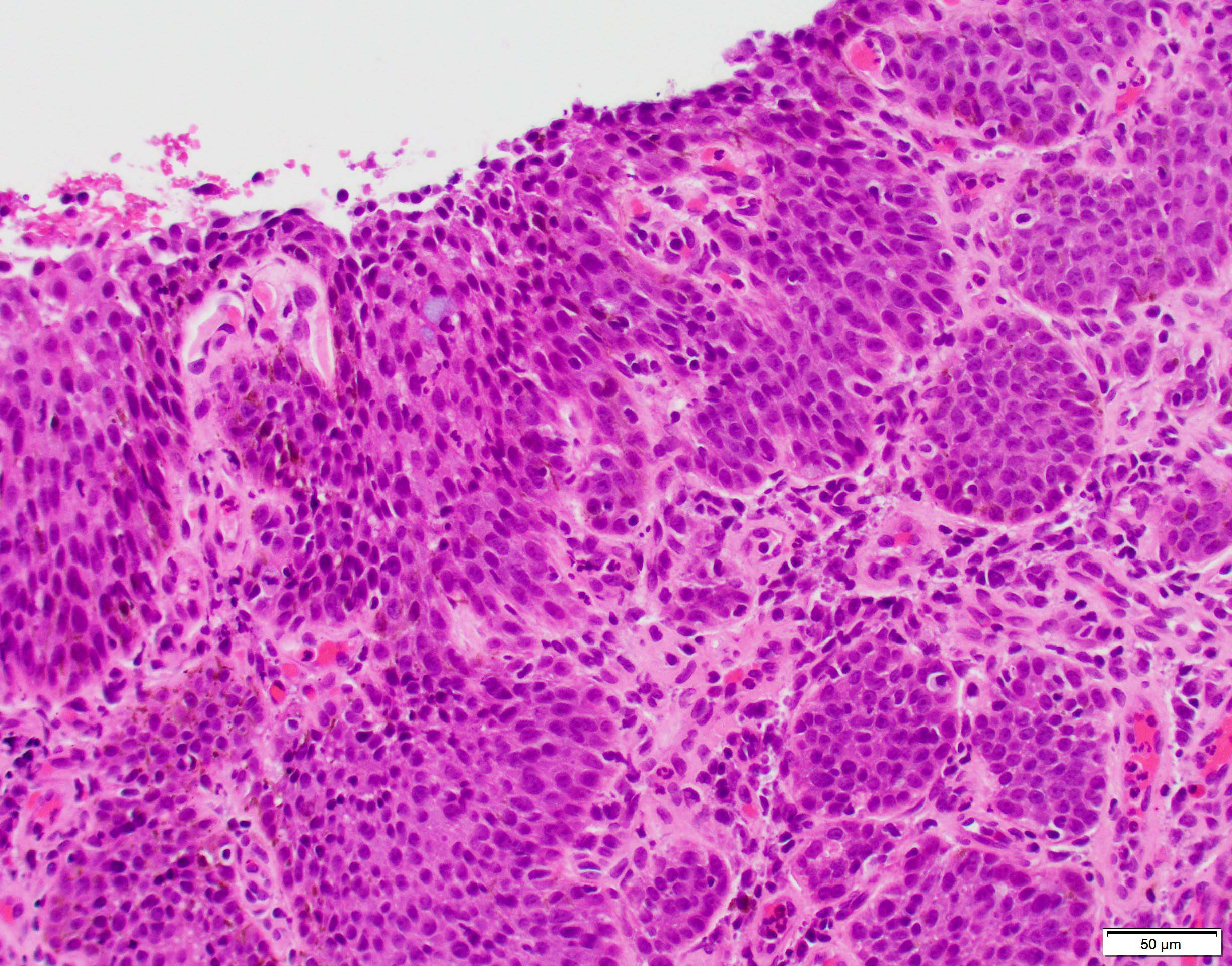
Microscopic (histologic) images
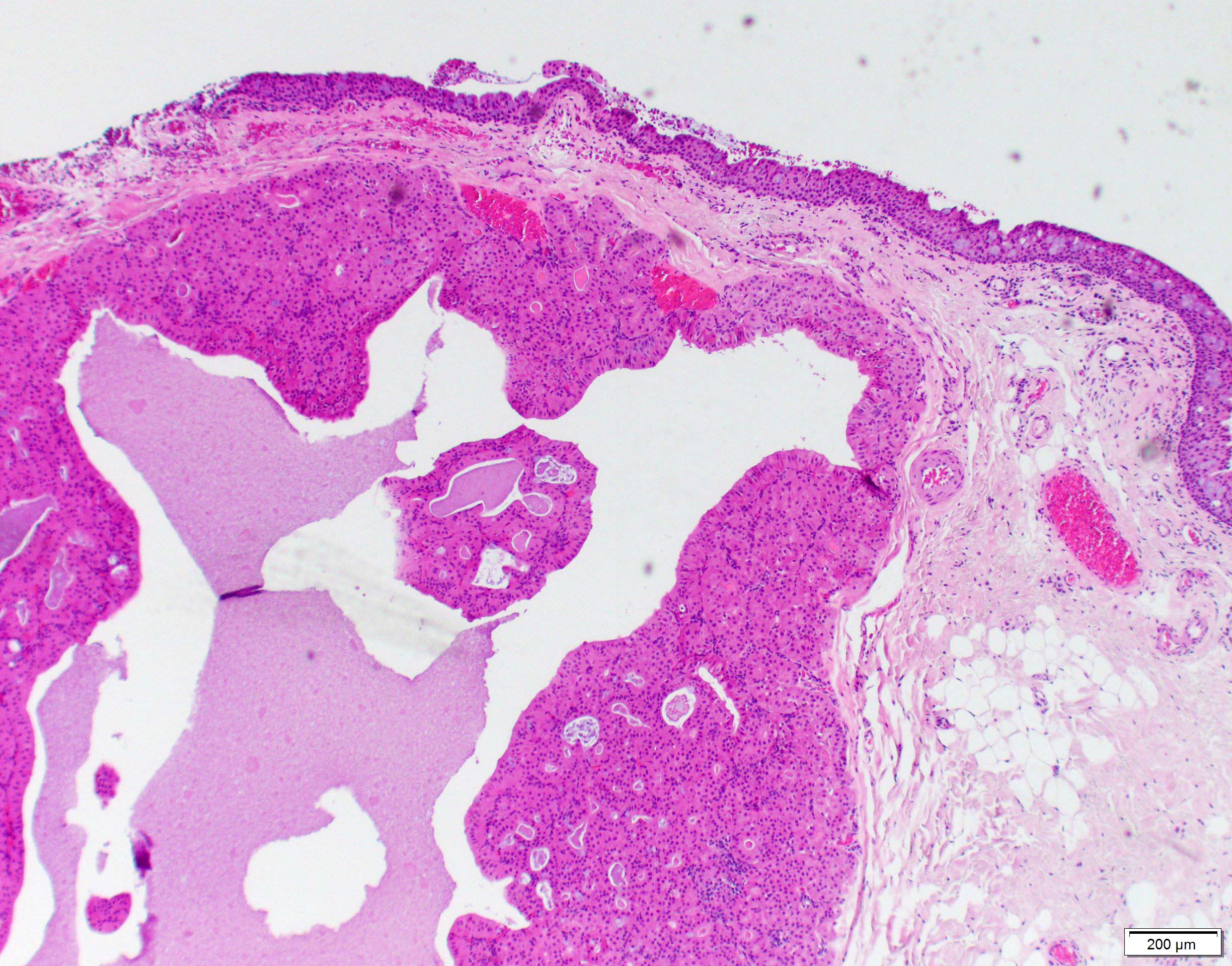
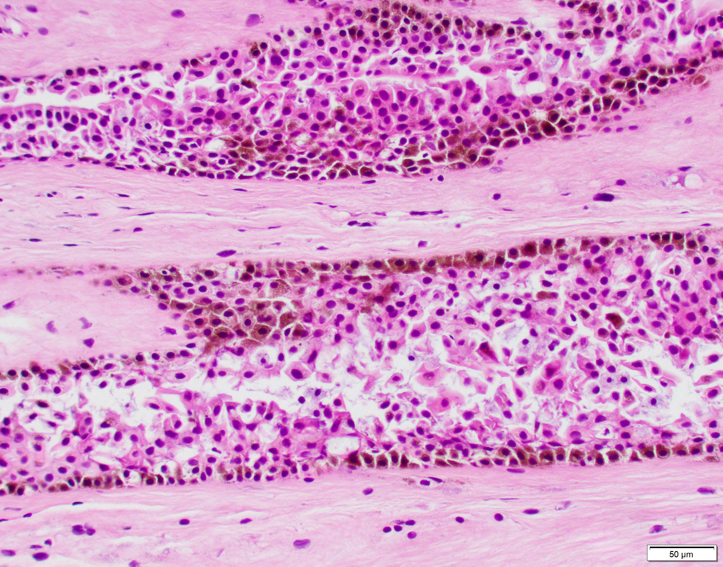
Contributed by Marta Couce, M.D., Ph.D.


Conjunctival oncocytoma
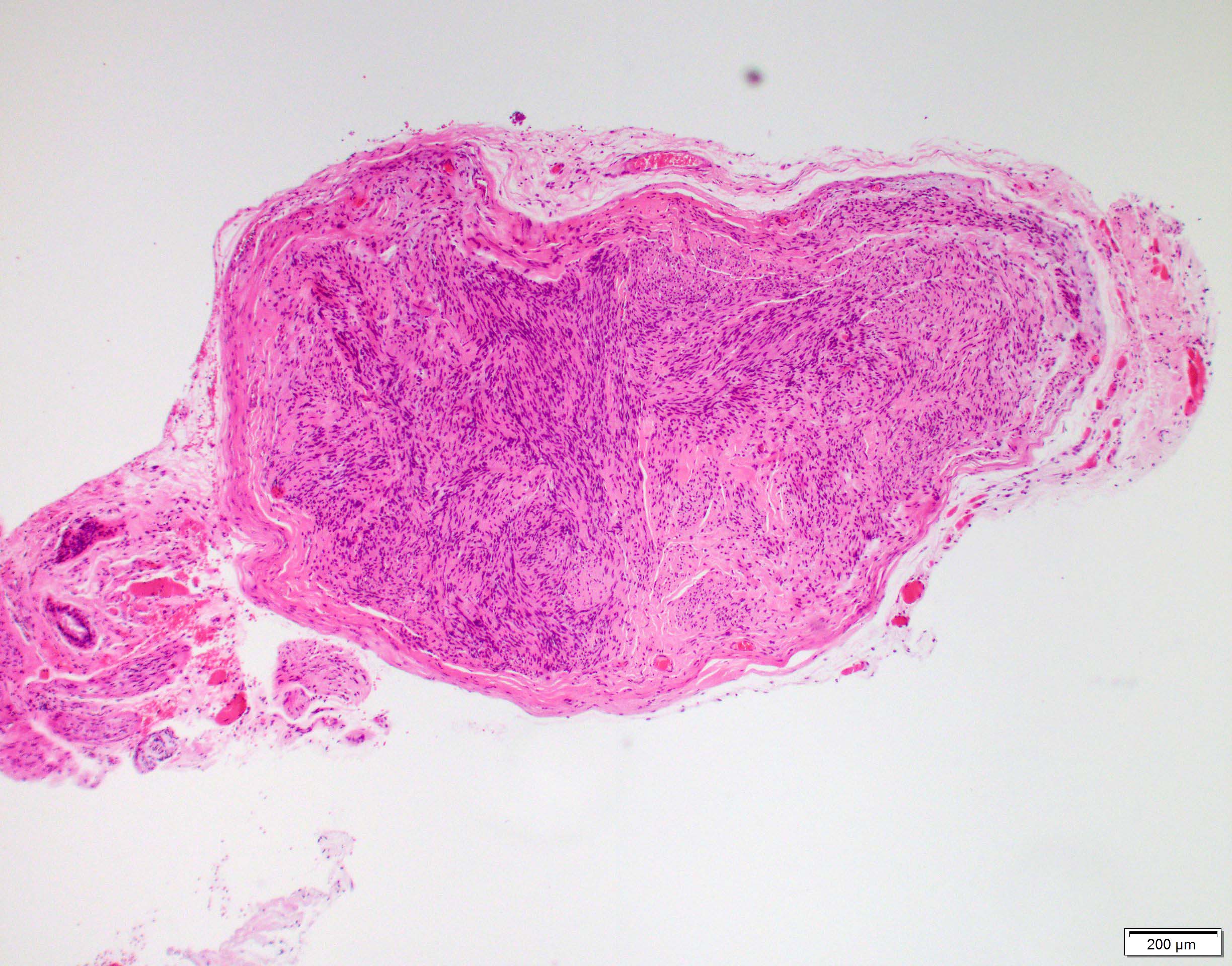
Conjunctival schwannoma


Cyst of the conjunctiva

Subepithelial conjunctival nevus

Compound conjunctival nevus
PAM of the conjunctiva
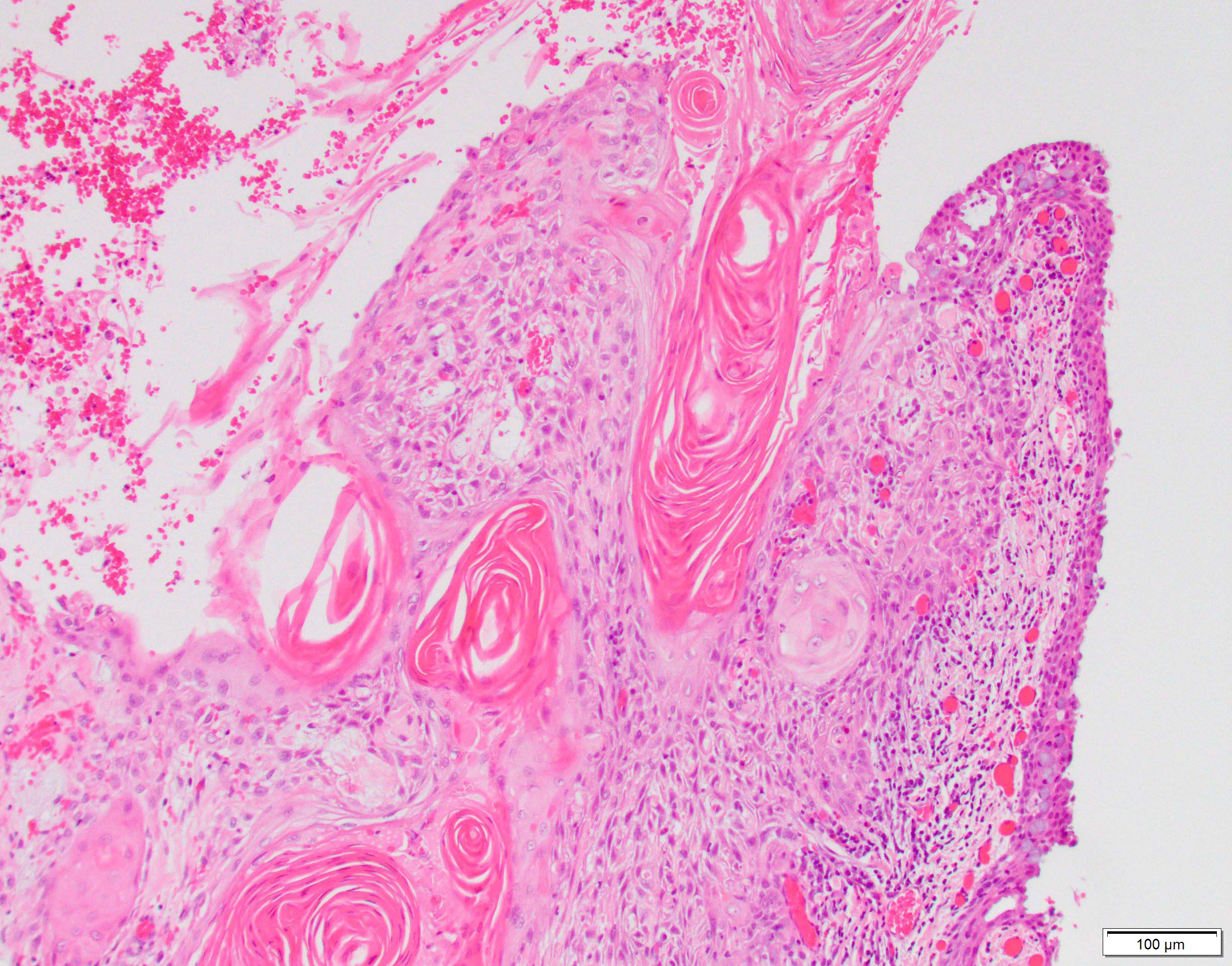
Conjunctival squamous cell carcinoma
Adenosquamous carcinoma

Conjunctival oncocytoma
Additional references
Practice question #1
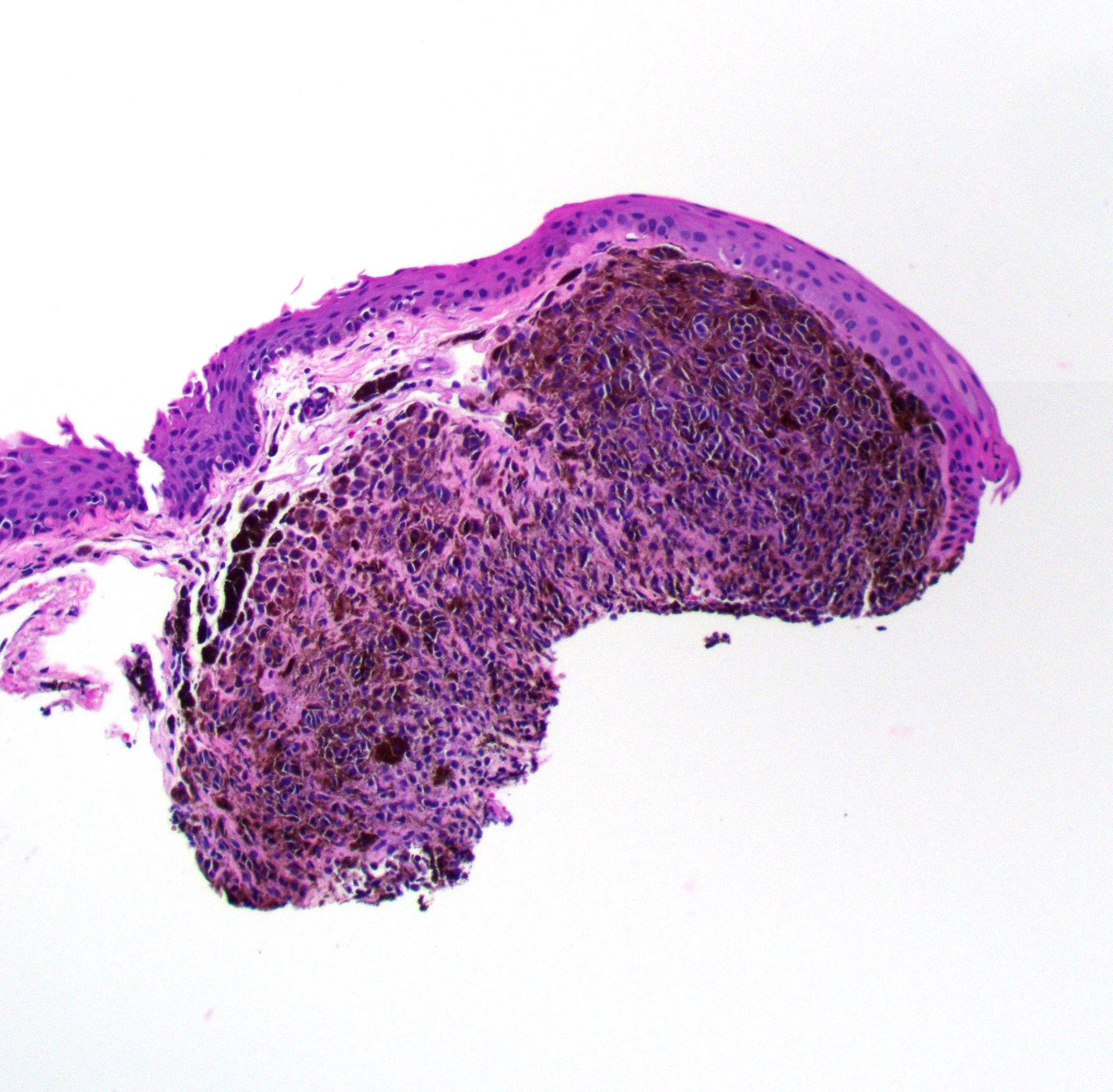
Among melanocytic lesions of the conjunctiva, which of the following statements is true about the lesion shown in the above image?
- It has a high risk for transformation into malignant melanoma
- It is best treated by enucleation
- It is commonly found as a poorly defined, pigmented mass
- It is most commonly found in ages 70 - 90 years
- It is the most common melanocytic lesion of the conjunctiva
Practice answer #1
E. It is the most common melanocytic lesion of the conjunctiva. This is a conjunctival compound nevus, which is the most common melanocytic lesion of the conjunctiva. They are benign. Differentials include primary acquired melanosis, racial melanosis, ocular melanocytosis and malignant melanoma. Answer A is incorrect because conjunctival nevi have a low rate of conversion to conjunctival melanoma (less than 1%). Answer B is incorrect because enucleation is a drastic treatment for a benign lesion. These are typically managed by periodic observation and serial photographic comparison. Removal is typically for cosmetic reasons or if there are concerning changes in color, shape or size. Answer C is incorrect because the morphology is usually a well defined, slightly elevated mass. The classic location is the interpalpebral bulbar conjunctiva, caruncle or plica semilunaris. Answer D is incorrect because, most often, these lesions are apparent within the first or second decade of life as a discrete, pigmented lesion.
Comment Here
Reference: WHO classification of tumors of the conjunctiva and caruncle
Comment Here
Reference: WHO classification of tumors of the conjunctiva and caruncle
