Nasal cavity, paranasal sinuses, nasopharynx
Inflammatory lesions
Inflammatory sinonasal polyp, including antrochoanal polyp
Author: Bin Xu, M.D., Ph.D.
Editorial Board Member: Molly Housley Smith, D.M.D.
Deputy Editor-in-Chief: Kelly Magliocca, D.D.S., M.P.H.

Last author update: 10 February 2022
Last staff update: 10 June 2025
Copyright: 2004-2025, PathologyOutlines.com, Inc.
PubMed search: Sinonasal inflammatory polyp
Table of Contents
Definition / general | Essential features | Terminology | ICD coding | Epidemiology | Sites | Pathophysiology | Etiology | Clinical features | Diagnosis | Radiology description | Radiology images | Prognostic factors | Case reports | Treatment | Clinical images | Gross description | Microscopic (histologic) description | Microscopic (histologic) images | Sample pathology report | Differential diagnosis | Practice question #1 | Practice answer #1 | Practice question #2 | Practice answer #2Cite this page: Xu B. Inflammatory sinonasal polyp, including antrochoanal polyp. PathologyOutlines.com website. https://www.pathologyoutlines.com/topic/nasalpolypinflammatory.html. Accessed August 25th, 2025.
Definition / general
- Benign, nonneoplastic inflammatory outgrowth of sinonasal mucosa
- Most common type of sinonasal polyp
- Most common space occupying lesion of the sinonasal tract (J Clin Diagn Res 2014;8:FC04)
- Antrochoanal polyp is an inflammatory sinonasal polyp that extends from the maxillary sinus through the ostium into the nasal cavity / nasopharynx
Essential features
- Benign, nonneoplastic polyp characterized by edematous stroma infiltrated by mixed inflammatory cells
Terminology
- Chronic rhinosinusitis with nasal polyps
- Nasal polyps
ICD coding
- ICD-10: J33.9 - nasal polyp, unspecified
Epidemiology
- Occurs in 2 - 10% of population (J Allergy Clin Immunol Pract 2021;9:4117, StatPearls: Nasal Polyps [Accessed 12 January 2022])
Sites
- Commonly bilateral and multifocal (Pak J Med Sci 2020;36:146)
- Most frequently involves nasal cavity and ethmoid sinus (J Allergy Clin Immunol Pract 2016;4:565)
Pathophysiology
- Defected epithelial cell barrier, increased exposure to bacteria and dysfunctioning host immune system may play a role in the pathogenesis (J Allergy Clin Immunol Pract 2016;4:565)
Etiology
- Most frequently occurs in the setting of chronic rhinosinusitis
- Other risk factors include allergy, systemic vasculitis, cystic fibrosis, sensitivity to aspirin and nonsteroid anti-inflammatory drugs (StatPearls: Nasal Polyps [Accessed 12 January 2022])
Clinical features
- Most common symptoms: obstruction, nasal congestion, rhinorrhea, facial pressure and pain (J Allergy Clin Immunol Pract 2016;4:565)
Diagnosis
- Diagnosis can be rendered based on the typical endoscopic or histologic findings
Radiology description
- Smooth soft tissue masses commonly originate in the ethmoid sinuses and protrude into the nasal cavity (Eur Radiol 2006;16:872)
- Hypodense but may be hyperdense due to concurrent fungal infection or increased protein content
- May demonstrate adjacent bony remodeling or erosion
Radiology images
Images hosted on other servers:

Coronal CT
Prognostic factors
- Recurrence rate is approximately 60% in patients followed for at least 10 years
- Presence of asthma is associated with increased risk of recurrence
- Reference: Am J Rhinol Allergy 2021;35:449
Case reports
- 21 and 32 year old men with infarcted angiomatous nasal polyps (Eur Arch Otorhinolaryngol 2005;262:225)
- 4 cases of angiomatous nasal polyps (Cureus 2020;12:e7642)
Treatment
- Medical treatments include nasal corticosteroid and high volume saline irrigation (JAMA 2015;314:926)
- Sinus surgery for patients failed medical management (J Allergy Clin Immunol Pract 2016;4:565)
Clinical images
Images hosted on other servers:

Translucent polyps with a smooth surface
Gross description
- Usually multiple and bilateral and involve nasal cavity and paranasal sinuses
- Have translucent, moist or edematous cut surface
- Broad base of attachment is present
- Usually not destructive
Microscopic (histologic) description
- Edematous, fibrotic or loosely myxoid stroma covered by respiratory epithelium
- Infiltrated by mixed inflammatory cells, including lymphocytes, plasma cells, eosinophils, neutrophils and mast cells
- Surface epithelium can show ulceration or squamous metaplasia
- May have bizarre stromal cells (large and pleomorphic)
- Submucosal glands are decreased or absent
- Concurrent fungal infection may be seen
- Rarely, osseous metaplasia may be present
Microscopic (histologic) images
Contributed by Bin Xu, M.D., Ph.D.
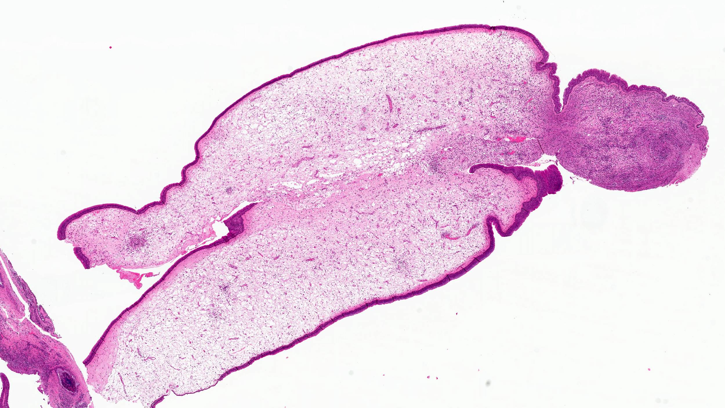
Edematous stroma

Mixed inflammatory infiltrate
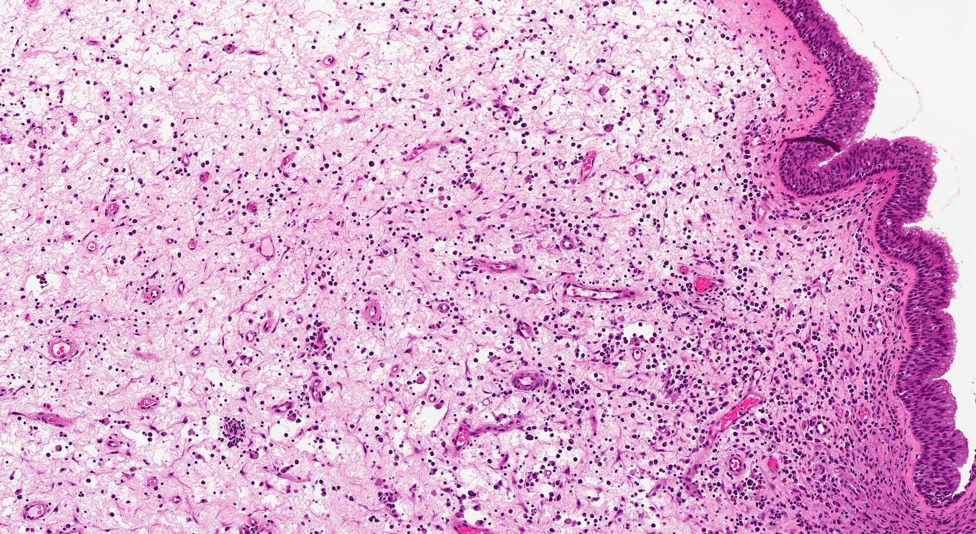
Respiratory epithelium lining
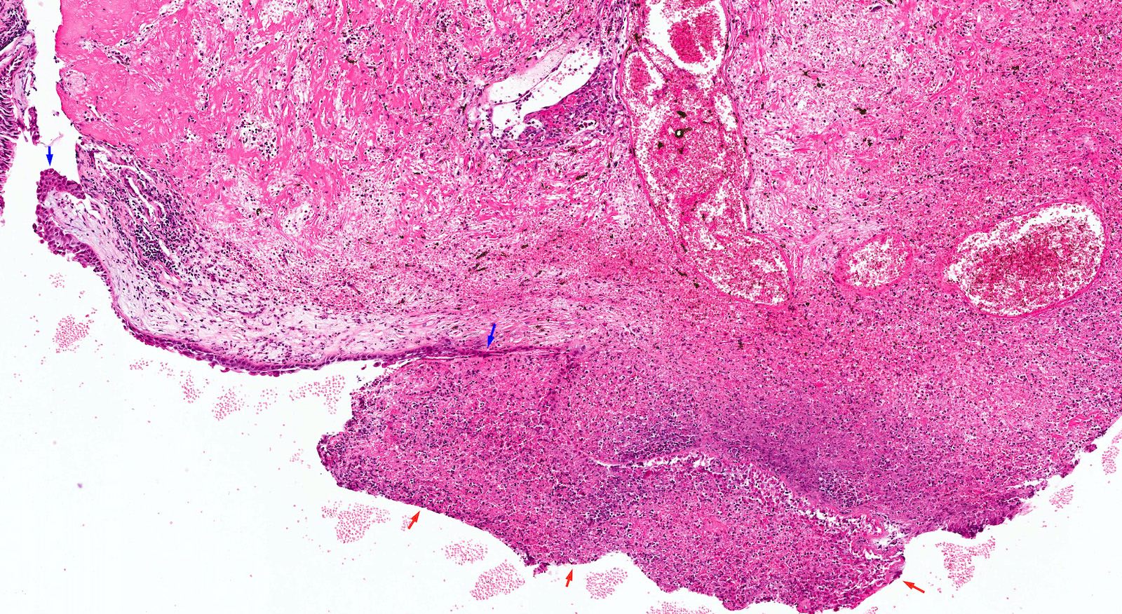
Surface ulceration, squamous metaplasia

Stroma with hemorrhage
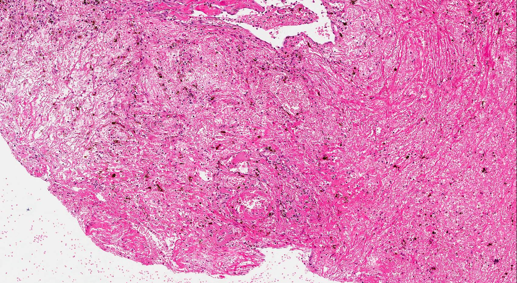
Infarction
Sample pathology report
- Nasal cavity, left, excision:
- Inflammatory sinonasal polyp
Differential diagnosis
- Sinonasal papilloma:
- Inflammatory polyp lacks the epithelial proliferation, the thickened epithelial lining or the inverted growth pattern seen in sinonasal papilloma
- Respiratory epithelial adenomatoid hamartoma:
- Inflammatory polyp lacks the proliferation of respiratory epithelium and thickened basement membrane typical of respiratory epithelial adenomatoid hamartoma
- Biphenotypic sinonasal sarcoma:
- Can be polypoid but characterized by cellular spindle cells in the stroma showing biphenotypic neural and myoid differentiation
- Rhabdomyosarcoma, embryonal (botryoid) type:
Practice question #1
What is the most common polypoid lesion in the sinonasal cavity?
- Angiofibroma
- Inflammatory sinonasal polyp
- Respiratory epithelial adenomatoid hamartoma
- Sinonasal papilloma
Practice answer #1
B. Inflammatory sinonasal polyp is the most common polypoid lesion and space occupying lesion in the sinonasal tract.
Comment Here
Reference: Inflammatory sinonasal polyp, including antrochoanal polyp
Comment Here
Reference: Inflammatory sinonasal polyp, including antrochoanal polyp
Practice question #2
What is the diagnosis of this lesion excised from the left nasal cavity of a 35 year old woman?
- Biphenotypic sinonasal sarcoma
- Inflammatory sinonasal polyp
- Inverted sinonasal papilloma
- Respiratory epithelial adenomatoid hamartoma
Practice answer #2
B. This is an inflammatory sinonasal polyp, a nonneoplastic benign lesion characterized by edematous stroma and inflammatory infiltrates.
Comment Here
Reference: Inflammatory sinonasal polyp, including antrochoanal polyp
Comment Here
Reference: Inflammatory sinonasal polyp, including antrochoanal polyp
