Skin nonmelanocytic tumor
Vascular tumors
Angiosarcoma
Author: Joel Tjarks, M.D.

Last author update: 1 October 2016
Last staff update: 23 September 2025 (update in progress)
Copyright: 2002-2025, PathologyOutlines.com, Inc.
PubMed Search: Angiosarcoma [title] skin "loattrfree full text"[sb]
Table of Contents
Definition / general | Essential features | Terminology | Epidemiology | Sites | Clinical features | Prognostic factors | Case reports | Treatment | Clinical images | Gross description | Microscopic (histologic) description | Microscopic (histologic) images | Positive stains | Negative stains | Molecular / cytogenetics description | Differential diagnosis | Additional referencesCite this page: Tjarks J. Angiosarcoma. PathologyOutlines.com website. https://www.pathologyoutlines.com/topic/skintumornonmelanocyticangiosarcoma.html. Accessed October 1st, 2025.
Definition / general
- Malignant neoplasm with vascular differentiation
Essential features
- Infiltrative vascular neoplasm with broad histologic profile ranging from a well differentiated neoplasm with frank vascular differentiation to a poorly differentiated tumor with epithelioid or spindled cells
- May mimic poorly differentiated carcinoma, inflammatory process, lymphoma or melanoma
Terminology
- Also known as hemangiosarcoma
Epidemiology
- Classically arises in one of three scenarios:
- Head and neck of the elderly
- Chronic lymphedema
- Postradiation (usually in the setting of breast cancer)
Sites
- Sun exposed skin of the elderly (head and neck); breast with history of lymphedema or radiation therapy
Clinical features
- Wide age range (most common in adults)
- Presents as purple nodules or plaques
- Highly aggressive
- Frequent recurrence and metastasis
Prognostic factors
- Poor prognosis - high mortality
- Epithelioid tumors are often more aggressive
Case reports
- 80 year old woman with secondary angiosarcoma postradiation and breast conserving therapy (J Clin Imaging Sci 2015;5:45)
Treatment
- Surgical resection with negative margins
- Chemotherapy is occasionally used
Clinical images
Images hosted on other servers:


Scalp, neck and breast lesions
Gross description
- Violet elevated nodules with ill defined margins
Microscopic (histologic) description
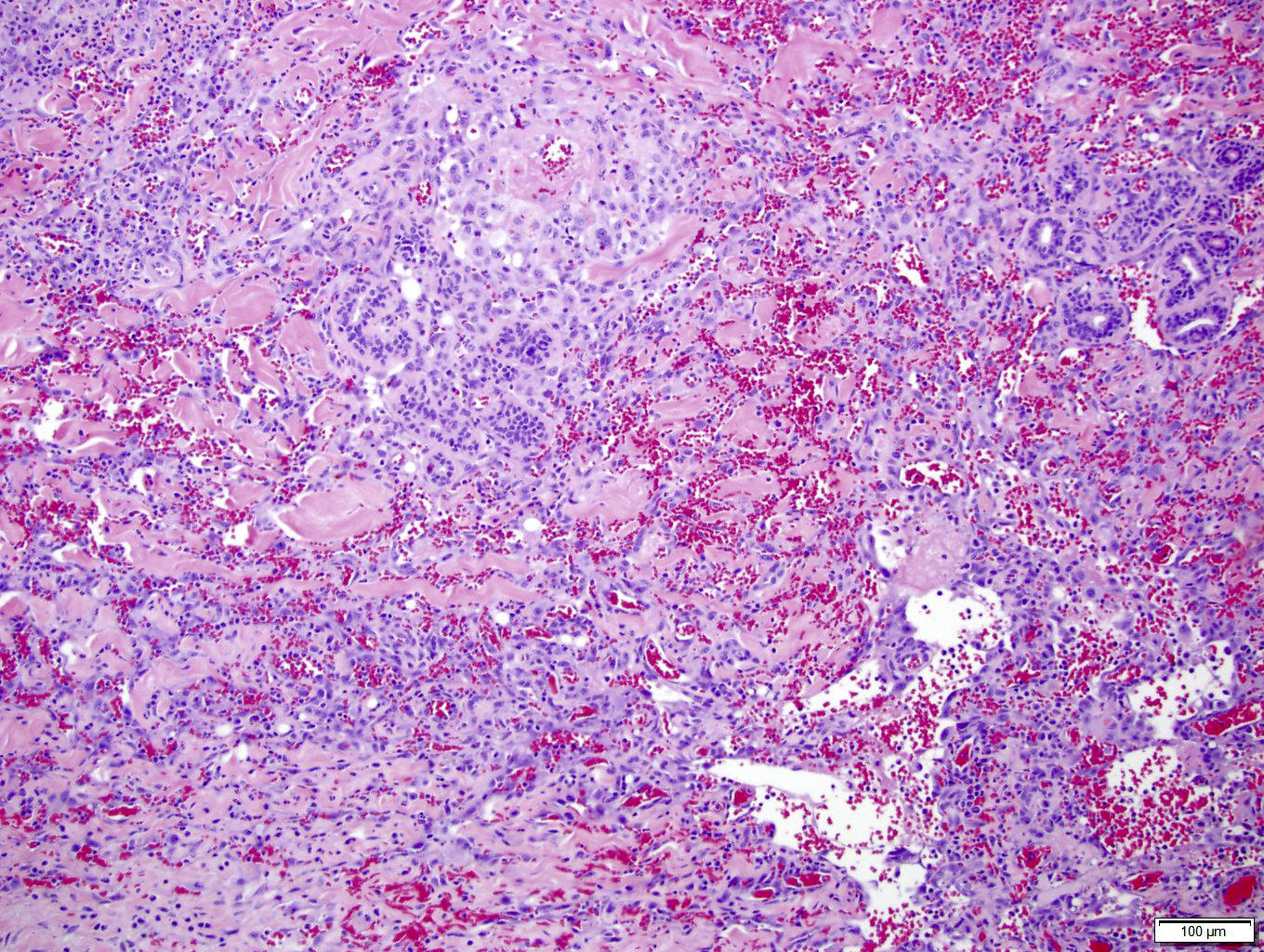
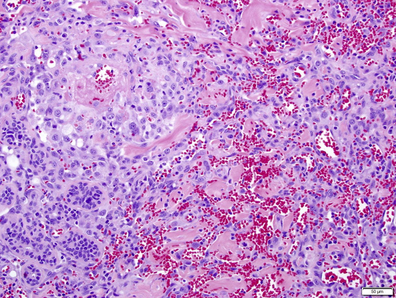
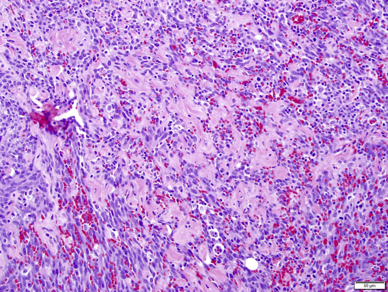
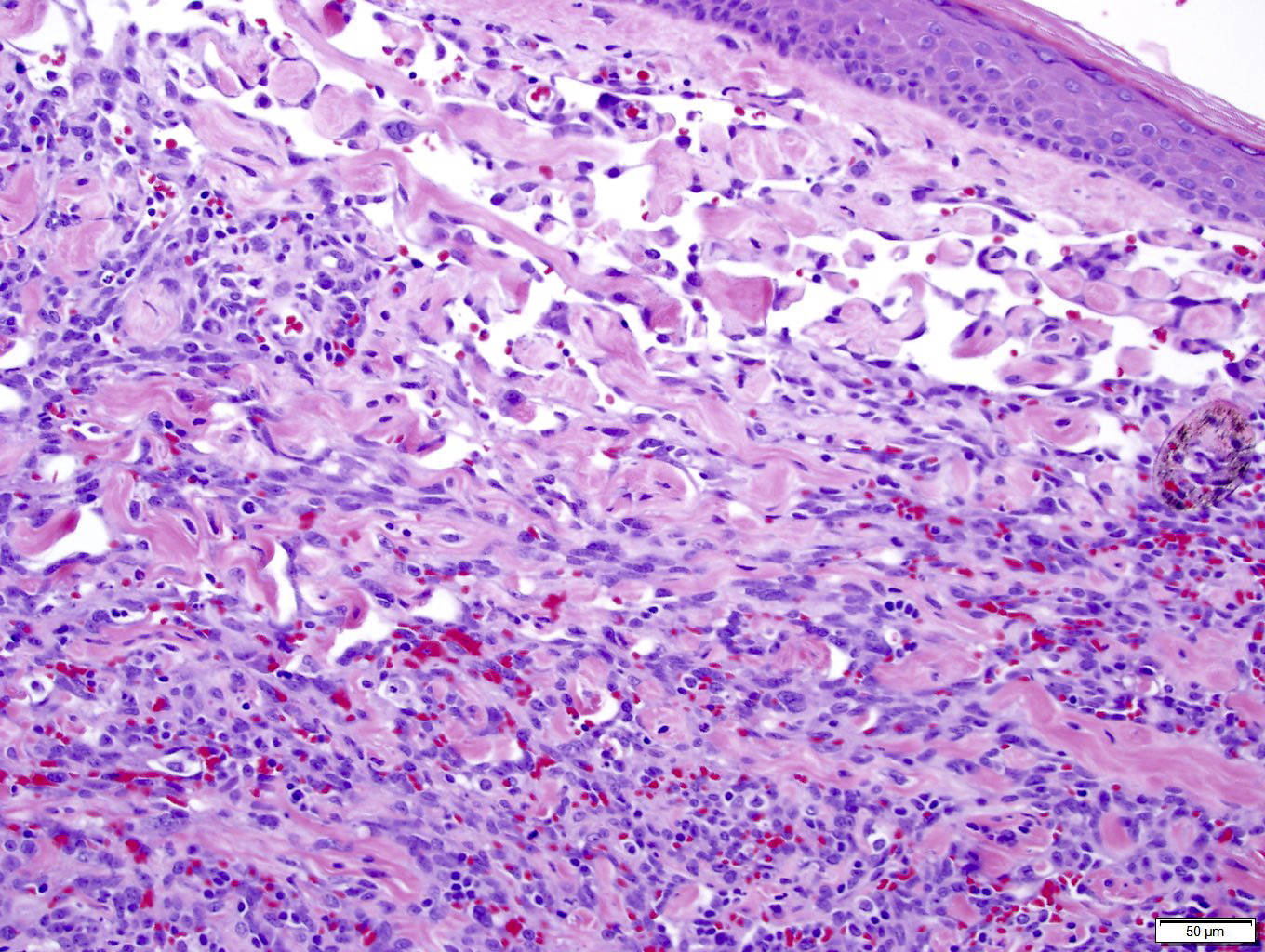
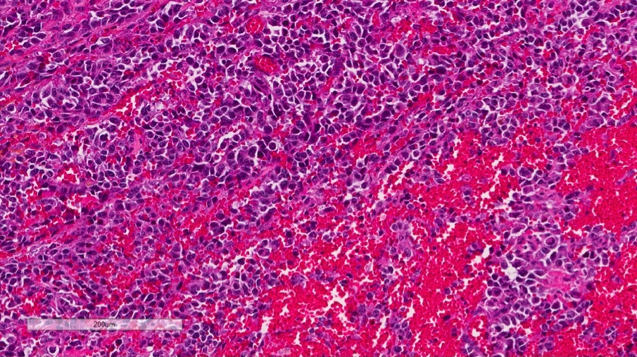
- Infiltrating, freely anastomosing channels lined by spindled to epithelioid endothelial cells with variable atypia, surrounding adnexae and dissecting dermal collagen
- Endothelial cells may have multilayered appearance
- May have free floating intraluminal endothelial cells (“fish in the creek”)
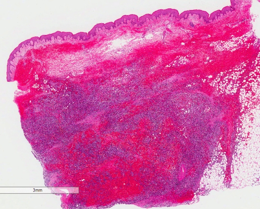
Microscopic (histologic) images
Contributed by Hillary Rose Elwood, M.D. and Joel Tjarks, M.D.

High grade atypical vascular tumor
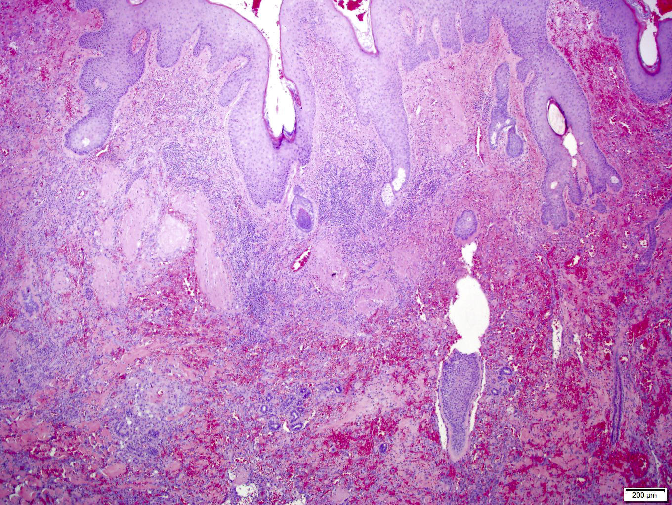
Atypical vascular tumor involving dermis and subcutis
Vascular proliferation dissecting throughout the dermal collagen
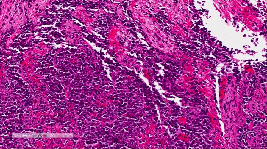
Angiosarcoma
Positive stains
Molecular / cytogenetics description
- MYC (8q24) amplification seen in great majority radiation / lymphedema associated tumors
Differential diagnosis
- Atypical fibroxanthoma
- Atypical vascular lesion
- Hemangioma
- Kaposi sarcoma
Additional references
