

Cytopathology
Lung
Benign


Copyright: 2022-2024, PathologyOutlines.com, Inc.
PubMed Search: Benign lung cytopathology
Lung
Benign
Editorial Board Member: Bonnie Choy, M.D.
Last staff update: 8 May 2024 (update in progress)
Copyright: 2022-2024, PathologyOutlines.com, Inc.
PubMed Search: Benign lung cytopathology

Table of Contents
Definition / general | Essential features | CPT coding | Sites | Case reports | Cytology description | Cytology images | Sample pathology report | Differential diagnosis | Additional references | Board review style question #1 | Board review style answer #1 | Board review style question #2 | Board review style answer #2 | Board review style question #3 | Board review style answer #3Cite this page: Chen-Yost HI, Jin X. Benign. PathologyOutlines.com website. https://www.pathologyoutlines.com/topic/cytopathologylungbenign.html. Accessed May 14th, 2024.
Definition / general
- Specimens categorized as benign demonstrate unequivocal benign cytopathological features, which may or may not be diagnostic of a specific process or benign neoplasm
Essential features
- The World Health Organization (WHO) Reporting System for Lung Cytopathology is the recommended system for reporting results (Acta Cytol 2023;67:80)
- Per the reporting system, benign includes benign bronchial elements, inflammatory or infectious processes (e.g., granulomatous inflammation) or a specific benign tumor (e.g., hamartoma)
- Rate of malignancy of cases categorized as benign is reported to be in the range of 20 - 50% (Acta Cytol 2022;66:124, Diagn Cytopathol 2016;44:399, Diagn Cytopathol 2018;46:725)
CPT coding
Sites
- Lung, bronchus
- Lung, parenchyma
Case reports
- 59 year old man with a left upper lobe lung cavitary lesion (Cytopathology 2023;34:158)
- 70 year old woman with a 3.3 cm soft tissue mass (J Cytol 2012;29:250)
- 72 year old man with ground class opacities and infiltrative shadows in bilateral lungs (Diagn Cytopathol 2021;49:E277)
Cytology description
Normal benign elements
Reactive elements
Infection
Benign neoplasms
- Bronchial cells
- Columnar cells
- Small nuclei
- Abundant apical cytoplasm
- Terminal bars and cilia frequent and prominent
- Alveolar macrophages
- Round cells with pale, irregular nuclei
- Abundant, foamy cytoplasm
- Pneumocytes (Diagn Cytopathol 2010;38:297)
- Appear similar to alveolar macrophages
- Few cohesive groups with scalloped borders, intercellular windows or gaps
Reactive elements
- Basal cell hyperplasia (Diagn Cytopathol 2010;38:297)
- Small, uniform cells
- Dark round or oval nuclei, smooth nuclear borders
- Scant basophilic cytoplasm
- Occasional molding; can mimic small cell carcinoma
- Squamous metaplasia (Diagn Cytopathol 2011;39:144)
- Smudgy chromatin
- Eosinophilic cytoplasm versus organophilic
- Preserved N:C ratio
- Creola bodies (Arerugi 1989;38:542)
- Clusters of ciliated bronchial epithelial cells
- Associated with asthma and eosinophils
- Curschmann spirals (Diagn Cytopathol 1998;19:349)
- Spiral shaped mucous plugs
- Nonspecific finding: can be seen in smokers, lung cancer, chronic bronchitis
- Treatment effect: radiation therapy (Pathologica 1991;83:317)
- Clinical context is helpful in this diagnosis
- Atypical nuclei with smudgy effect
- Abundant cytoplasm, often vacuolated
- Anthracosis
- Dark pigment within macrophages
- Finer than melanin
Infection
- Bacterial
- Actinomycosis (Monaldi Arch Chest Dis 2022;92:1641)
- Dark cotton ball-like mass
- Spider leg-like projections
- Branches at acute angles
- Sulfur granules
- Nocardiosis (Diagn Cytopathol 2017;45:1105)
- Narrower than Actinomyces
- Right angle branching
- Associated with neutrophils
- Tuberculosis (Diagn Cytopathol 2014;42:993)
- Strongly associated with necrotizing granulomas
- Acid fast positive
- Actinomycosis (Monaldi Arch Chest Dis 2022;92:1641)
- Fungal
- Blastomycosis (Acta Cytol 2020;64:532)
- Thick walled, double contoured spores (8 - 15 μm)
- Broad based bud
- Histoplasmosis (Acta Cytol 2020;64:532)
- Histiocytes filled with small spores (2 - 4 μm)
- Narrow based budding with capsule
- Pneumocystis (Cancer Cytopathol 2018;126:643)
- Curved, cup shaped cysts (4 - 8 μm)
- Central dot with GMS staining
- Associated with frothy pink exudates
- Aspergillus (Cancer Cytopathol 2018;126:643)
- Septate hyphae
- 45 degree acute angle branches
- Background often has abundant neutrophils and necrosis
- Cryptococcosis (Acta Cytol 2020;64:532)
- Narrow based budding (4 - 12 μm)
- Mucin rich capsule
- Positive for mucicarmine
- Coccidioidomycosis (Semin Diagn Pathol 2017;34:530)
- Thick walled, large spherules (10 - 80 μm)
- Can be filled with endospores
- Mucormycosis (Semin Diagn Pathol 2017;34:530)
- Exclusively in diabetic or immunocompromised patients
- Broad hyphae with thin walls
- Twisted and folded walls
- Irregular branching
- Candida (Semin Diagn Pathol 2017;34:530)
- Can occur as endobronchial growth or in abscess
- Oval yeasts
- Pseudohyphae: elongated and pinched at attachment
- Blastomycosis (Acta Cytol 2020;64:532)
- Viral cytopathic effects
- Cytomegalovirus (Cancer Cytopathol 2018;126:643)
- Nuclear enlargement with owl eye nucleoli
- Intranuclear inclusion with surrounding halo
- Nuclear enlargement with owl eye nucleoli
- Adenovirus (Diagn Cytopathol 2017;45:614)
- Smudged nuclei
- Intranuclear basophilic inclusions surrounded by a small halo
- Herpes (Cancer Cytopathol 2018;126:643)
- Triad: multinucleation, margination, molding
- Can have significant acute inflammation and necrosis
- Cytomegalovirus (Cancer Cytopathol 2018;126:643)
- Parasitic infections
- Strongyloidiasis (Cancer Cytopathol 2018;126:643)
- Larval form in lung: filariform
- May be present in sputum
- Echinococcus (Paediatr Respir Rev 2022;43:11)
- Thick walled hydatid cyst
- 2 layers: outer PAS positive layer and endocyst layer
- Inner layer shows protoscoleces (2 circular rows of hooklets and suckers)
- Strongyloidiasis (Cancer Cytopathol 2018;126:643)
Benign neoplasms
- Hamartoma (Diagn Cytopathol 2011;39:144)
- Can have varying proportions of epithelial cells and mesenchymal tissue
- Recognition of cartilage and fibromyxoid stroma is key to diagnosis
- Meningioma (Acta Cytol 1998;42:1424, Neurosurg Rev 2022;45:2671)
- Spindled shaped cells with elongated or fusiform nuclei
- Arranged in parallel or whorled formations
- Psammoma bodies
- Positive IHC: EMA, vimentin, SSTR2
- Negative IHC: S100
- Granular cell tumor (Int J Clin Exp Pathol 2014;7:5186)
- Polygonal cells with eccentric nuclei
- Eosinophilic granular cytoplasm
- Indistinct borders
- Positive IHC: S100
- Endobronchial lipoma (Ther Adv Respir Dis 2014;8:162)
- Abundant adipose tissue
- Typically polypoid and smooth on bronchoscopy
- Langerhans cell histiocytosis (N Engl J Med 2022;387:2449)
- Langerhans cells
- Foamy cytoplasm, large coffee bean shaped nuclei
- Positive IHC: S100, CD1a
- Eosinophils
- Langerhans cells
Cytology images
Contributed by Xiaobing Jin, M.D., Ph.D. and Heather I-Hsuan Chen-Yost, M.D.
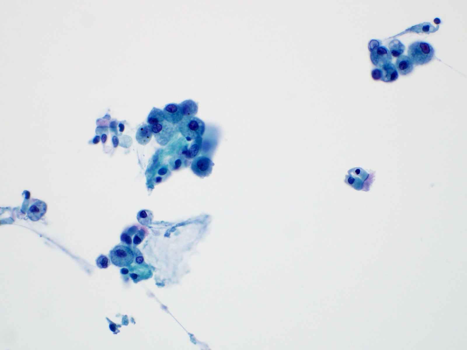

Bronchial cells

Bronchial cells cross section
Macrophages (ThinPrep)
Anthracosis

Hamartoma
(Diff-Quik)

Meningioma (Diff-Quik)
Images hosted on other servers:

Herpes inclusions

Cytomegalovirus (CMV)

Creola body
Sample pathology report
- Lung, right upper lobe, fine needle aspiration:
- Negative for carcinoma
- Granulomatous inflammation present
- Lung, left upper lobe, bronchoalveolar lavage:
- No malignant cells identified
- Predominately pulmonary macrophages and chronic inflammation
- Lung, right lower lobe, fine needle aspiration:
- Adipose, cartilage and benign reactive bronchial cells (see comment)
- No malignant cells identified
- Comment: The combination of these findings is suggestive of a hamartoma but clinicoradiographic correlation and surgical biopsy are recommended for definitive diagnosis.
Differential diagnosis
Additional references
Board review style question #1
This image is from a 63 year old man who presented with a right upper lobe mass. Which of the following is the best diagnosis?
- Atypical cells present
- Benign: bronchial cells only
- Benign: favor hamartoma
- Nondiagnostic
- Positive for non-small cell carcinoma
Board review style answer #1
C. Benign: favor hamartoma. This FNA image comes from a 63 year old man who presented with a right upper lobe lobulated mass. A Diff-Quik smear shows benign, matrix-like material with adjacent chronic inflammation. This case was signed out as having neoplastic cells that are compatible with pulmonary hamartoma. Correlation with the imaging findings was helpful in rendering this diagnosis. Answers A and D are incorrect because the matrix material and adipose tissue are hallmarks for hamartoma; therefore, the categories of nondiagnostic and atypical are not appropriate in this context. Answer B is incorrect because there is an abundance of matrix material and a lack of bronchial cells. Answer E is incorrect because there are no small cell carcinoma cells in this sample.
Comment Here
Reference: Cytopathology - Benign (lung)
Comment Here
Reference: Cytopathology - Benign (lung)
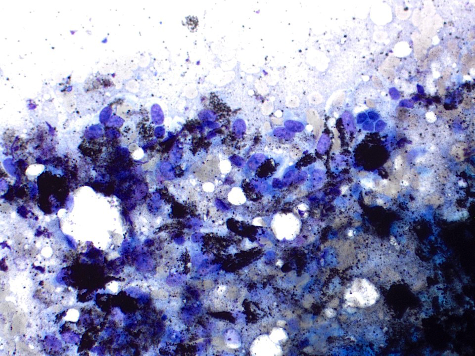
Board review style question #2

This image is from a 35 year old woman that has a history of lung transplant and underwent a bronchial washing. Which of the following is the best diagnosis?
- Atypical cells present
- Benign: cytomegalovirus (CMV) present
- Nondiagnostic
- Positive for non-small cell carcinoma
- Suspicious for adenocarcinoma
Board review style answer #2
B. Benign: cytomegalovirus (CMV) present. This ThinPrep slide shows cells with owl eye inclusions characteristic of CMV. There is an enlarged nucleus with a surrounding halo, consistent with viral cytopathic changes of CMV. Answer A is incorrect because while the nuclei are atypically large, the nuclear halo helps with the diagnosis of CMV infection in benign cells. Answer C is incorrect because the cells present are diagnostic of CMV. Answers D and E are incorrect because the sample does not show neoplastic cells. The nuclei are enlarged but the changes seen, such as the nuclear halo and the preserved N:C ratio, are more supportive of CMV infection.
Comment Here
Reference: Cytopathology - Benign (lung)
Comment Here
Reference: Cytopathology - Benign (lung)
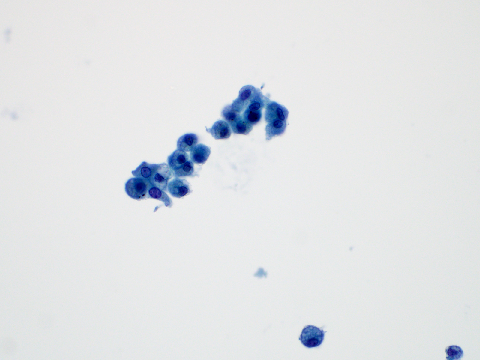
Board review style question #3
This image is from a 57 year old man that underwent a bronchial washing. Which of the following is the best diagnosis?
- Atypical cells present
- Benign: bronchial cells only
- Nondiagnostic
- Positive for metastatic colorectal adenocarcinoma
- Positive for non-small cell carcinoma
Board review style answer #3
B. Benign: bronchial cells only. The cells in the image show a low N:C ratio and are columnar. Prominent cilia are present, supporting the diagnosis of benign bronchial cells. Answer A is incorrect because the cells do not show nuclear atypia. In addition, there is appropriate N:C ratio and terminal bars with cilia. Overall, these features exclude these cells from the atypical category. Answer C is incorrect because this is a bronchial washing and the presence of bronchial cells and macrophages are considered normal elements in such a specimen. Answers D and E are incorrect because the background columnar cells show preserved N:C ratio and terminal bars with cilia. The nuclei are not overlapping. Overall, these features are diagnostic of benign bronchial cells.
Comment Here
Reference: Cytopathology - Benign (lung)
Comment Here
Reference: Cytopathology - Benign (lung)

