Appendix
Adenocarcinoma and related tumors
LAMN and HAMN
Authors: Michael Feely, D.O., Raul S. Gonzalez, M.D.
Editorial Board Member: Catherine E. Hagen, M.D.


Last author update: 28 April 2021
Last staff update: 31 January 2024
Copyright: 2002-2025, PathologyOutlines.com, Inc.
PubMed Search: Mucinous neoplasms appendix[title] free full text[sb]
Table of Contents
Definition / general | Essential features | Terminology | Epidemiology | Clinical features | Radiology description | Prognostic factors | Treatment | Gross description | Gross images | Microscopic (histologic) description | Microscopic (histologic) images | Positive stains | Negative stains | Molecular / cytogenetics description | Sample pathology report | Differential diagnosis | Practice question #1 | Practice answer #1Cite this page: Feely M, Gonzalez RS. LAMN and HAMN. PathologyOutlines.com website. https://www.pathologyoutlines.com/topic/appendixmucinousneoplasm.html. Accessed August 25th, 2025.
Definition / general
- Evolving nomenclature with considerable controversy, although recent consensus terminology has been established (Am J Surg Pathol 2016;40:14)
- Variable clinical consequences depending on the location of neoplastic epithelium and associated mucin
- By definition, must lack infiltrative invasion, which would be termed mucinous adenocarcinoma
Essential features
- Low grade appendiceal mucinous neoplasm (LAMN) is a low grade noninvasive epithelial proliferation that can cause pseudomyxoma peritonei if the appendix ruptures
- Similar, rare lesions with high grade nuclear dysplasia are termed high grade appendiceal mucinous neoplasm (HAMN)
Terminology
- Low grade appendiceal mucinous neoplasm (LAMN): lesion arising in appendix with low grade epithelial features in the absence of infiltrative growth
- High grade appendiceal mucinous neoplasm (HAMN): lesion arising in appendix with high grade epithelial features in the absence of infiltrative growth
- Pseudomyxoma peritonei: strictly clinical term for apparent mucinous ascites or peritoneal mucin deposition
- Mucocele: strictly clinical term for dilated, mucin filled appendix
- Cystadenoma: outdated diagnostic term that should no longer be used
Epidemiology
- Incidence may be higher in interval appendectomy specimens from adults (JAMA Surg 2013;148:703)
Clinical features
- Typically occurs in patients during their sixth decade of life, although age range is broad; more common in women (Am J Surg Pathol 2009;33:1425)
- Most patients with disease restricted to the appendix present with acute appendicitis-like symptoms, while those with disseminated disease may present with abdominal or ovarian masses or pseudomyxoma peritonei
Radiology description
- Appendix with a diameter of more than 15 mm, a soft tissue mass or wall thickening may raise the possibility of a mucinous neoplasm (Cancer Imaging 2013;13:14)
Prognostic factors
- Mucinous lesions confined to appendix largely considered cured by resection
- Lesions with extra-appendiceal acellular mucin considered to be low risk for recurrence or progression, occurring in about 4% of cases (Am J Surg Pathol 2009;33:248)
- If extra-appendiceal mucin contains neoplastic epithelium, patient is at high risk for recurrence or dissemination, which occurs in 33 - 75% of these cases (Am J Surg Pathol 2009;33:248, Am J Surg Pathol 2009;33:1425)
Treatment
- Simple appendectomy considered sufficient for lesions limited to appendix
- Close surveillance for patients with localized periappendiceal disease following initial surgery
- Disseminated peritoneal disease may be treated with hyperthermic intraperitoneal chemotherapy (HIPEC) following cytoreductive surgery
Gross description
- Typically, appendix appears dilated with luminal mucin, although diameter may appear unremarkable
- Serosa appears smooth when appendiceal wall is intact
- Adhesions or extra-appendiceal mucin are concerning for underlying rupture
Gross images
Contributed by @Andrew_Fltv on Twitter

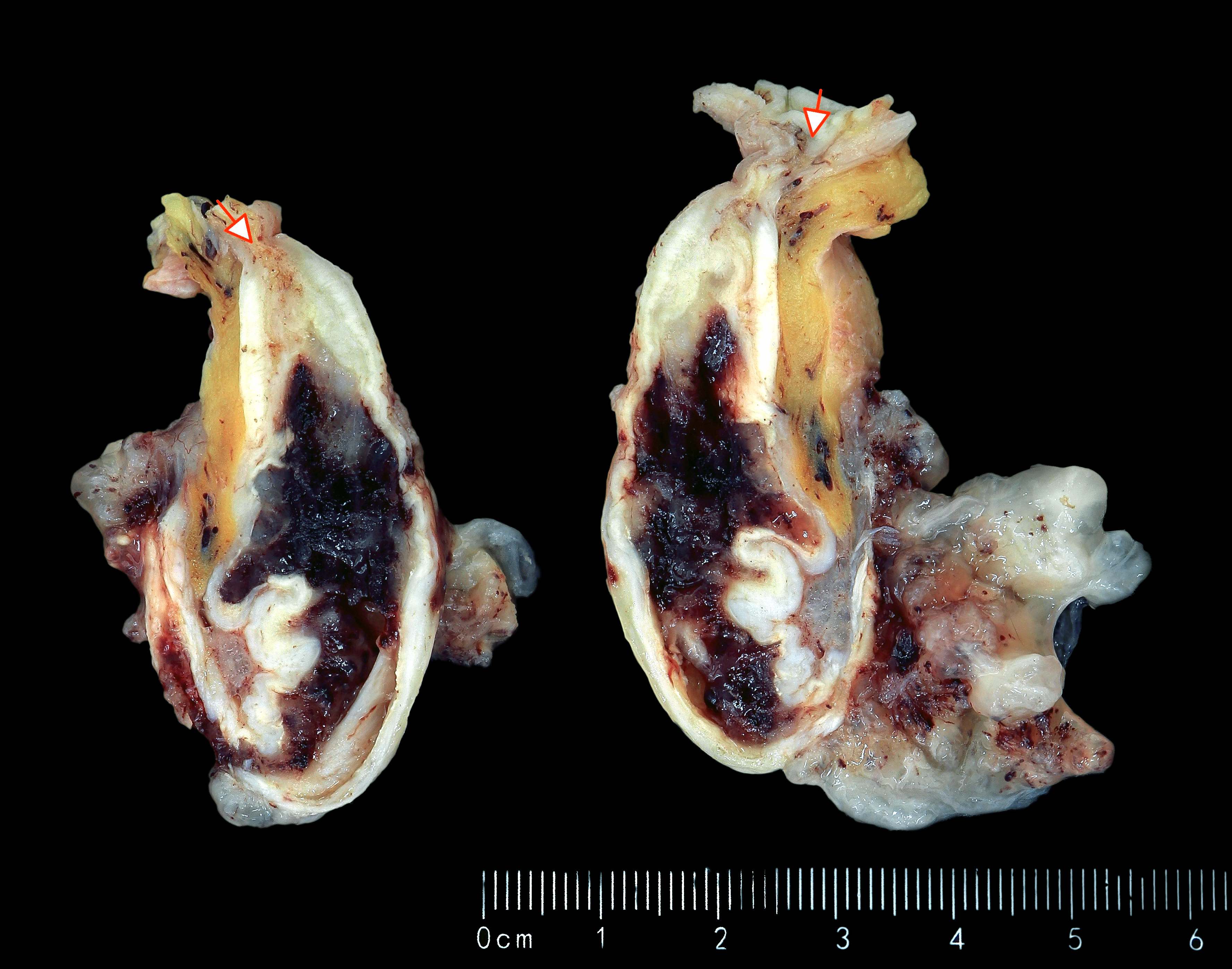
LAMN and HAMN
Images hosted on other servers:

Mucocele: dilated appendix
Microscopic (histologic) description
- Villous or occasionally flat proliferation of mucinous epithelial cells originating from appendiceal lumen
- Lesional cells typically demonstrate abundant apical mucin with elongated nuclei and low grade nuclear atypia (LAMN); however, nuclei may appear compressed or rarely high grade (HAMN)
- HAMN may show convoluted architecture, including micropapillary or cribriform features (Histopathology 2020;76:461)
- Often associated with atrophy of underlying lymphoid tissue, crypt loss and effacement of muscularis mucosae
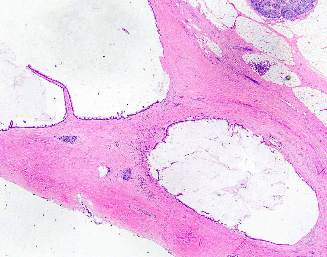
- Broad dissection of mucin, epithelium or both may occur with potential involvement of extra-appendiceal surface, an important finding affecting prognosis
- Extra-appendiceal mucin incites a serosal reaction and may contain neovascularization, assisting in differentiation from benign transfer of mucin during gross examination
Microscopic (histologic) images
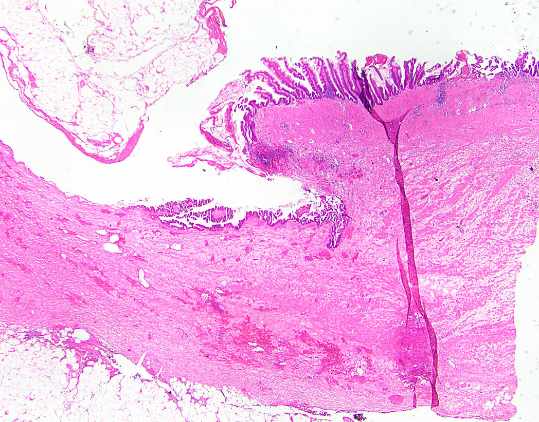
Contributed by Raul S. Gonzalez, M.D. and Michael Feely, D.O.
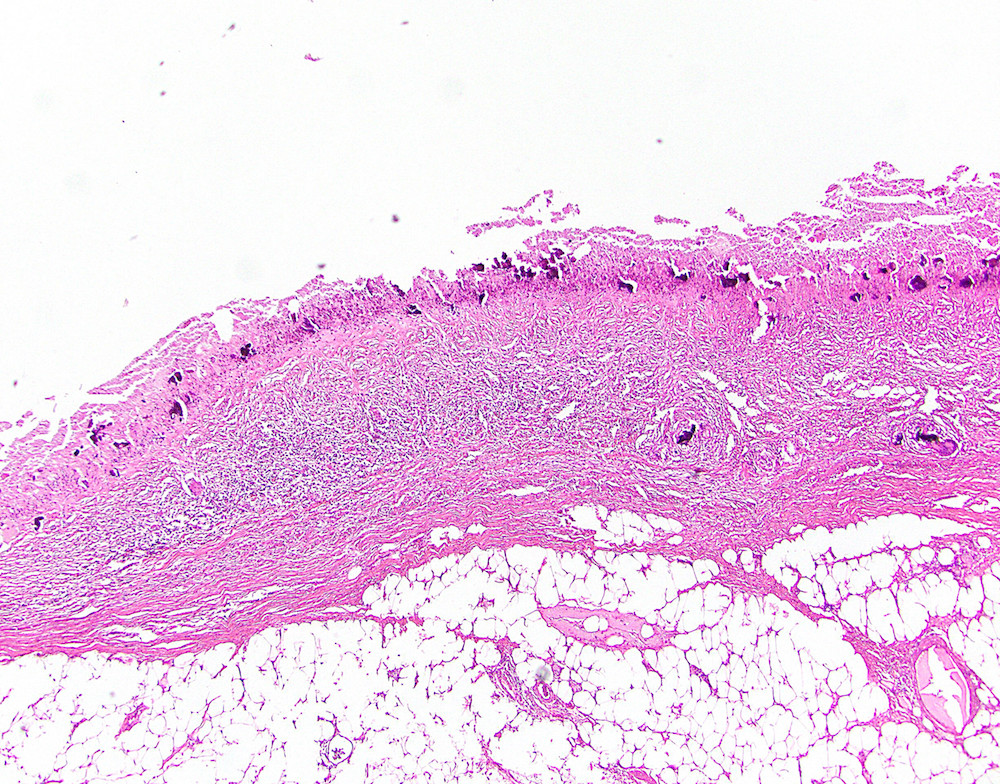
Denuded LAMN
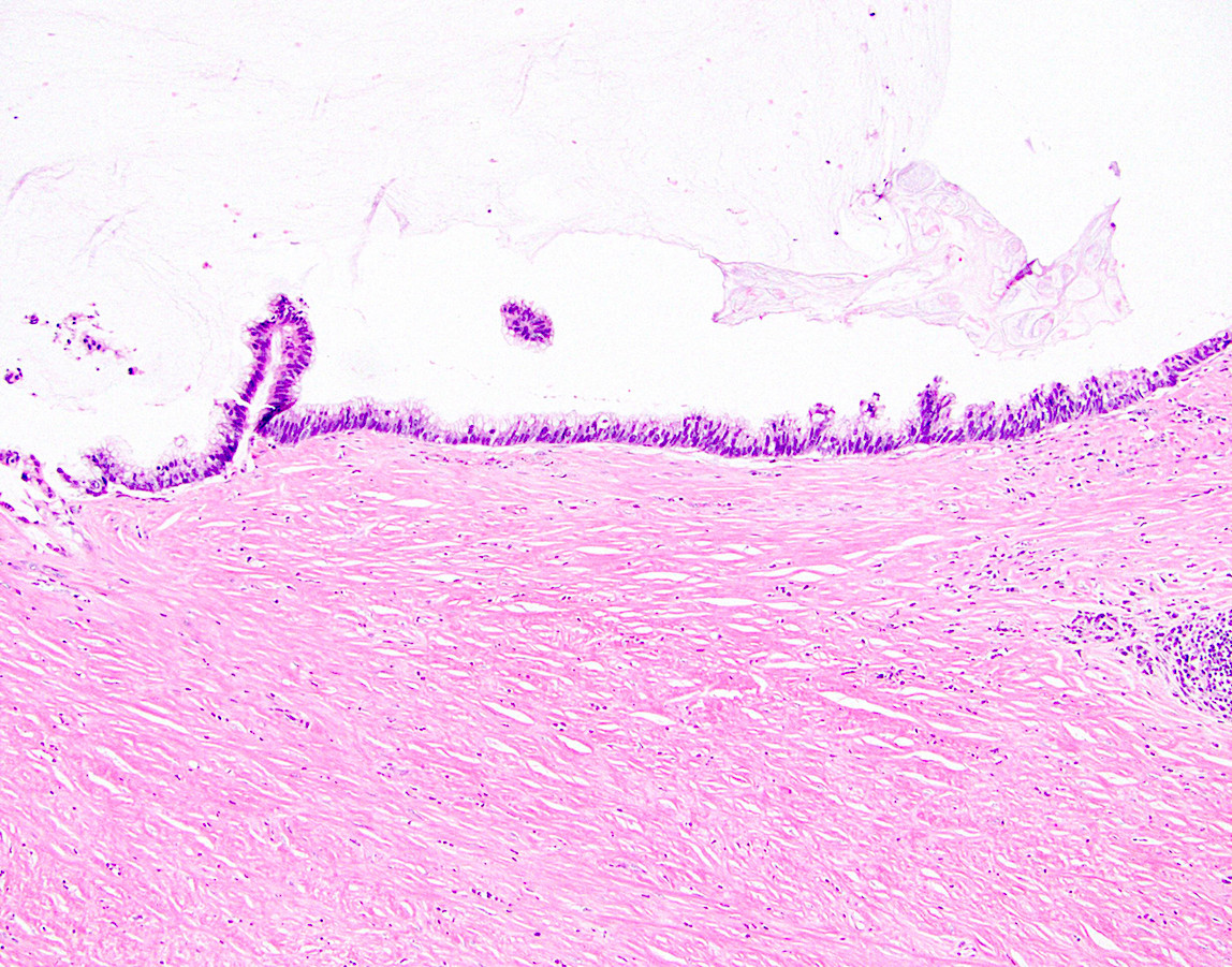
LAMN with loss of muscularis mucosae
LAMN with pushing invasion
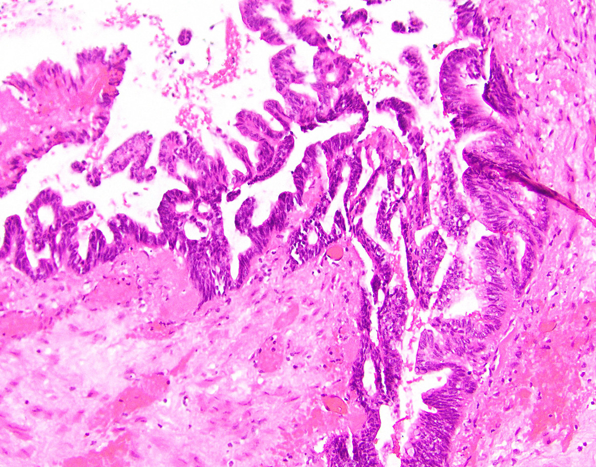
HAMN

LAMN in cross section

Villous architecture in LAMN
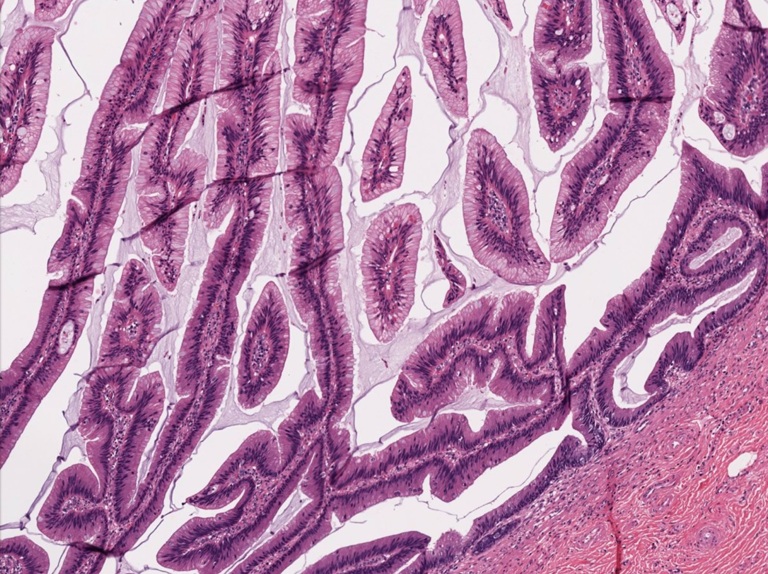
Low grade epithelium in LAMN

LAMN with extra-appendiceal mucin

Extra-appendiceal mucin with serosal reaction

Cellular extra-appendiceal mucin
Positive stains
Negative stains
Molecular / cytogenetics description
- Frequently harbor KRAS mutations and loss of chromosome 5q has been reported (Am J Pathol 1999;154:1849)
- GNAS alterations have been reported to occur in 50% of cases (Br J Cancer 2013;108:951)
- HAMN may also have TP53 or ATM mutations (Histopathology 2020;76:461)
- Microsatellite instability and BRAF mutations have not been demonstrated
Sample pathology report
- Appendix, appendectomy:
- Low grade appendiceal neoplasm, confined to appendix (see synoptic report)
Differential diagnosis
- Mucinous adenocarcinoma of the appendix:
- Infiltrative pattern of invasion with high grade cytologic features, desmoplasia or single cell infiltration
- Mucinous tumors of the ovary:
- Frequently nonreactive for SATB2 (Histopathology 2016;68:977)
- Ruptured appendiceal diverticulum:
- Reactive as opposed to neoplastic epithelial changes, preserved lamina propria
- May be associated with neural or Schwann cell proliferations (Am J Surg Pathol 2009;33:1515)
- Serrated polyp:
- Localized serrated epithelial lesion within the luminal appendix, with retention of the muscularis mucosae
- Villous adenoma:
- More striking dysplasia, focally tubulovillous architecture, retention of muscularis mucosae
- Endometriosis:
- Appendiceal cases with intestinal metaplasia may mimic LAMN (Am J Surg Pathol 2014;38:698)
Practice question #1
Which finding associated with an appendiceal mucinous neoplasm has the greatest risk of progression?
- Acellular mucin outside the right lower quadrant of abdomen
- Acellular mucin restricted to right lower quadrant of abdomen
- Extra-appendiceal mucin containing low grade epithelium
- High grade epithelium confined to appendix
- Low grade epithelium confined to appendix
Practice answer #1
C. Extra-appendiceal mucin containing low grade epithelium
Comment Here
Reference: LAMN and HAMN (mucinous neoplasms)
Comment Here
Reference: LAMN and HAMN (mucinous neoplasms)
