Oral cavity & oropharynx
Soft tissue tumors & proliferations
Giant cell fibroma
Author: Sarah H. Glass, D.D.S.
Editorial Board Member: Molly Housley Smith, D.M.D.
Deputy Editor-in-Chief: Kelly Magliocca, D.D.S., M.P.H.

Last author update: 21 February 2022
Last staff update: 21 February 2022
Copyright: 2002-2025, PathologyOutlines.com, Inc.
PubMed Search: Giant cell fibroma [TI]
Table of Contents
Definition / general | Essential features | ICD coding | Epidemiology | Sites | Pathophysiology | Etiology | Clinical features | Diagnosis | Prognostic factors | Case reports | Treatment | Clinical images | Gross description | Gross images | Microscopic (histologic) description | Microscopic (histologic) images | Positive stains | Negative stains | Sample pathology report | Differential diagnosis | Practice question #1 | Practice answer #1 | Practice question #2 | Practice answer #2Cite this page: Glass SH. Giant cell fibroma. PathologyOutlines.com website. https://www.pathologyoutlines.com/topic/oralcavitygiantcellfibroma.html. Accessed October 2nd, 2025.
Definition / general
- Benign fibrous proliferation of the oral cavity mucosa with distinct stellate fibroblasts
Essential features
- Fibrous nodule that most commonly presents on the gingiva and can have a papillary appearance
- Histologic features include large, plump stellate fibroblasts in the superficial connective tissue
- Treatment is conservative excision and recurrence is rare
ICD coding
Epidemiology
- 2 - 5% of all oral benign fibrous growths (Neville: Oral and Maxillofacial Pathology, 4th Edition, 2015)
- Increased frequency in (Case Rep Dent 2014;2014:864512):
- Patients' first to third decade
- Caucasians
Sites
- Most common location is the gingiva followed by the tongue, palate, buccal mucosa and lip (Oral Surg Oral Med Oral Pathol 1982;53:582)
Pathophysiology
- Fibrous proliferation generally considered to be reactive process (Braz J Otorhinolaryngol 2019;85:399, Case Rep Dent 2014;2014:864512)
- Giant cell fibroma shows limited growth potential, local recurrence would be considered exceptional and microscopic appearance is indistinguishable from nonneoplastic retrocuspid papilla; on balance, some authors have suggested the possibility that the lesion represents a neoplasm (Acta Histochem 2016;118:451, Head Neck Pathol 2019;13:71)
Etiology
- Unclear etiology but less likely to be associated with chronic mild trauma (Head Neck Pathol 2019;13:71)
Clinical features
- Dome shaped sessile or pedunculated fibrous nodule
- Surface may be smooth, pebbly, bosselated, nodular or papillary
- Most lesions are less than 1 cm in diameter (Head Neck Pathol 2019;13:71)
- Asymptomatic unless secondarily traumatized
Diagnosis
- Definitive diagnosis is made on histopathology
Prognostic factors
- Excellent prognosis and recurrence is rare
Case reports
- 7 year old boy with a pedunculated soft tissue nodule on the mandibular gingiva (J Oral Maxillofac Res 2013;4:e5)
- 19 year old man with a small growth on the tip of the tongue (Case Rep Dent 2014;2014:864512)
- 30 year old man with a growth on gingiva in the retromolar area (J Oral Maxillofac Pathol 2017;21:429)
- 40 year old man with a sessile mass of the buccal mucosa (J Dent Sci 2021;16:552)
Treatment
- Conservative surgical excision
Clinical images
Contributed by Sarah H. Glass, D.D.S.
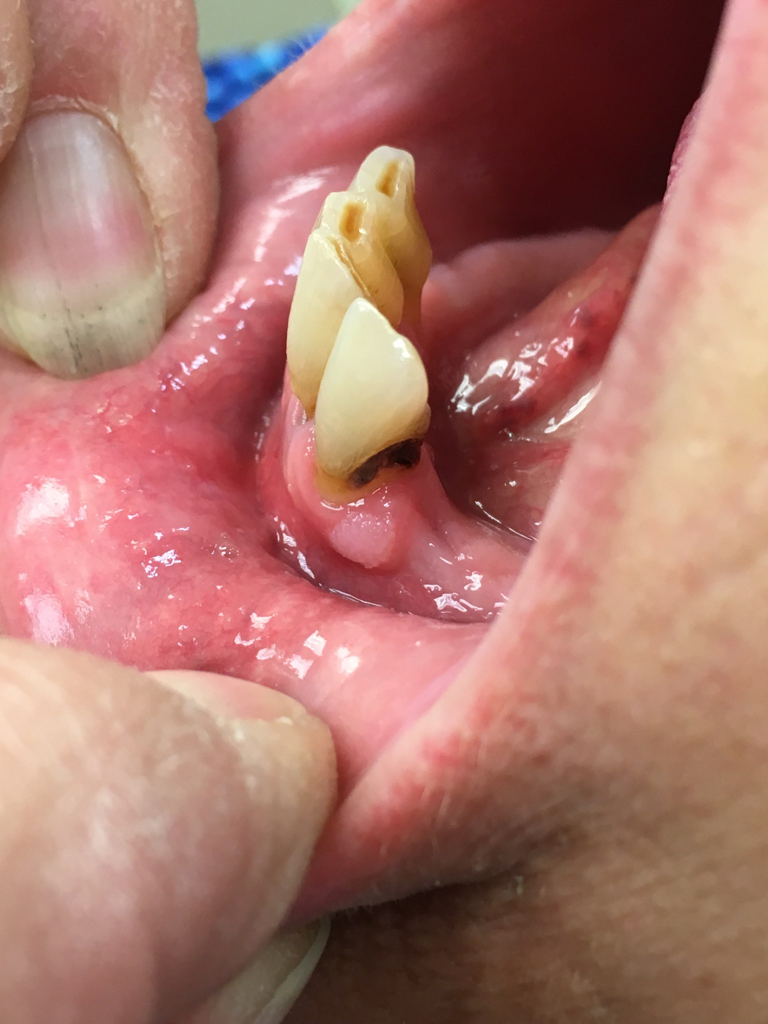
Bosselated gingival mass
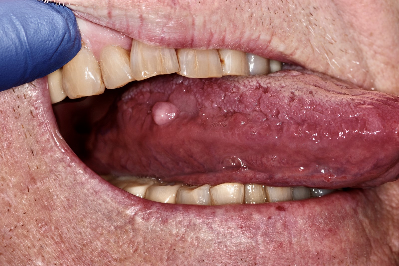
Small tongue mass
Gross description
- Firm tan nodule with white cut surface
Gross images
Contributed by Sarah H. Glass, D.D.S.
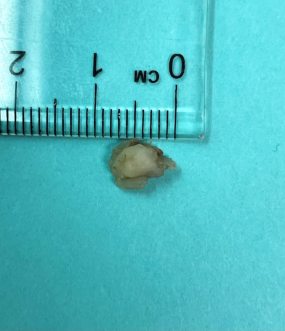
Dome shaped mass
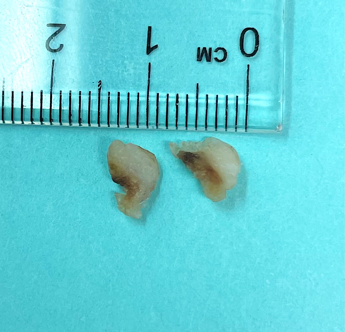
White cut surface
Microscopic (histologic) description
- Varying numbers of large, plump angular and stellate fibroblasts that may demonstrate multiple nuclei in the superficial connective tissue (Oral Surg Oral Med Oral Pathol 1974;37:374)
- Proliferation of collagen bundles
- Overlying surface epithelium with narrow and elongated epithelial rete ridges
- Absence of inflammation
Microscopic (histologic) images
Contributed by Sarah H. Glass, D.D.S.
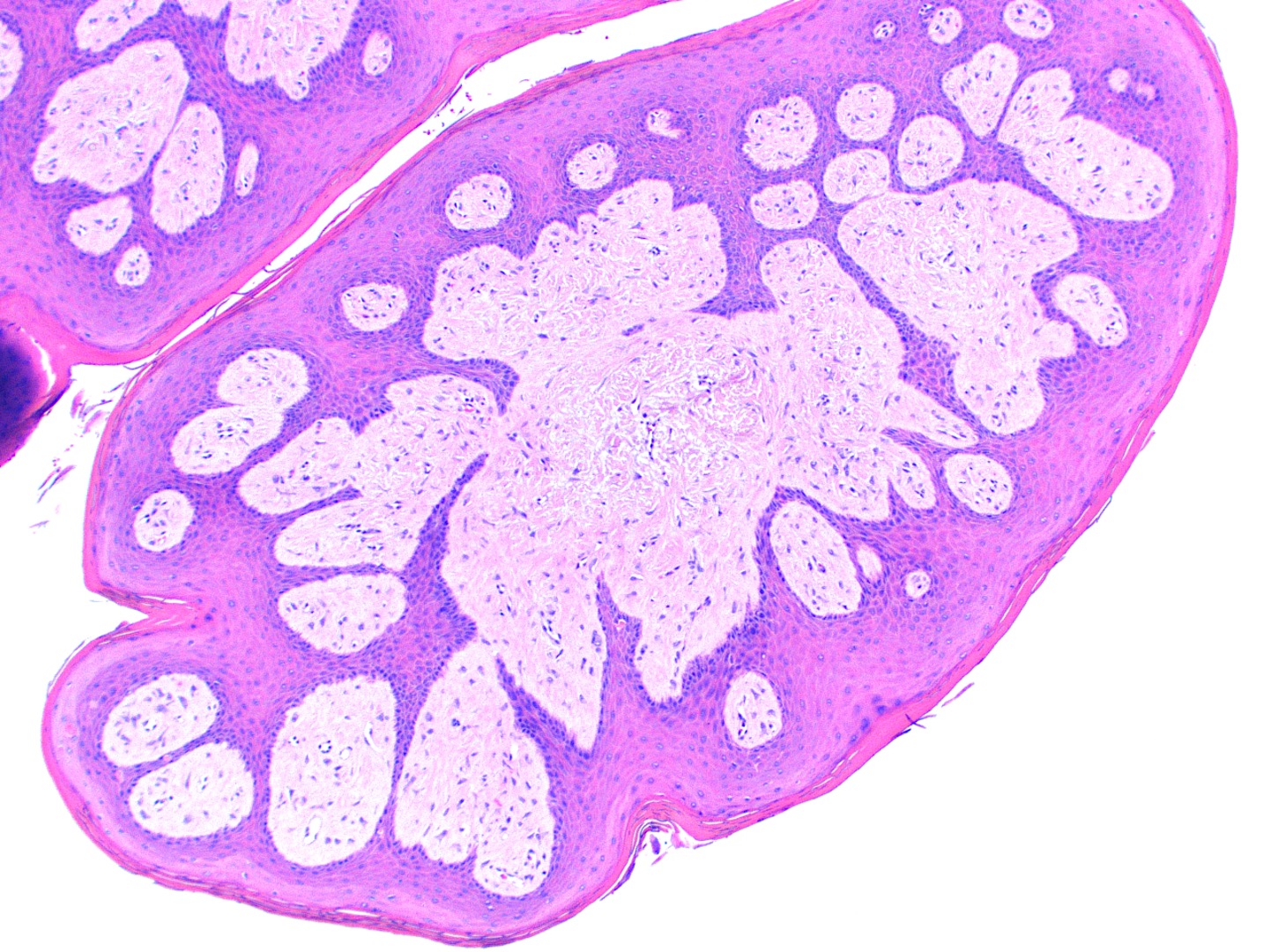
Nodular fibrous proliferation
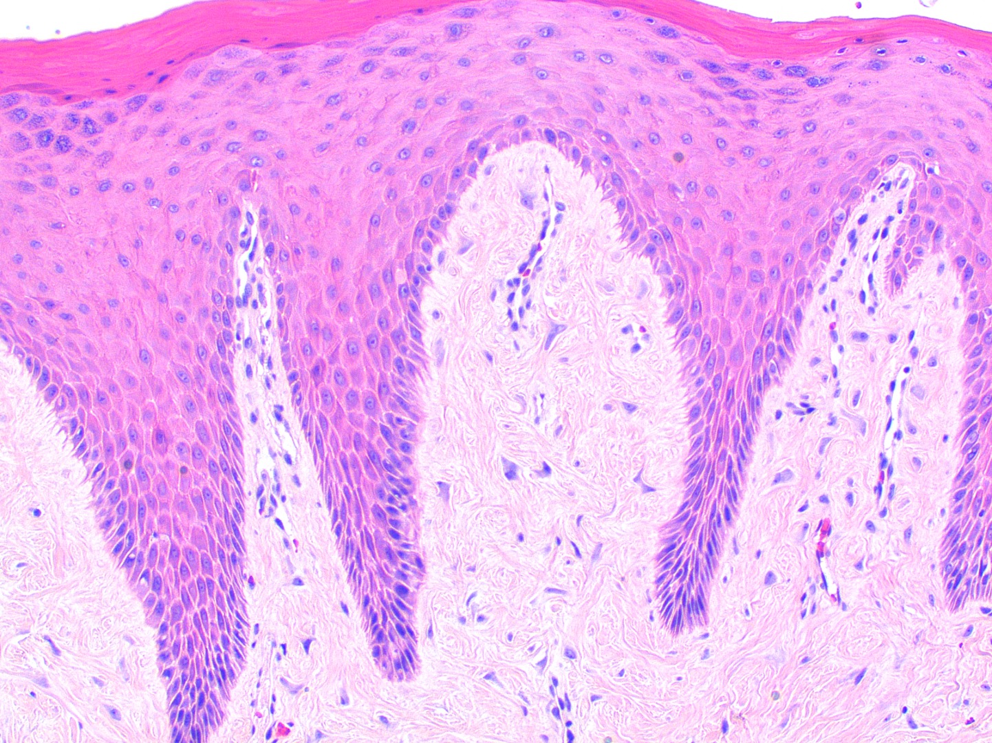
Elongated rete ridges
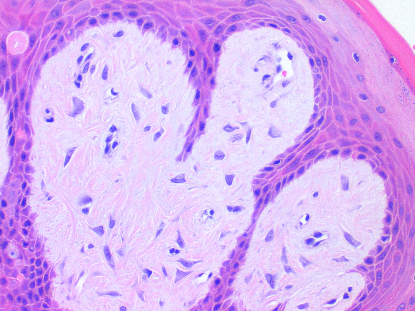
Stellate fibroblasts
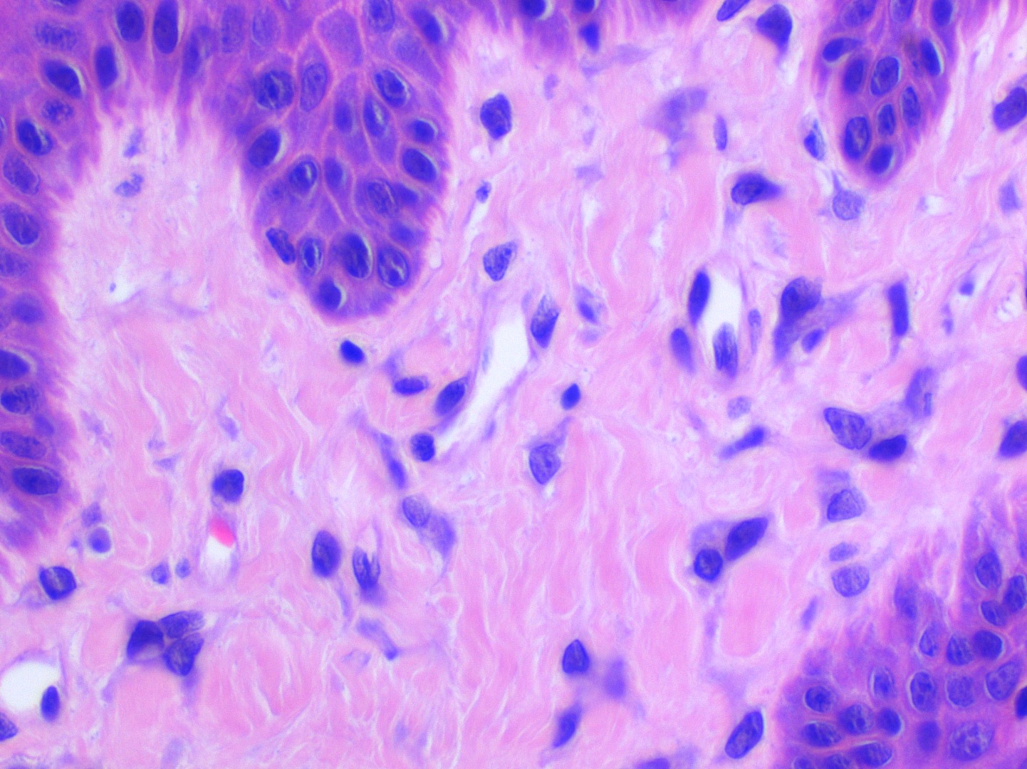
Multinucleated fibroblasts
Positive stains
Negative stains
Sample pathology report
- Oral cavity, excisional biopsy:
- Giant cell fibroma
Differential diagnosis
- Retrocuspid papillae (Head Neck Pathol 2019;13:71):
- Normal anatomic structure, therefore clinicopathologic correlation required
- Histopathologically identical to giant cell fibroma
- Oral squamous papilloma:
- Epithelial proliferation with koilocytes related to low risk human papillomavirus
- Verruciform xanthoma:
- Foam cells in the superficial connective tissue with overlying clefts of orange keratin
- Irritation fibroma:
- Reactive fibrous proliferation with chronic inflammation and without large stellate fibroblasts
- Other clinically noted common masses of the gingiva include pyogenic granuloma, peripheral ossifying fibroma and peripheral giant cell granuloma:
- Diagnosis made with histopathologic examination
Practice question #1
A 30 year old patient presents with a 5 mm bosselated soft tissue mass on the mandibular gingiva. Histopathology shows a benign fibrous proliferation with stellate fibroblasts in the superficial connective tissue with no inflammation. There is no history of trauma. What is the diagnosis?
- Giant cell fibroma
- Irritation fibroma
- Papilloma
- Pyogenic granuloma
Practice answer #1
A. Giant cell fibroma. All of the answer choices could be included on a clinical differential diagnosis of a gingival mass. Giant cell fibromas demonstrate stellate fibroblasts on microscopic exam. These lesions are not associated with trauma and do not routinely demonstrate inflammation unless secondarily traumatized.
Comment Here
Reference: Giant cell fibroma
Comment Here
Reference: Giant cell fibroma
Practice question #2
Which of the following is true regarding giant cell fibromas of the oral cavity?
- Features of human papillomavirus in the epithelium
- History of repeated trauma to the site
- Majority of lesions are larger than 1 cm
- Stellate fibroblasts in the superficial connective tissue
Practice answer #2
D. Stellate fibroblasts in the superficial connective tissue. Defining histologic feature of a giant cell fibroma is the presence of large, plump angular and stellate fibroblasts that may demonstrate multiple nuclei in the superficial connective tissue. These lesions generally have limited growth potential, are not associated with trauma and are not related to human papillomavirus.
Comment Here
Reference: Giant cell fibroma
Comment Here
Reference: Giant cell fibroma
