Ovary
Other nonneoplastic lesions
Ectopic decidual reaction
Editorial Board Member: Kyle Devins, M.D.
Deputy Editor-in-Chief: Gulisa Turashvili, M.D., Ph.D.


Last author update: 19 April 2024
Last staff update: 19 April 2024
Copyright: 2003-2025, PathologyOutlines.com, Inc.
PubMed Search: Ectopic decidual reaction
Table of Contents
Definition / general | Essential features | Terminology | Epidemiology | Sites | Pathophysiology | Diagnosis | Radiology description | Case reports | Treatment | Clinical images | Microscopic (histologic) description | Microscopic (histologic) images | Positive stains | Negative stains | Sample pathology report | Differential diagnosis | Additional references | Practice question #1 | Practice answer #1 | Practice question #2 | Practice answer #2Cite this page: Busca A, Parra-Herran C. Ectopic decidual reaction. PathologyOutlines.com website. https://www.pathologyoutlines.com/topic/ovarynontumorectopicdecidual.html. Accessed August 26th, 2025.
Definition / general
- Presence of ectopic decidualized uterine stromal cells in the ovary
Essential features
- Presence of ectopic decidualized uterine stromal cells in the ovary
- Usually an incidental finding associated with pregnancy
- Typically transient, will regress 4 - 6 weeks postpartum
Terminology
- Ectopic decidua or ovarian deciduosis
Epidemiology
- Associated with pregnancy
- In a study of 307 consecutive caesarean sections, macroscopic deciduosis was found in 31 (10.1%) cases (Eur J Obstet Gynecol Reprod Biol 2016;197:54)
- Can also develop as a result of exogenous progesterone effect
Sites
- Ectopic decidua have been described in the cervix, ovary and fallopian tube; also peritoneal surface, appendix, bladder, small and large intestine, mesentery, lymph nodes
Pathophysiology
- High levels of estrogen and progesterone during pregnancy induce mesenchymal fibroblast differentiation into decidual cells (Eur J Obstet Gynecol Reprod Biol 2016;197:54, Pathol Res Pract 1993;189:352, Korean J Obstet Gynecol 2011;54:373)
Diagnosis
- Often an incidental finding in surgical specimens or a discrete nodule / mass discovered during caesarean section
Radiology description
- When the ovarian deciduosis is mass forming, it may be difficult to differentiate from a neoplastic process (Case Rep Obstet Gynecol 2015;2015:217367)
Case reports
- 31 year old woman with decidualized ovarian mass mimicking malignancy (Case Rep Obstet Gynecol 2015;2015:217367)
- 32 year old woman with ovarian deciduosis in pregnancy (Korean J Obstet Gynecol 2011;54:373)
- 33 year old woman with ovarian and peritoneal deciduosis in pregnancy (Diagn Histopathol 2011;17:313)
Treatment
- Benign entity, no further treatment necessary; usually regresses 4 - 6 weeks postpartum
- If mass forming and showing worrisome features for malignancy on imaging, the lesion is excised to establish the correct diagnosis
- Reference: Case Rep Obstet Gynecol 2015;2015:217367
Clinical images
Images hosted on other servers:

Macroscopic appearance
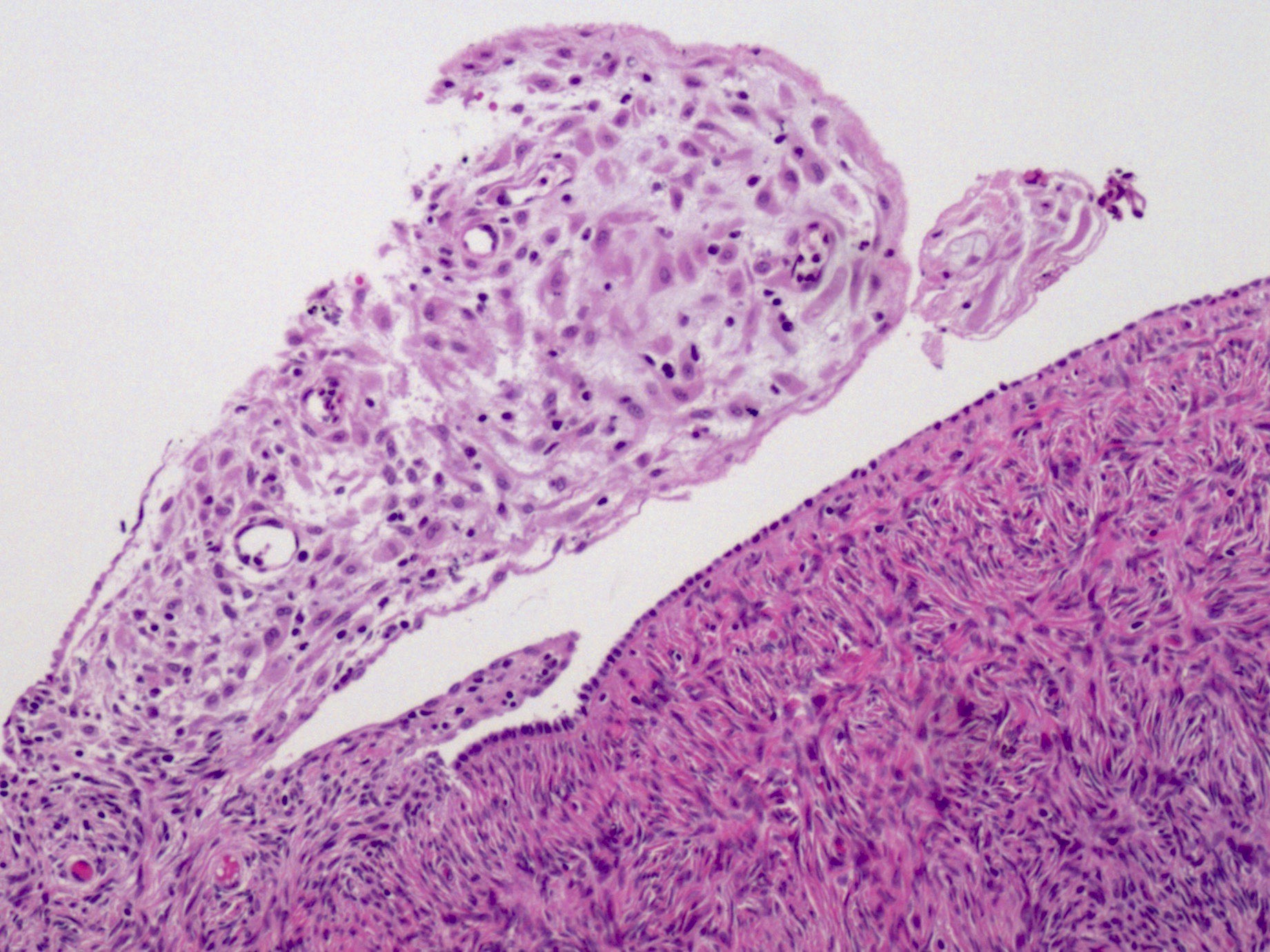
Microscopic (histologic) description
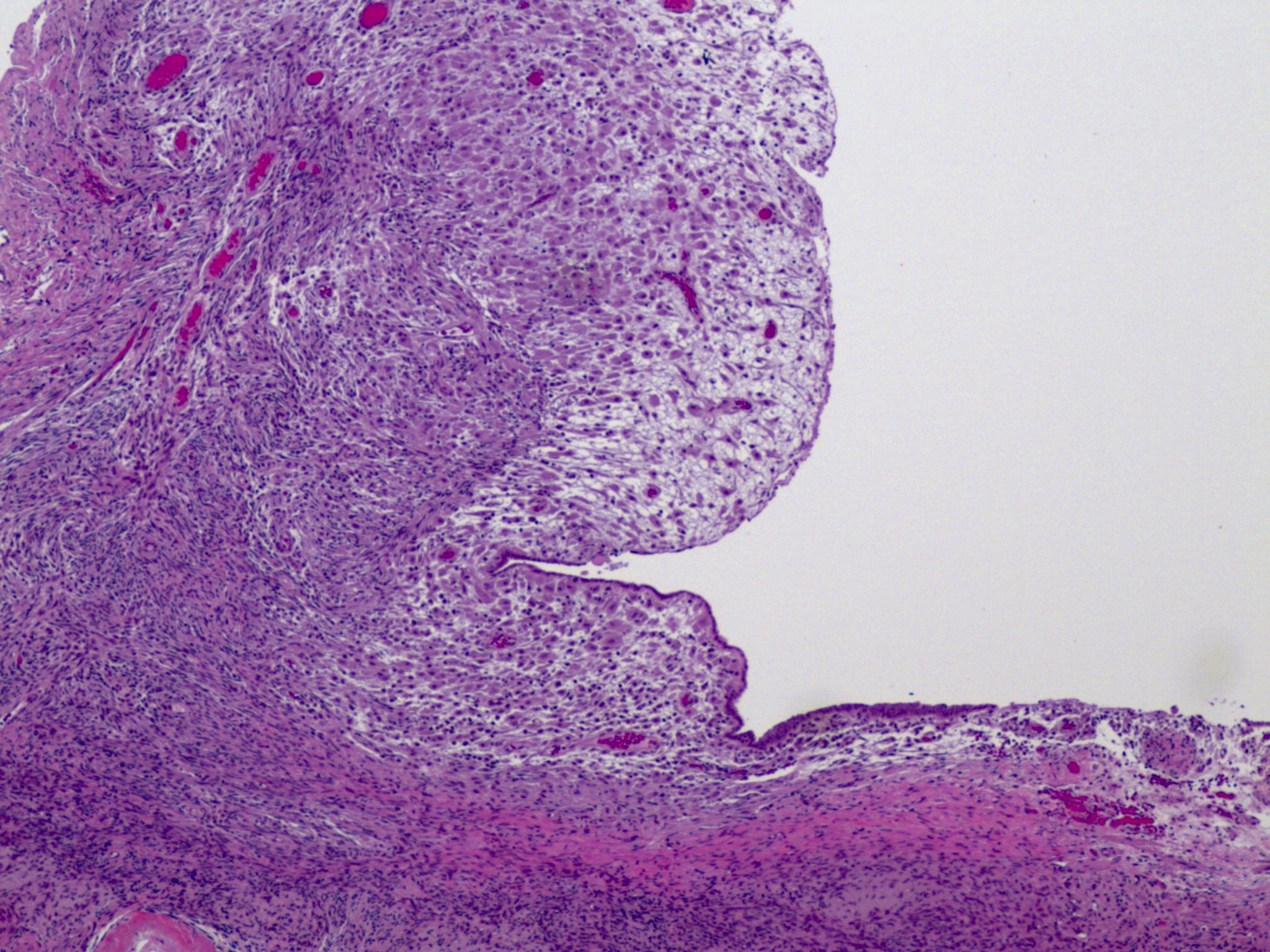
- Large polygonal cells with abundant eosinophilic cytoplasm, bland nuclei and visible nucleoli
- No glands present (to differentiate it from decidualized endometriosis)
- Reference: Case Rep Obstet Gynecol 2015;2015:217367
Microscopic (histologic) images
Contributed by Aurelia Busca, M.D., Ph.D.
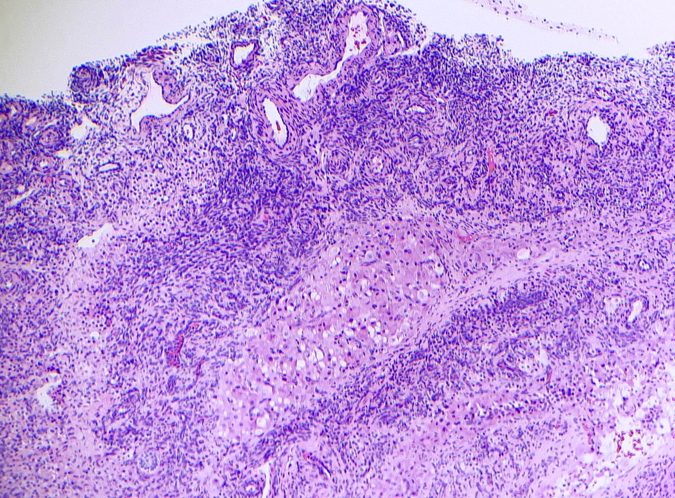
Deciduosis of ovarian stroma

Areas of deciduosis
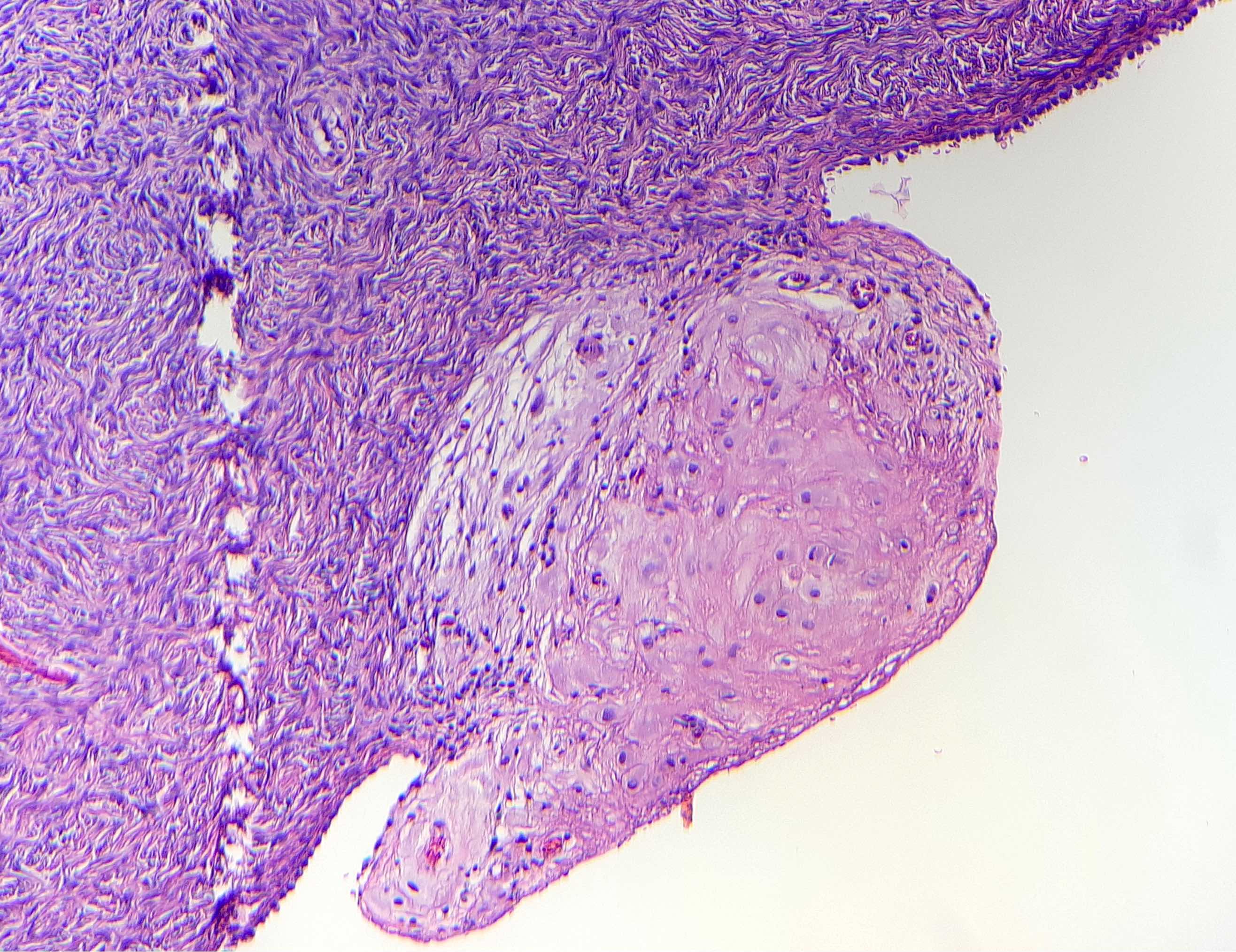
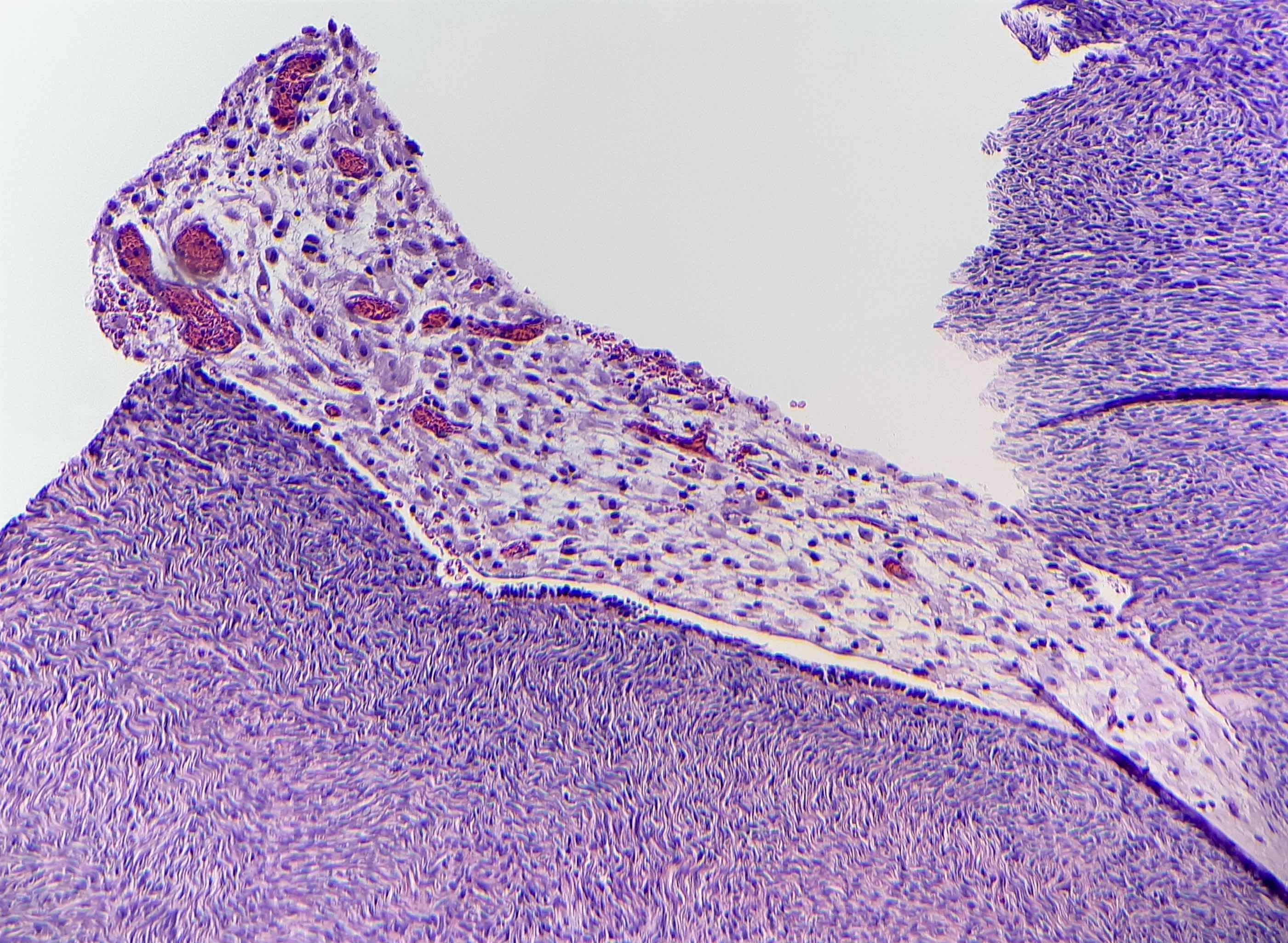
Ovarian surface adhesions with deciduosis
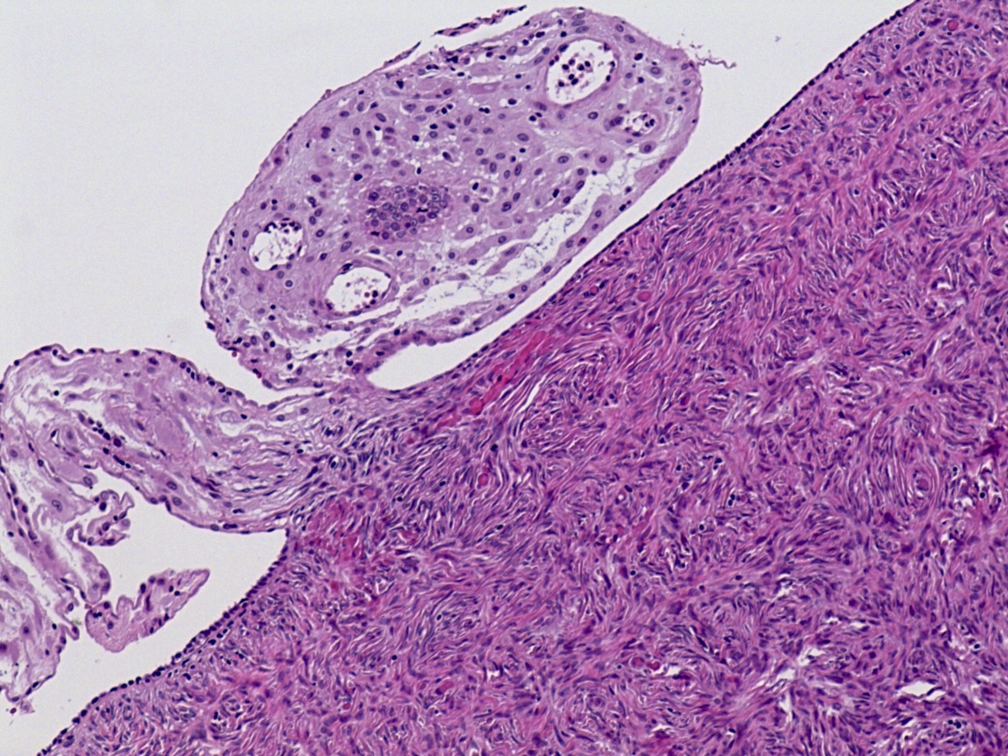
Ovarian surface adhesions with deciduosis
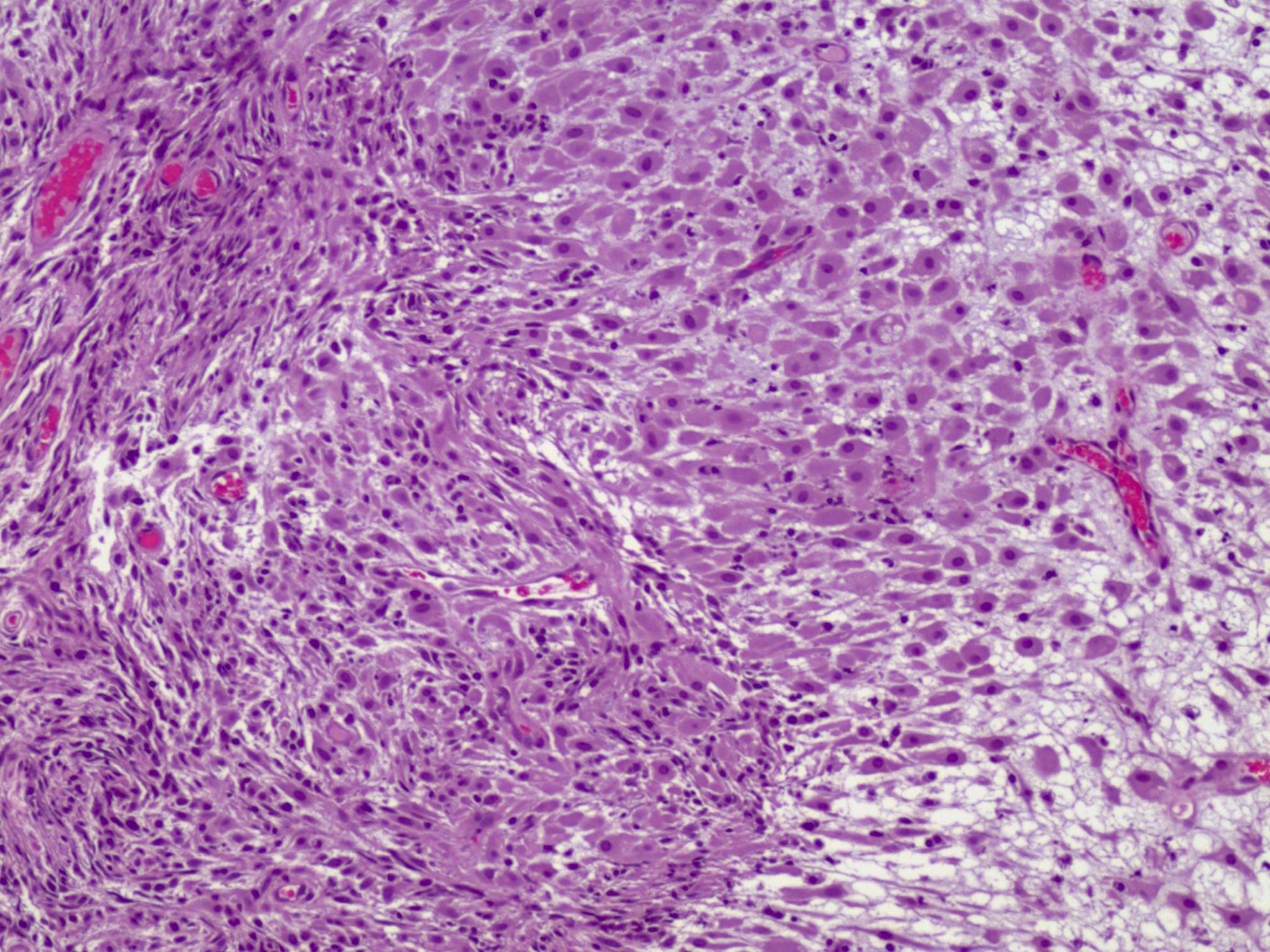
Ovarian deciduosis in a patient on progestin therapy
Positive stains
Negative stains
- Cytokeratin
- Mesothelial markers (WT1, calretinin)
Sample pathology report
- Ovary, oophorectomy:
- Benign ovary with focal deciduosis
Differential diagnosis
- Endometriosis:
- Can also undergo decidualization during pregnancy
- Main difference is that ectopic decidua have no glands (Gynecol Obstet Invest 2008;66:241)
- Epithelial ovarian neoplasm when mass forming:
- Histologic features are bland and mitotic activity is low in deciduosis
- Absence of glands in deciduosis
- Epithelial proliferation with variable atypia and architectural complexity in epithelial neoplasms
- Peritoneal mesothelioma or peritoneal carcinomatosis when there is additional involvement of peritoneal surface:
- Histologic features are bland in deciduosis; also, context of pregnancy is helpful
- Increased atypia, proliferation and architectural complexity in carcinomatosis and mesothelioma
- Metastatic squamous cell carcinoma with keratinization:
- More atypical
- Presence of necrosis
- Mitotically active
- Cytokeratin+
Additional references
Practice question #1
Which of the following is true about the ovarian change illustrated in the figure shown above?
- It is a malignant process
- It is only seen in the ovary
- It requires lymph node sampling and full staging for management
- More sampling is required to demonstrate the presence of glands
- Typically associated with pregnancy
Practice answer #1
E. Typically associated with pregnancy. The image shows ovarian deciduosis, which is associated with pregnancy, is typically transient and resolves postpartum. Answer A is incorrect because ovarian deciduosis is a benign process typically associated with pregnancy. Answer D is incorrect because it consists of stromal tissue, without glands. Answer C is incorrect because it typically regresses after pregnancy and does not require treatment. Answer B is incorrect because deciduosis can be seen at other sites (cervix, peritoneal cavity, etc.).
Comment Here
Reference: Ectopic decidual reaction
Comment Here
Reference: Ectopic decidual reaction
Practice question #2
Which of the following is true about ovarian deciduosis?
- It can be mistaken for endometriosis
- It is CD10 negative by immunohistochemistry
- Presence of nuclear atypia indicates malignant potential
- Responds to chemotherapy
- Treatment includes resection with or without chemotherapy
Practice answer #2
A. It can be mistaken for endometriosis with stromal decidualization but unlike endometriosis, it does not contain glands. Answer B is incorrect because both have CD10 positive stroma. Answers C, D and E are incorrect because ovarian deciduosis is a benign incidental process and does not require treatment.
Comment Here
Reference: Ectopic decidual reaction
Comment Here
Reference: Ectopic decidual reaction
