Soft tissue
Fibroblastic / myofibroblastic
Calcifying aponeurotic fibroma
Resident / Fellow Advisory Board: Erna Forgó, M.D.
Deputy Editor-in-Chief: Borislav A. Alexiev, M.D.


Last author update: 18 May 2022
Last staff update: 1 February 2023
Copyright: 2002-2025, PathologyOutlines.com, Inc.
PubMed Search: Calcifying aponeurotic fibroma
Table of Contents
Definition / general | Essential features | Terminology | ICD coding | Epidemiology | Sites | Pathophysiology | Etiology | Clinical features | Diagnosis | Radiology description | Radiology images | Prognostic factors | Case reports | Treatment | Clinical images | Gross description | Gross images | Microscopic (histologic) description | Microscopic (histologic) images | Cytology description | Positive stains | Negative stains | Molecular / cytogenetics description | Molecular / cytogenetics images | Videos | Sample pathology report | Differential diagnosis | Additional references | Practice question #1 | Practice answer #1 | Practice question #2 | Practice answer #2Cite this page: Ashfaq Z, Anjum S, Ud Din N. Calcifying aponeurotic fibroma. PathologyOutlines.com website. https://www.pathologyoutlines.com/topic/softtissuecalcifying.html. Accessed September 16th, 2025.
Definition / general
- Rare tumor of children and adolescents, that involves the distal extremities, in association with aponeuroses, tendons and fascia
- Characterized by bland spindle cells and less cellular zones of calcifications that have epithelioid to plump fibroblasts
Essential features
- Infiltrative lesion composed of bland spindle cells within a collagenous matrix
- Calcified areas that contain epithelioid fibroblasts or scattered giant cells
Terminology
- Juvenile aponeurotic fibroma
ICD coding
- ICD-O: 8816/0 - calcifying aponeurotic fibroma
Epidemiology
- Rare benign fibroblastic tumor
- Usually occurs in children < 15 years old
- Cases in adults have been reported (Medicine (Baltimore) 2021;100:e26803)
Sites
- Most commonly occurs on the palmar aspect of hands and fingers, followed by plantar aspect of feet and toes (Cancer 1970;26:857)
- Wrists and ankles are less commonly involved
- Unusual locations include the proximal extremities and trunk (Hum Pathol 1998;29:1504)
- Rare examples are documented in head and trunk regions
Pathophysiology
- Unknown
Etiology
- Unknown
Clinical features
- Presents as painless, poorly circumscribed soft tissue swelling of prolonged duration
- Propensity to recur
Diagnosis
- Appropriate clinical, radiological and histological examination
Radiology description
- Xray and ultrasound may show nonspecific soft tissue mass with variable extent of fine stippled calcifications (Radiographics 2009;29:2143)
- CT scan: optimal for evaluation of calcified areas
- MRI: superficial, ill defined, subcutaneous soft tissue mass with a tendency to infiltrate or adhere to surrounding tissues
Radiology images
Contributed by Nazmul Baqui, M.B.B.S., M.D.
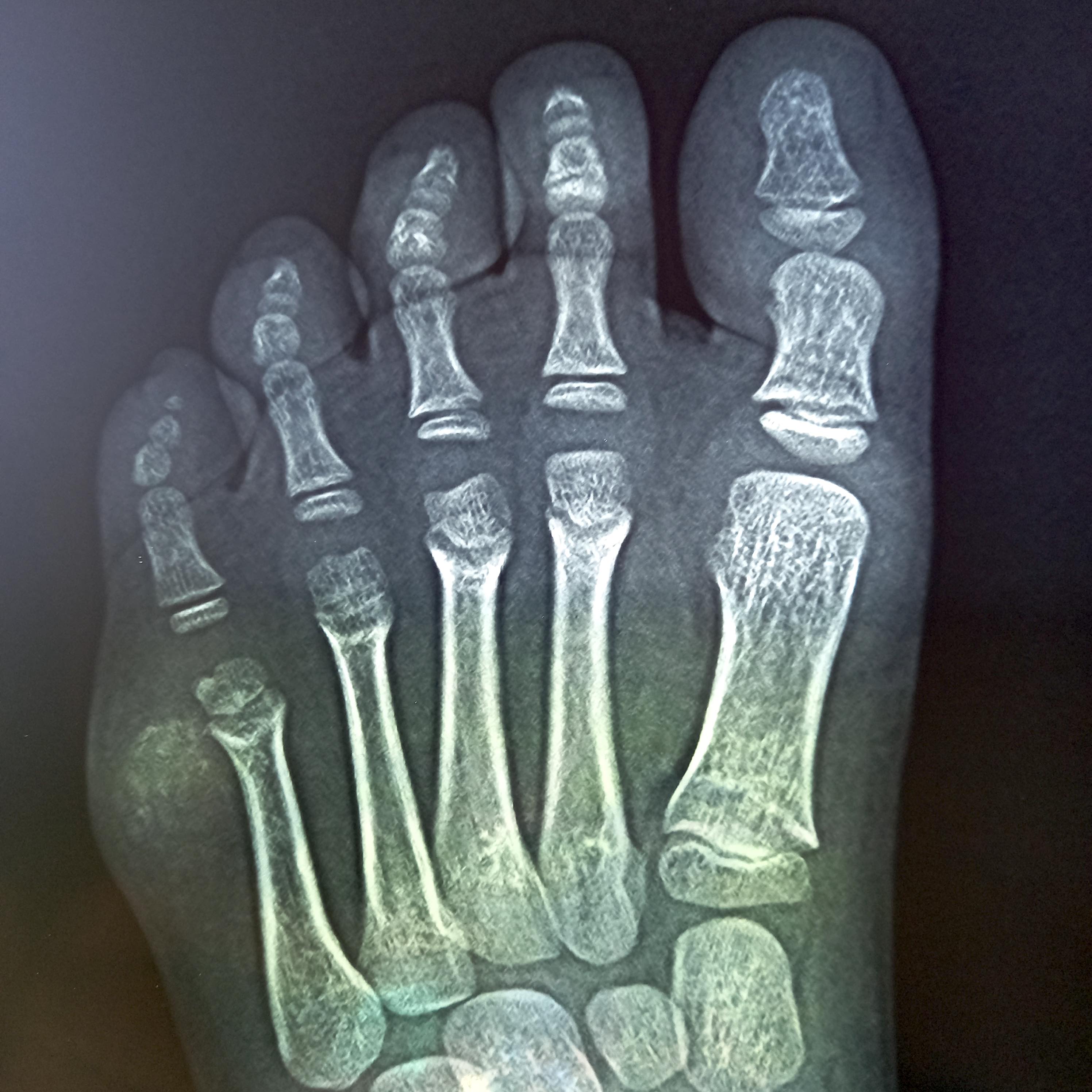
Xray of left foot
Images hosted on other servers:

Plain Xray of foot and axial CT

CT of lesion on medial aspect of foot

46 year old woman


36 year old woman with distal phalangeal bone involvement
Prognostic factors
- Benign but locally aggressive
- Due to infiltrative nature, local recurrences may occur
Case reports
- 4 year old girl with calcifying aponeurotic fibroma of Achilles tendon (Radiol Case Rep 2020;15:753)
- 8 year old boy with calcifying aponeurotic fibroma of right elbow (J Med Invest 2011;58:159)
- 8 year old girl with calcifying aponeurotic fibroma of right thigh (Int J Surg Case Rep 2020;69:96)
- 44 year old man with calcifying aponeurotic fibroma of distal phalanx (J Plast Reconstr Aesthet Surg 2013;66:e47)
- 52 year old man with multiple calcifying aponeurotic fibromas in the aponeuroses and fascia of the head, neck, trunk, upper and lower extremities (Ter Arkh 2018;90:91)
- 74 year old woman with calcifying aponeurotic fibroma involving the posterior tibialis tendon (Medicine (Baltimore) 2021;100:e26803)
Treatment
- Complete surgical excision is warranted
Clinical images
Images hosted on other servers:

Lesion in foot, intraoperative

Lesion at tip of index finger
Gross description
- Ill defined firm mass with variable grittiness
- Usually ≤ 3 cm
Gross images
Contributed by Nazmul Baqui, M.B.B.S., M.D.
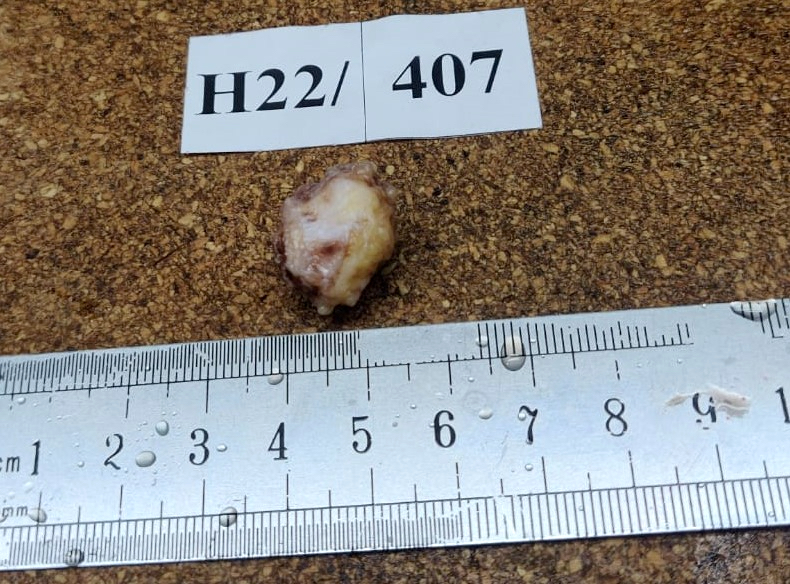
Vaguely nodular mass
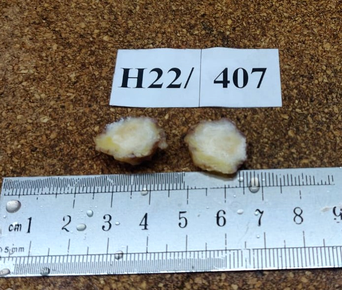
Firm gray cut surface
Images hosted on other servers:

Soft tissue mass with calcification

Gritty lesion resected from thigh
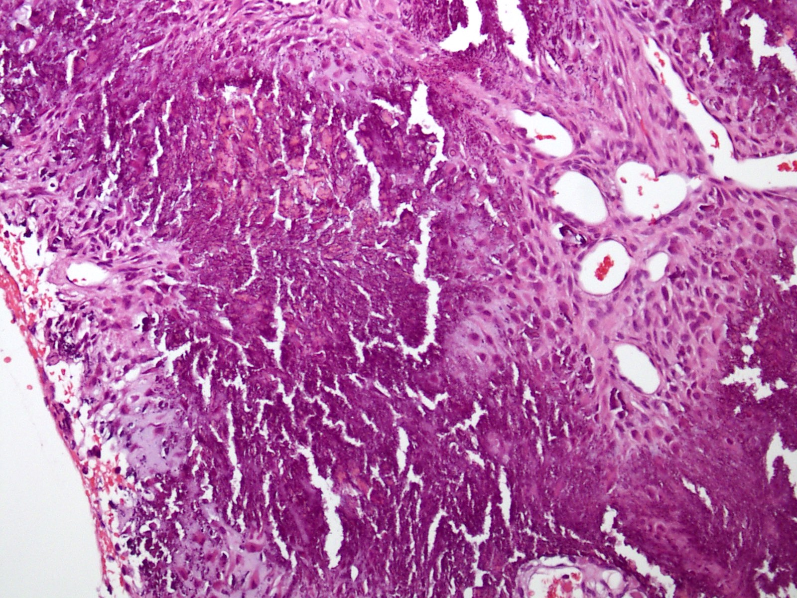
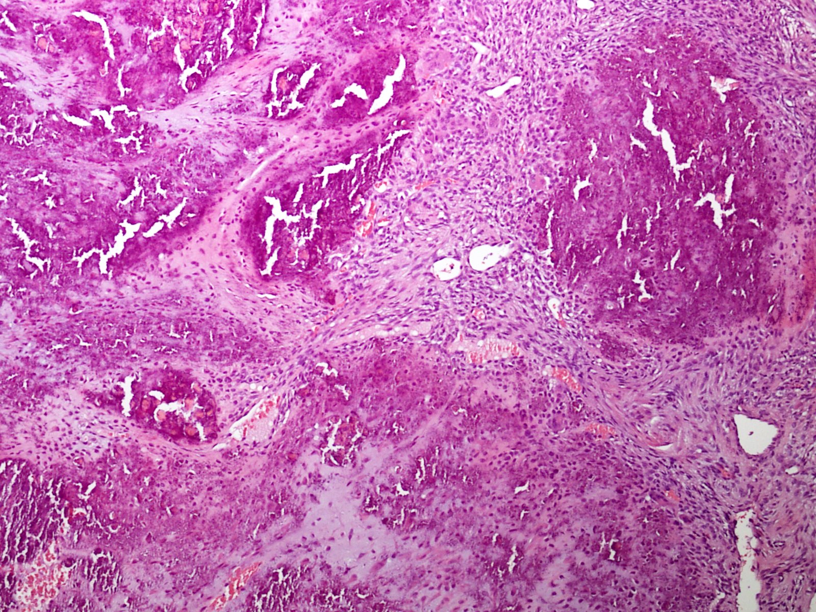
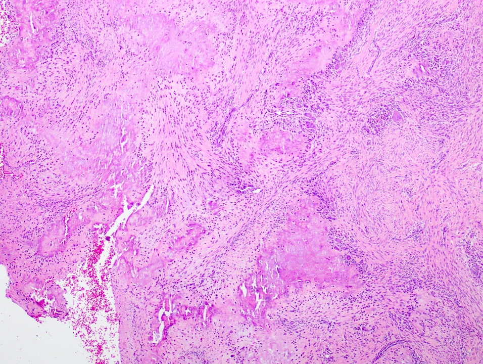
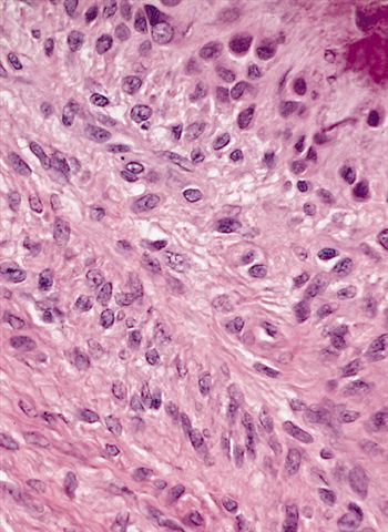
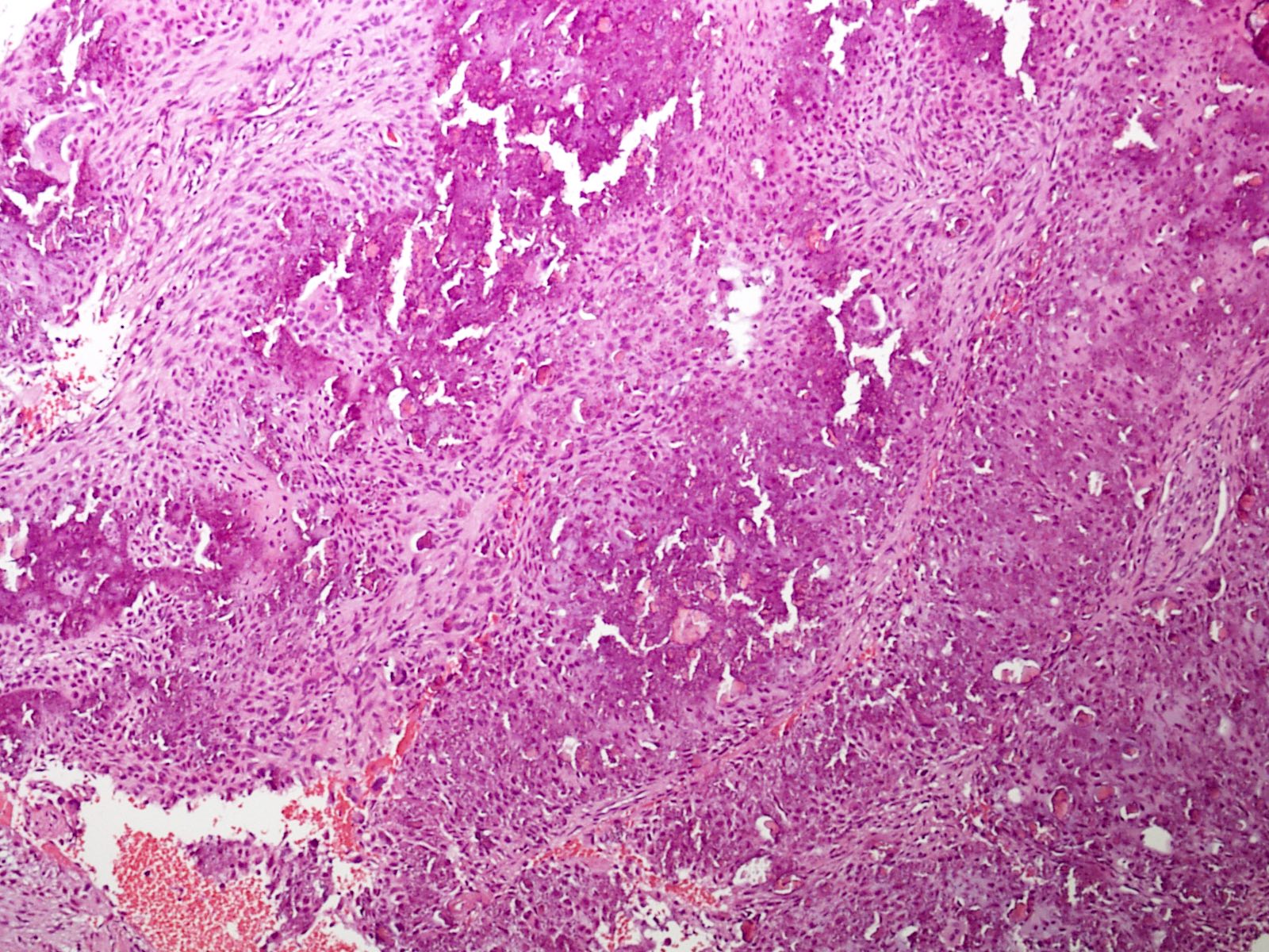
Microscopic (histologic) description
- Fibromatosis-like, infiltrative and nodular calcified components
- Infiltrative cellular component is composed of uniform plump spindle cells
- No significant nuclear atypia or mitoses are seen
- Calcified hypocellular component is either hyalinized or shows chondrocytes
- Osteoclast-like giant cells are usually present
- Lesion infiltrates the surrounding soft tissue
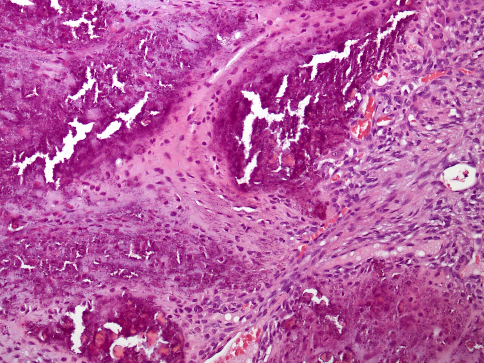
Microscopic (histologic) images
Contributed by Nasir Ud Din, M.B.B.S., Nazmul Baqui, M.B.B.S., M.D. and AFIP
Circumscribed calcified lesion
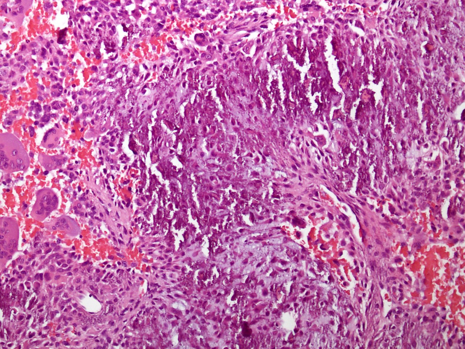
Giant cells
Spindle and giant cells
Prominent calcified area
Chondroid nodules
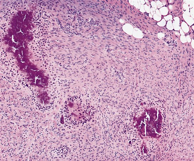
Poorly circumscribed fibroproliferative process

Amorphous
calcification
surrounded by
palisading cells
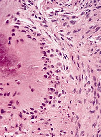
Typical zonation pattern
Rounded cells adjacent to hyalinized layer
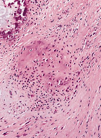
Chondroid area adjacent to calcification
Cytology description
- Cytologic examination reveals benign appearing spindled cells, chondroid cells, multinucleated giant cells and calcific debris (Diagn Cytopathol 2001;24:336)
Negative stains
Molecular / cytogenetics description
- In cases lacking calcification, FN1::EGF gene fusion is detected (J Pathol 2016;238:502)
Molecular / cytogenetics images
Images hosted on other servers:

Schematic diagrams of genes and RT PCR images
Videos
Calcifying aponeurotic fibroma
Sample pathology report
- Left hand, swelling, excision:
- Calcifying aponeurotic fibroma (see comment)
- Comment: Histology showed a lesion composed of bland spindle cells and less cellular zones of calcifications that have epithelioid to plump fibroblasts. These tumors are prone to recur if incompletely excised.
Differential diagnosis
- Inclusion body fibromatosis:
- Myofibroblastic tumor that usually occurs on the digits of children
- Composed of bland spindle cells with characteristic intracytoplasmic inclusions
- Ganglion cyst:
- Most common soft tissue lesion observed in the hand
- Frequently located in the dorsal wrist
- Histologically hypocellular cystic fibrocollagenous tissue fragments with no definite lining (J Orthop Surg (Hong Kong) 2019;27:2309499019840736)
- Tenosynovial giant cell tumor, localized type:
- Mostly occurs on the volar aspect of the first 3 fingers
- Usually occurs in patients aged 30 - 50 years
- Moderately cellular with abundant mononuclear cells
- Scattered multinucleated osteoclast-like giant cells, hemosiderin pigment, foamy histiocytes and collagenized stroma (Medicine (Baltimore) 2021;100:e26445)
- Schwannomas and neurofibromas:
- Both tumors occur less frequently in the hands and are composed of spindle shaped cells with serpentine nuclei
- Scwannomas typically show Verocay bodies
- Neurofibromas have a uniform spindle cell population within a collagenous to myxoid background and may show nerves at the periphery
- Both usually lack calcifications and usually are not circumscribed (J Orthop Surg (Hong Kong) 2019;27:2309499019840736)
- Rheumatoid arthritis:
- Usually affects older individuals and affects the synovium of the wrist
- Proliferative synovitis with villous hypertrophy and fibrinoid necrosis
- Extensive lymphoplasmacytic and histiocytic infiltration and lymphoid follicle / germinal center formation (Medicine (Baltimore) 2021;100:e26445)
Additional references
Practice question #1
A 2 year old boy presented with mass lesion of the hand. Radiology shows a soft tissue mass near finger tendons with specks of calcifications. What is the most appropriate diagnosis with respect to age and findings?
- Calcifying aponeurotic fibroma
- Epidermal inclusion cyst
- Ganglion cyst
- Inclusion body fibromatosis
- Rheumatoid arthritis
Practice answer #1
Practice question #2
This lesion, as shown in the photomicrograph above, was resected from the hand of a 2 year old boy. What is the most likely diagnosis?
- Calcifying aponeurotic fibroma
- Epidermal inclusion cyst
- Fibroma of tendon sheath
- Fibromatosis
- Nuchal type fibroma
Practice answer #2
