Soft tissue
Extraskeletal bone tumors
Soft tissue chondroma
Editorial Board Member: Josephine Kam Tai Dermawan, M.D., Ph.D.
Deputy Editor-in-Chief: Borislav A. Alexiev, M.D.


Last author update: 6 November 2023
Last staff update: 17 November 2023
Copyright: 2002-2025, PathologyOutlines.com, Inc.
PubMed Search: Soft tissue chondroma
Table of Contents
Definition / general | Essential features | Terminology | ICD coding | Epidemiology | Sites | Clinical features | Diagnosis | Radiology description | Radiology images | Prognostic factors | Case reports | Treatment | Clinical images | Gross description | Gross images | Frozen section description | Frozen section images | Microscopic (histologic) description | Microscopic (histologic) images | Cytology images | Positive stains | Negative stains | Molecular / cytogenetics description | Sample pathology report | Differential diagnosis | Additional references | Practice question #1 | Practice answer #1 | Practice question #2 | Practice answer #2Cite this page: Beshah FN, Velez Torres JM. Soft tissue chondroma. PathologyOutlines.com website. https://www.pathologyoutlines.com/topic/softtissueeskchondroma.html. Accessed September 9th, 2025.
Definition / general
- Benign, cartilage forming tumor that usually arises in the vicinity of joints or tendons in the hands and feet of adults
Essential features
- Arises from soft tissue of fingers, hands and feet
- Not connected to the underlying bone
- Well circumscribed, cartilaginous proliferation
Terminology
- Soft tissue chondroma, extraskeletal chondroma, chondroma of soft parts
ICD coding
- ICD-10: D21.9 - benign neoplasm of connective and other soft tissue, unspecified
Epidemiology
- Age: 30 - 60 years (Surg Pathol Clin 2015;8:419)
- Gender: slight male predominance
Sites
- Mostly occurs in the fingers
- Hands, toes, feet and trunk are less frequently affected
Clinical features
- Occurs in soft tissue of hands and feet
- Solitary, slowly enlarging nodule; occasionally causes pain or tenderness
- Reference: Histopathology 1986;10:147
Diagnosis
- Clinical presentation
- Solitary nodule
- Involves fingers, toes and hands
- < 3 cm
- Imaging
- Computed tomography
- Magnetic resonance imaging
- Reference: Histopathology 1986;10:147
Radiology description
- Well demarcated
- Does not involve bone, although some tumors cause compression deformities or bone erosion
- Discrete, irregular, ring-like or curvilinear calcifications
- Reference: AJR Am J Roentgenol 1985;144:1263
Radiology images
Contributed by Andrew E. Rosenberg, M.D.
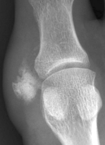
Calcified lesion
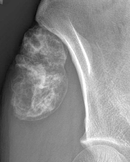
Well circumscribed, calcified soft tissue mass
Prognostic factors
- Favorable prognosis
- 15 - 20% recurrence rate (Surg Pathol Clin 2015;8:419)
Case reports
- 14 year old boy with plantar swelling (Int J Surg Case Rep 2022;90:106688)
- 51 year old man with posterior mediastinal mass (J Int Med Res 2021;49:3000605211053557)
- 84 year old man with palmar mass (Cureus 2021;13:e19467)
Treatment
- Simple excision
Clinical images
Contributed by Andrew E. Rosenberg, M.D.
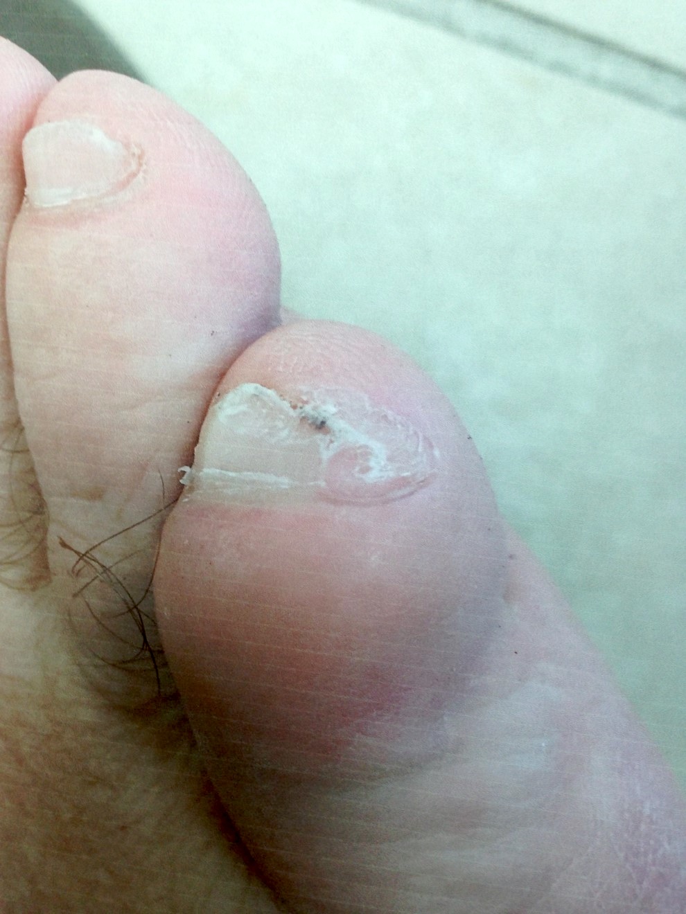
Swelling over the toe
Images hosted on other servers:

Foot tumor
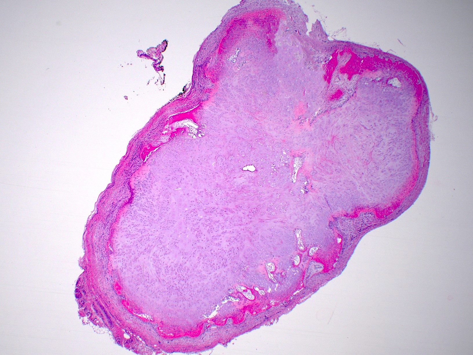
Gross description
- Well demarcated, oval - round
- < 3 cm in size
Gross images
Images hosted on other servers:

Well circumscribed mass


Foot tumor
Frozen section description
Frozen section images
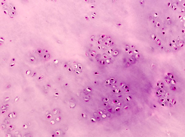
Contributed by Fireneh N. Beshah, M.D. and Jaylou M. Velez Torres, M.D.
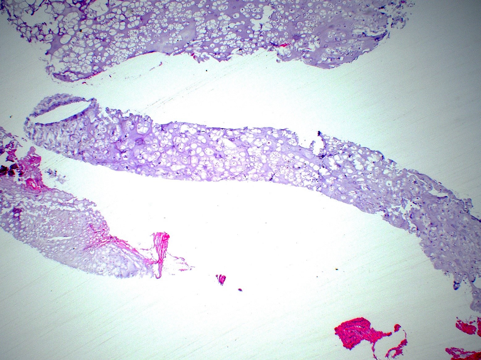
Hypocellular cartilage
Chondrocytes with small nuclei
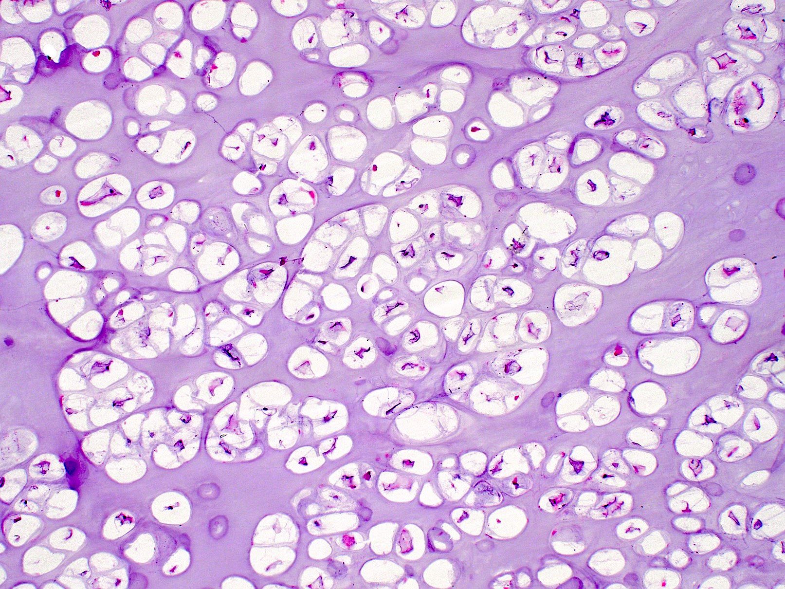
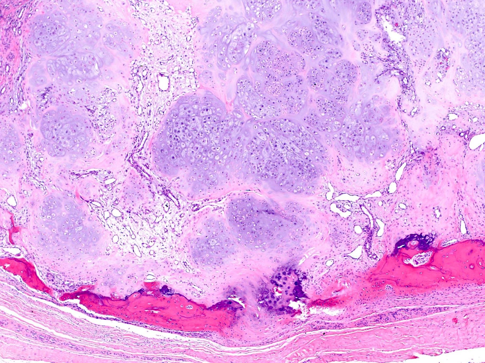
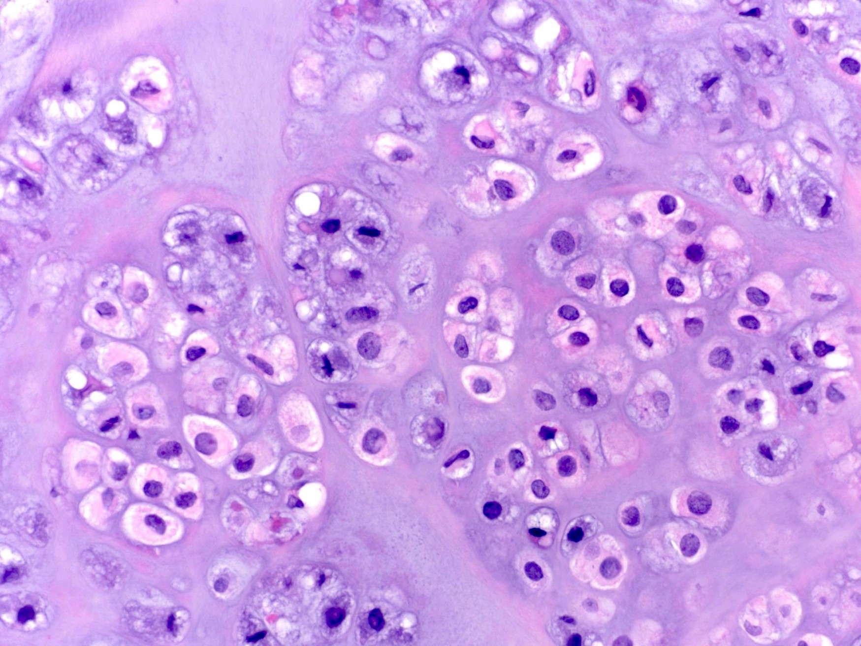
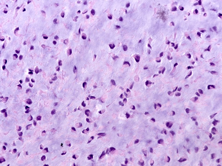
Microscopic (histologic) description
- Mature hyaline cartilage arranged in lobules with sharp borders
- Chondrocytes in lacunae, arranged diffusely or in small clusters
- Rarely can have moderate pleomorphism
- 33% show focal or diffuse calcifications
- Chondroblastoma-like soft tissue chondroma shows hypercellular areas with cells resembling chondroblasts, recently reclassified as calcified chondroid mesenchymal tumor (Mod Pathol 2021;34:1373)
Microscopic (histologic) images
Contributed by Andrew E. Rosenberg, M.D.
Well circumscribed nodule
Peripheral ossification
Enlarged nuclei
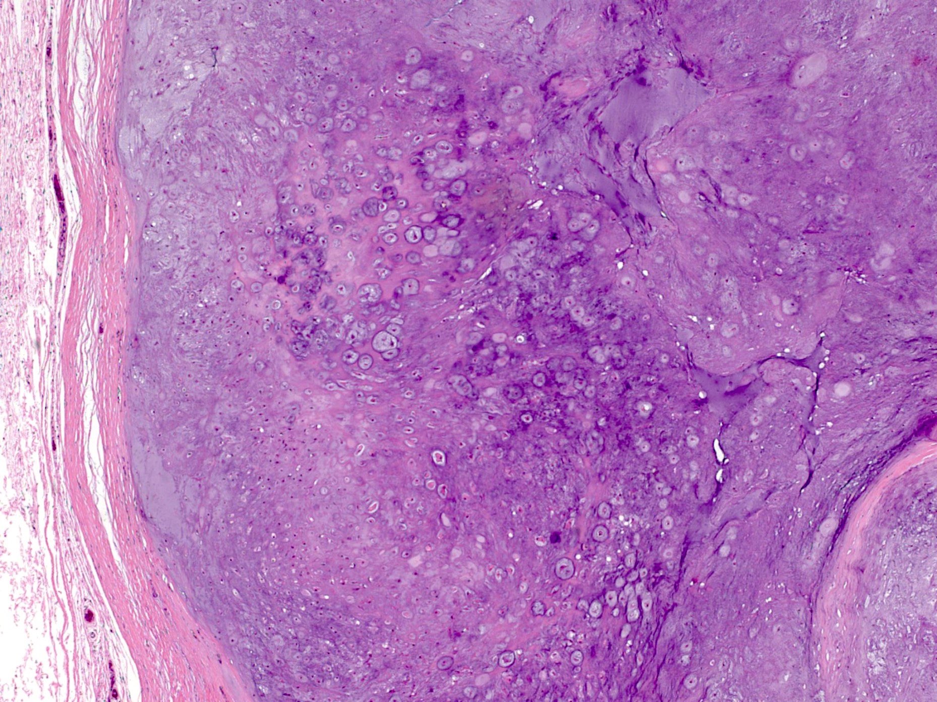
Fibrous pseudocapsule
Cellular and myxoid areas
Fibrocartilage
Cytology images
Images hosted on other servers:

Foot tumor
Negative stains
Molecular / cytogenetics description
- 12q13-q15 rearrangements involving HMGA2 have been reported (Cancer Genet Cytogenet 1993;69:79, Cancer Genet Cytogenet 2000;118:144, J Pathol 2002;196:194, Mod Pathol 2003;16:1132, Clin Neuropathol 2015;34:13)
- Also FN1 rearrangements (Mod Pathol 2019;32:1762)
- Soft tissue chondromas harbor the FN1::FGFR1 fusion (Mod Pathol 2019;32:1762)
Sample pathology report
- Right hip mass, excision:
- Soft tissue chondroma
Differential diagnosis
- Synovial chondromatosis:
- Large joints, multiple small nodules attached to synovial membrane
- Cloning / clustering of chondrocytes
- FN1 gene rearrangement (Mod Pathol 2019;32:1762)
- The mutual exclusivity of ACVR2A rearrangements observed in synovial chondromatosis and FGFR1 / 2 in soft tissue chondromas suggests these represent separate entities (Mod Pathol 2019;32:1762)
- Calcifying aponeurotic fibroma:
- Islands of calcification surrounded by palisaded epithelioid fibroblasts (resembling chondrocytes), poorly circumscribed and infiltrative
- Juxtacortical chondroma:
- Attached to surface of bone
- Acral fibrochondromyxoid tumor:
- Lobular growth pattern with fibrovascular septa (Mod Pathol 2020;33:1360)
- More myxoid stroma
- Calcified chondroid mesenchymal tumor:
- Multinodular architecture with increased cellularity towards the periphery of the nodules (Mod Pathol 2021;34:1373)
- Matrix frequently shows coarse, grungy to lacy calcifications, which are refractive rhomboid crystals under polarized light
- Osteoclast-like giant cells are frequently identified
Additional references
Practice question #1
A 42 year old man presented with a painless soft tissue nodule on the right index finger. Magnetic resonance imaging showed a 2 cm well circumscribed nodule (T1 hypointense) with no connection to the underlying bone. What is the diagnosis?
- Chondrosarcoma
- Osteochondroma
- Soft tissue chondroma
- Synovial chondromatosis
Practice answer #1
C. Soft tissue chondroma. The histology shows a nodule of cartilage surrounded by fibrous tissue, which is characteristic of soft tissue chondroma. Answer D is incorrect because synovial chondromatosis is characterized by multinodular mass with superficial synovial lining. Answer A is incorrect because chondrosarcomas tend to be hypercellular and have cytologic atypia. Answer B is incorrect because the tumor is not connected to the underlying bone and does not have the characteristic trabecular bone with cartilage cap.
Comment Here
Reference: Soft tissue chondroma
Comment Here
Reference: Soft tissue chondroma
Practice question #2
Which of the following is a feature of soft tissue chondroma?
- Arises from big joints like hip and knee joint
- Size is usually > 3 cm
- Tends to be a solitary nodule
- Usually arises from the underlying bones
Practice answer #2
C. Tends to be a solitary nodule. Soft tissue chondroma usually presents as a single nodule. Answer B is incorrect because soft tissue chondromas are < 3 cm in size in the majority of cases. Answer D is incorrect because soft tissue chondromas have no connection to the underlying bone. Answer A is incorrect because soft tissue chondromas usually involve fingers, toes and hands.
Comment Here
Reference: Soft tissue chondroma
Comment Here
Reference: Soft tissue chondroma
