Soft tissue
General
Histology-smooth muscle
Authors: Rajaguru Paramaguru, M.D., Rola H. Ali, M.D.
Editorial Board Member: Josephine Kam Tai Dermawan, M.D., Ph.D.
Deputy Editor-in-Chief: Borislav A. Alexiev, M.D.


Last author update: 7 November 2023
Last staff update: 14 December 2023
Copyright: 2023, PathologyOutlines.com, Inc.
PubMed Search: Histology smooth muscle
Table of Contents
Definition / general | Essential features | Terminology | Embryogenesis | Functional anatomy | Physiology | Diagrams / tables | Microscopic (histologic) description | Microscopic (histologic) images | Virtual slides | Positive stains | Negative stains | Electron microscopy description | Electron microscopy images | Videos | Practice question #1 | Practice answer #1 | Practice question #2 | Practice answer #2Cite this page: Paramaguru R, Ali RH. Histology-smooth muscle. PathologyOutlines.com website. https://www.pathologyoutlines.com/topic/softtissuesmoothmuscle.html. Accessed September 25th, 2025.
Definition / general
- Nonstriated muscle that serves diverse functions throughout the body and is responsible for involuntary movements
- 1 of 3 types of muscle tissue alongside cardiac and skeletal muscle
Essential features
- Smooth muscle is widely distributed throughout the body and serves diverse functions
- Physiologically divided into single unit and multi unit fibers
- Intracellular actin and myosin filaments generate contractile forces by a sliding filament mechanism
- Contractile filaments are not arranged into sarcomeres, thus giving it a nonstriated (smooth) appearance
- Awareness of the distribution of smooth muscle helps avoid microscopic misinterpretation
Terminology
- Synonyms: nonstriated muscle, involuntary muscle
- Smooth pertains to the lack of striations
- Muscle fiber is the basic functional unit (also known as myocyte, myofiber, muscle cell)
- Myofilaments are the contractile apparatus of smooth muscle cells consisting of thick myosin filaments and thin actin filaments along with regulatory proteins
Embryogenesis
- Smooth muscle is derived from mesoderm and neural crest cells
- Heterogeneity of smooth muscle origins in vascular development provides insights into the pathophysiology and site specific localization of some vascular disorders (e.g., atherosclerosis)
- References: StatPearls: Physiology, Smooth Muscle [Accessed 3 July 2023], Cell Mol Life Sci 2014;71:2271
Functional anatomy
- Smooth musculature is ubiquitous in viscera and blood vessels
- At certain sites, smooth and skeletal muscles come into contact coordinating the contractile force and direction with each other (e.g., esophagus, rectum and pelvic floor) (Anat Sci Int 2023;98:407)
- Muscle cell spatial arrangements (J Smooth Muscle Res 2021;57:19)
- Circumferential ring arrangement (e.g., arterioles)
- Less circumferential and not as tightly packed bundles (e.g., ureter, bile duct)
- Orthogonal layers (e.g., intestine, vas deferens, vessels)
- Cord-like (e.g., teniae coli)
- Bundles running seemingly in all directions (e.g., urinary bladder, myometrium)
- Laminar sheet (e.g., trachea)
- Short straight bundles (e.g., arrector pili)
- Contractile properties serve diverse functions based on
- Dimensions of the muscle fibers
- Spatial arrangement of the fibers (above)
- Types of stimuli
- Characteristics of innervation
- Types of intracellular filaments
- Thin actin, contractile function
- Thick myosin, contractile function
- Intermediate desmin and vimentin, maintain structure
- Visceral smooth muscle: gamma smooth muscle actin and desmin predominate
- Vascular smooth muscle: abundant alpha smooth muscle actin, vimentin > desmin (Proc Natl Acad Sci U S A 1981;78:298)
Skin
| Arrector pili muscles |
|
| Nipple |
|
| Scrotum |
|
| Vulva |
|
Eye
| Iris |
|
| Ciliary body |
|
| Müller muscle of eyelid |
|
| Orbit |
|
Cardiovascular
| Elastic arteries |
|
| Muscular arteries |
|
| Arterioles |
|
| Capillaries |
|
| Venules |
|
| Veins |
|
| Capillary pericytes |
|
| Lymphatics |
|
| Endocardium |
|
Lung
| Conducting airways |
|
| Respiratory airways |
|
Gastrointestinal
| Muscularis mucosae |
|
| Muscularis propria (externa) |
|
| Anal canal |
|
| Pelvic floor and perineum |
|
Biliary system
| Gallbladder |
|
| Extrahepatic biliary tract |
|
| Sphincter of Oddi |
|
Genitourinary
| Bladder |
|
| Ureter |
|
| Renal pelvis |
|
| Male ducts & seminal vesicles |
|
| Penis |
|
| Prostate |
|
| Urethra |
|
Female reproductive
| Uterus |
|
| Vagina |
|
| Fallopian tube |
|
| Ovary |
|
Physiology
- 2 major types of smooth muscle
- Single unit
- Hundreds to thousands of muscle fibers contract together as a single unit
- Action potential spreads through gap junctions between fibers
- Controlled by nervous and nonnervous stimuli
- Examples: walls of viscera such as gastrointestinal tract, blood vessels
- Multi unit
- Each fiber is innervated by a single nerve ending and operates independently for much finer control
- Controlled by nervous stimuli
- Examples: ciliary muscle of the eye, arrector pili muscles
- Single unit
- Types of stimuli
- Nervous: autonomic
- Nonnervous: hormones, local tissue chemical factors, stretch of the fibers (mechanical)
- Mechanism of contraction
- Initiated by an increase in intracellular Ca2+ ions upon stimulation (as in skeletal muscle)
- Mediated by actin myosin filament interaction (as in skeletal muscle)
- Does not involve troponin (unlike skeletal muscle)
- Source of Ca2+ ions
- Entry through channels in the caveolae of the cell membrane
- Release of sequestered Ca2+ from the sarcoplasmic (endoplasmic) reticulum
- Ca2+ binds to calmodulin (regulatory protein in place of troponin)
- Calmodulin calcium complex → activation of myosin light chain kinase (MLCK) → phosphorylation of myosin light chain → myosin actin interaction → contraction
- Some differences from skeletal muscle
- Involuntary contractions
- Prolonged and slower yet stronger contractions
- Less energy consumption
- Nervous and nonnervous stimuli
- References: Hall: Guyton and Hall Textbook of Medical Physiology, 13th Edition, 2015, StatPearls: Physiology, Smooth Muscle [Accessed 3 July 2023]
Diagrams / tables
Contributed by Rola H. Ali, M.D.

Smooth muscle fiber structure
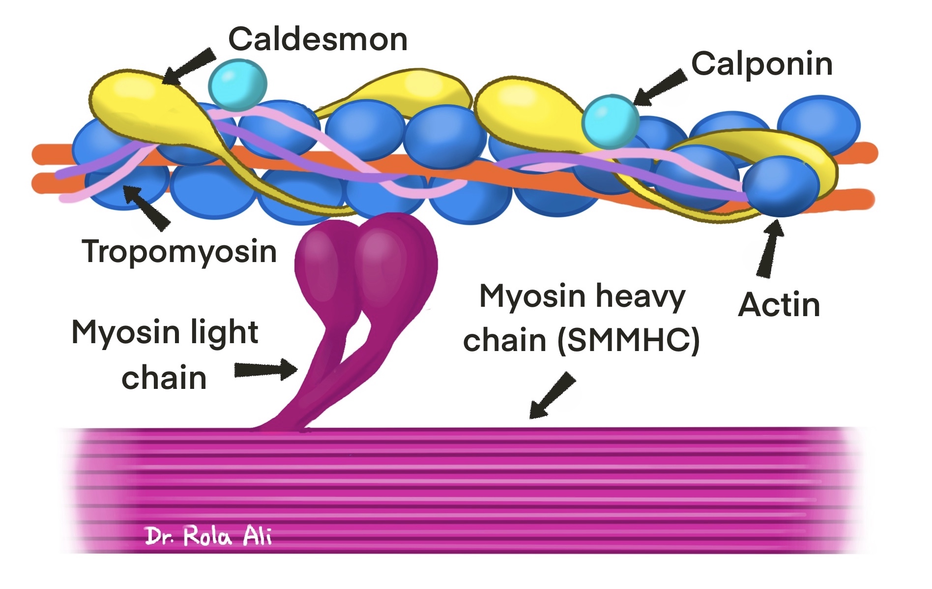
Contractile proteins relevant to IHC
Microscopic (histologic) description
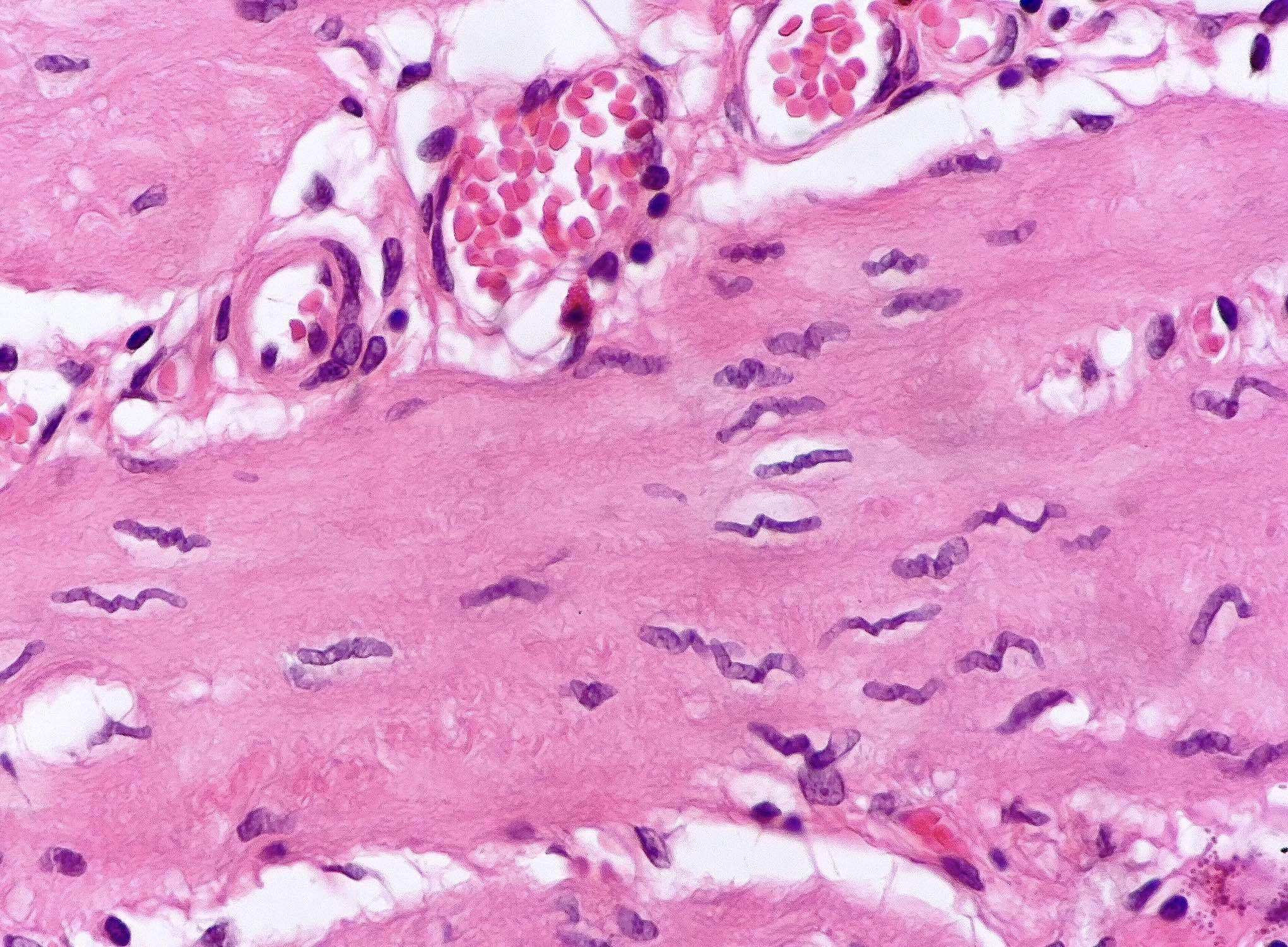
- Cell size: diameter between 1 - 5 μm and length between 20 - 500 μm (much smaller than skeletal muscle)
- Cell shape: spindle shaped or fusiform with tapered ends
- Cytoplasm: eosinophilic fibrillar with distinct cell borders and no cross striations
- Nucleus
- Central elongated cigar shaped nucleus with blunted ends
- Spiral corkscrew appearance in the contracted state
- Appear round on cross section
- Spindle cell mimics
- Fibroblasts in dense regular connective tissue
- Myofibroblasts: spindle to stellate, amphophilic cytoplasm, single elongated nucleus with 1 or 2 small nucleoli, collagen in between the cells
- Schwann cells: cells show slightly undulated buckled nuclei, often with 1 blunt and 1 pointed end, described as S shaped, serpentine, comma shaped, bullet shaped or boomerang-like
- Reference: Young: Wheater's Functional Histology, 6th Edition, 2013
Microscopic (histologic) images
Contributed by Rola H. Ali, M.D.
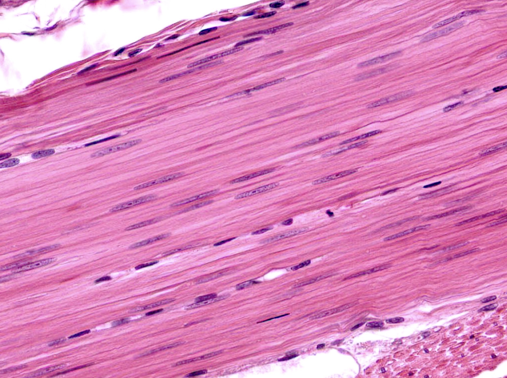
Relaxed smooth muscle fibers
Contracted smooth muscle
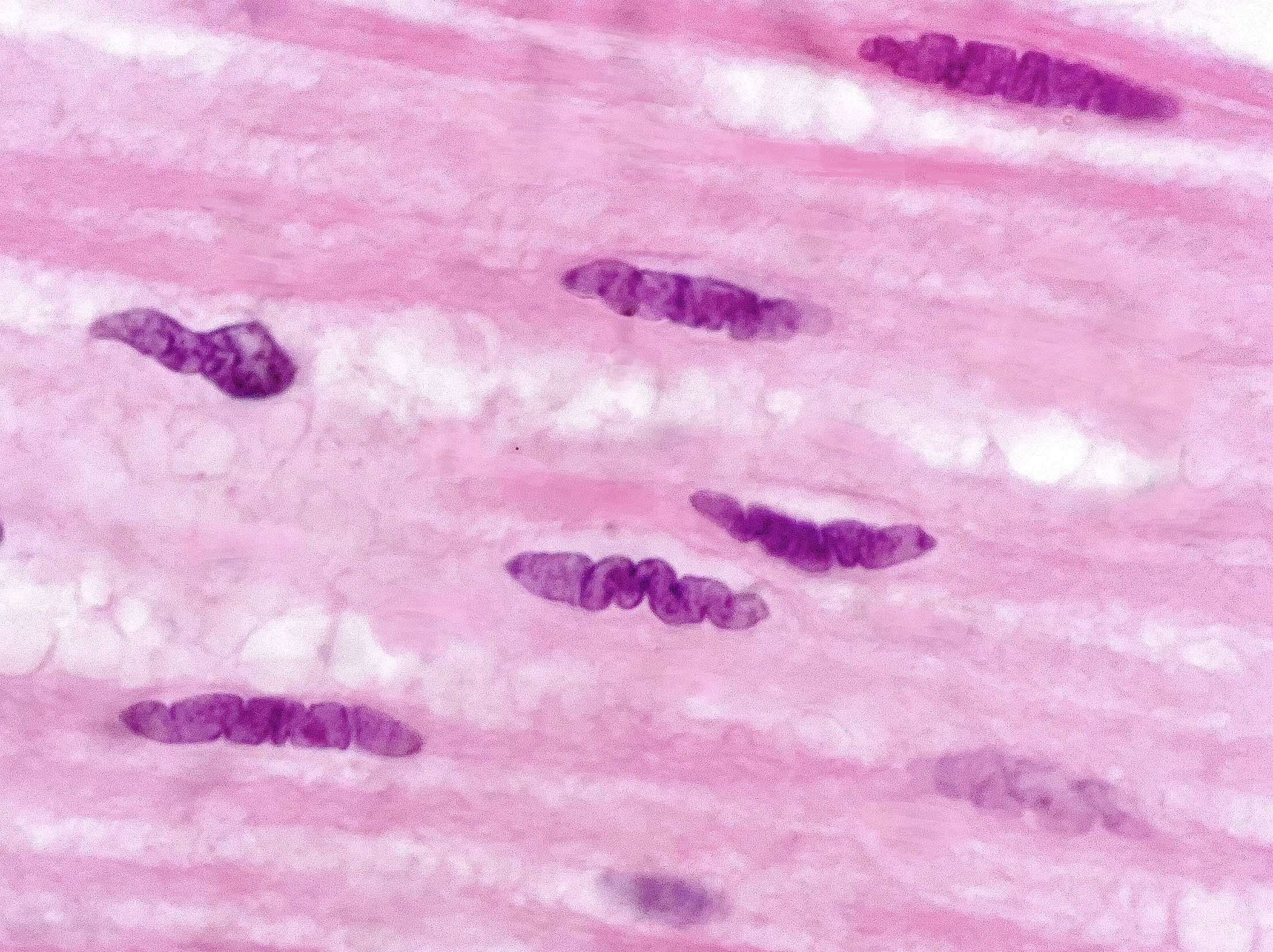
Spiral corkscrew nuclei
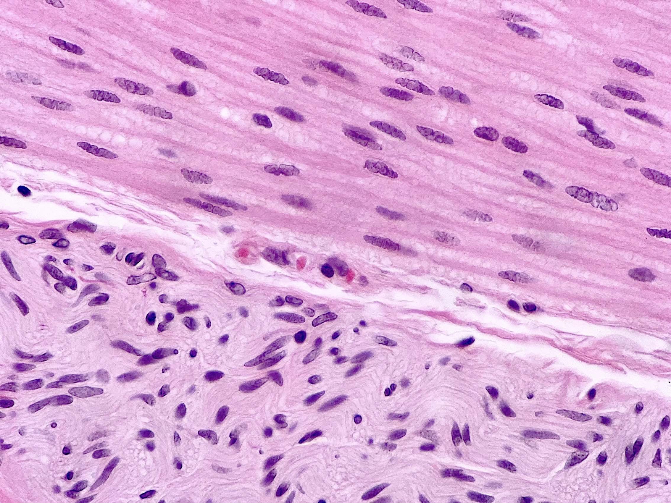
Cross versus longitudinal sections
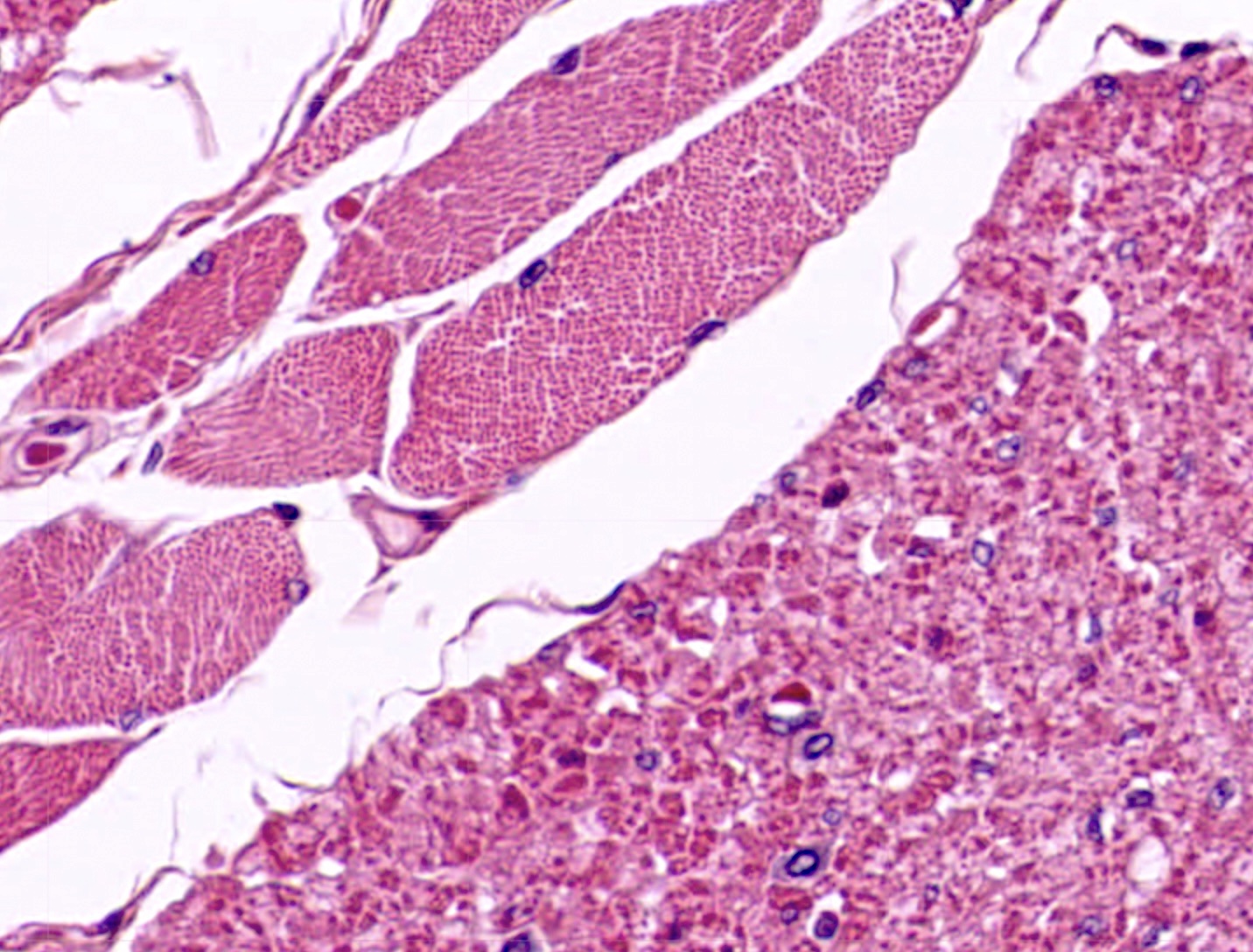
Smooth versus skeletal muscle
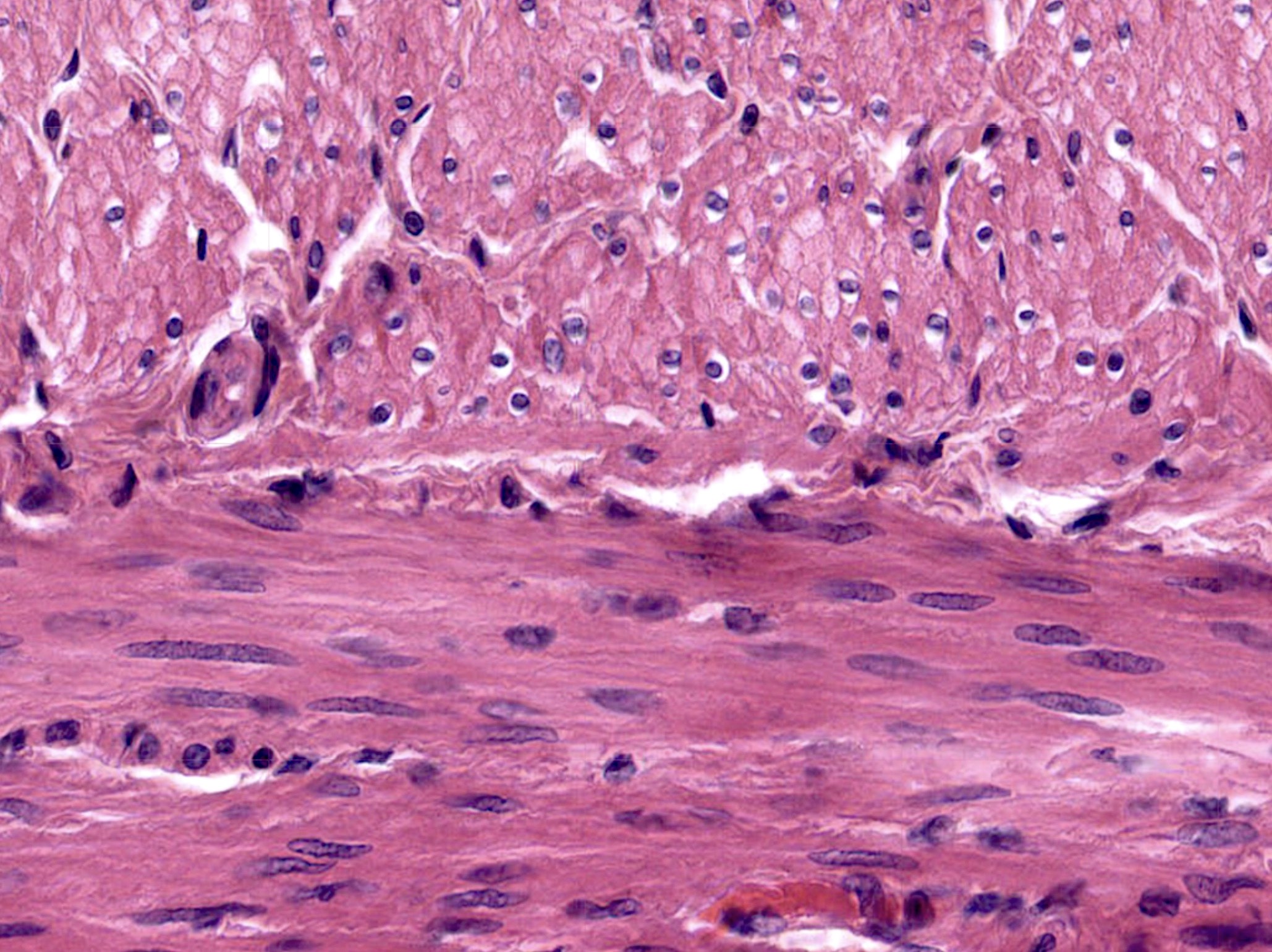
Smooth muscle versus nerve
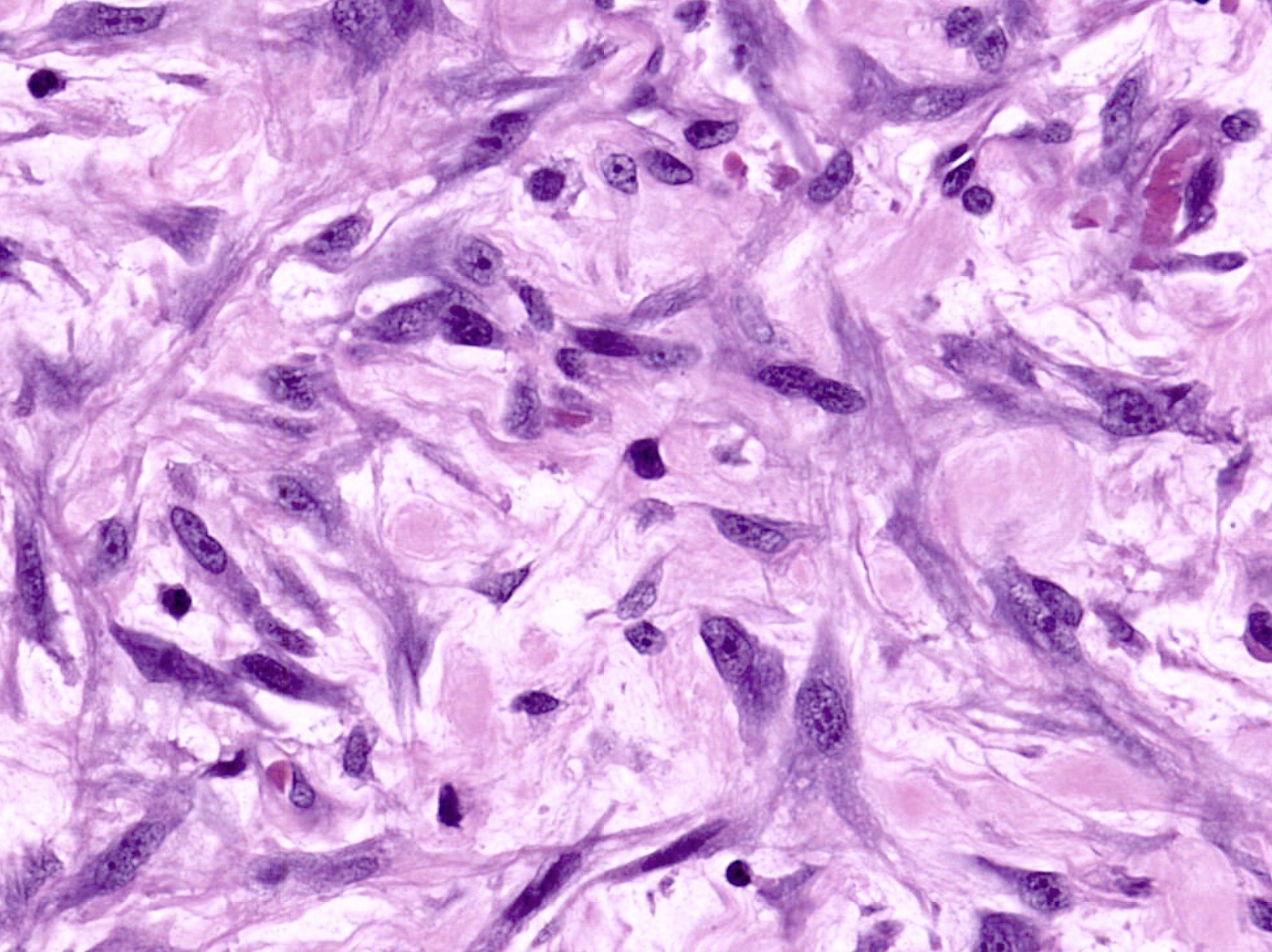
Myofibroblasts for comparison
Virtual slides
Images hosted on other servers:

Gallbladder muscularis beneath lamina

Juxtaposition of smooth and skeletal esophagus
Positive stains
- Smooth muscle actin (SMA): diffuse cytoplasmic staining (as opposed to membranous tram track in myofibroblasts)
- Muscle specific actin (HHF35): panmuscle marker
- Desmin: panmuscle marker
- h-caldesmon: actin binding protein
- Calponin: actin binding protein
- Smooth muscle myosin heavy chain (SMMHC)
- Smoothelin: terminally differentiated smooth muscle cells (Am J Surg Pathol 2009;33:1795)
- Masson trichrome: smooth muscle stains red
Electron microscopy description
- Cytoplasm
- Filaments
- Thin (actin) and thick (myosin) filaments
- Intermediate filaments
- Dense bodies
- Electron opaque structures floating freely within the cytoplasm → analogous to Z lines of striated muscle anchoring actin and myosin
- Organelles
- Include sarcoplasmic reticulum, mitochondria and Golgi apparatus
- Filaments
- Cell membrane
- Membrane bound dense bodies
- Pinocytotic vesicles or caveolae → Ca2+ movement across the membrane
- Gap junctions → consist of connexins facilitating the transfer of metabolites, ions and signaling molecules
- References: Edwin: Methods in Pharmacology - Smooth Muscle, 1st Edition, 1975, Motta: Ultrastructure of Smooth Muscle, 1st Edition, 1990
Electron microscopy images
Images hosted on other servers:

Smooth muscle longitudinal section

Smooth muscle cross section

Wall of muscular artery

Intestinal muscularis propria (externa)

Smooth muscle cross section
Videos
Smooth muscle versus myofibroblasts
Practice question #1

Practice answer #1
A. Cytoplasmic staining with h-caldesmon. Smooth muscle cells contain cytoplasmic caldesmon molecules bound to actin filaments. Answers B and E are incorrect because myogenin and MyoD1 both encode nuclear proteins expressed in skeletal muscle. Answer C is incorrect because membranous staining with smooth muscle actin is seen in myofibroblasts. Answer D is incorrect because S100 staining is seen in Schwann cells.
Comment Here
Reference: Histology-smooth muscle
Comment Here
Reference: Histology-smooth muscle
Practice question #2
Which of the following statements pertaining to the physiology of smooth muscle contraction is true?
- Actin filaments pull the Z lines inward in the contracted state
- Ca2+ binding to calmodulin stimulates myosin actin interaction
- Consumption of tremendous amounts of ATP is required
- Contraction is stimulated by somatic motor neurons
- Strong affinity of troponin for Ca2+ ions initiates contractions
Practice answer #2
B. Ca2+ binding to calmodulin stimulates myosin actin interaction. In smooth muscle, calmodulin acts in place of troponin. Answers A and C - E are incorrect because these statements are true for skeletal muscle.
Comment Here
Reference: Histology-smooth muscle
Comment Here
Reference: Histology-smooth muscle
