

Table of Contents
Definition / general | Essential features | Terminology | ICD coding | Epidemiology | Sites | Pathophysiology | Etiology | Clinical features | Diagnosis | Radiology description | Radiology images | Prognostic factors | Case reports | Treatment | Gross description | Gross images | Microscopic (histologic) description | Microscopic (histologic) images | Positive stains | Negative stains | Molecular / cytogenetics description | Sample pathology report | Differential diagnosis | Board review style question #1 | Board review style answer #1 | Board review style question #2 | Board review style answer #2Cite this page: Chami R. Metanephric adenofibroma. PathologyOutlines.com website. https://www.pathologyoutlines.com/topic/kidneytumormetaadenofibroma.html. Accessed April 26th, 2024.
Definition / general
- Biphasic neoplasm composed of epithelial and stromal components
- Member of metanephric tumor family including metanephric adenoma (purely epithelial) and metanephric stromal tumor (purely stromal)
Essential features
- Biphasic neoplasm composed of varying proportions of stromal and epithelial components
- Typically solitary and nonencapsulated
- No nuclear atypia in both components
- Mitotic figures are absent or rare in epithelial component
- Epithelial component is identical to metanephric adenoma
- Stromal / mesenchymal component is identical to metanephric stromal tumor
Terminology
- Previously known as nephrogenic adenofibroma
ICD coding
- 9013/0 - adenofibroma, NOS
Epidemiology
- Rare
- Age range 5 months - 36 years (Am J Surg Pathol 1992;16:325, Am J Surg Pathol 2001;25:433)
Sites
- Metanephric adenofibroma occurs in renal cortex and medulla
Pathophysiology
- Unknown, however, several cases have been reported to harbor BRAF V600E mutations; genomic alterations in BRAF oncogene appear at an early stage in tumorigenesis and are preserved through the tumor progression and therefore are considered as drivers of oncogenesis (Hum Pathol 2015;46:1153, Am J Surg Pathol 2015;39:1301)
Etiology
- Uncommon renal neoplasm; unknown etiology
Clinical features
- Present with polycythemia, hematuria, hypertension or abdominal pain (Am J Surg Pathol 1992;16:325, Am J Surg Pathol 2001;25:433)
- Associated with papillary renal cell carcinoma and Wilms tumor (Am J Surg Pathol 2001;25:433)
Diagnosis
- The diagnosis is made by microscopic examination of resected tissue
Radiology description
- The imaging appearance is nonspecific, with the tumor often resembling Wilms tumor (Radiol Case Rep 2018;13:610)
Radiology images
Images hosted on other servers:

Homogenous solid mass, no calcification or necrosis
Prognostic factors
- Benign
- One case of metanephric adenofibroma in combination with Wilms tumor and renal papillary carcinoma has been reported in literature (Pediatr Dev Pathol 2012;15:65)
Case reports
- 5 year old boy with incidental finding of an abdominal mass (Radiol Case Rep 2018;13:610)
- 5 year old girl with progressively increasing abdominal swelling for 7 months (APSP J Case Rep 2016;7:37)
- 10 year old girl with a history of recurrent urinary tract infections (Am J Surg Pathol 2015;39:1301)
- 10 year old boy with a history of abdominal pain (Int J Clin Exp Pathol 2015;8:3250)
- 10 year old boy with history of left flank pain following a fall (Can J Urol 2013;20:6737)
- 29 year old woman (Int Braz J Urol 2017;43:563)
Treatment
- Excision of the tumor is adequate therapy
Gross description
- Solitary, unencapsulated, vaguely circumscribed / indistinct borders
- Tan / gray / white-yellow cut surface
- Partially cystic
- Reference: Am J Surg Pathol 2001;25:433
Gross images
Images hosted on other servers:

Largely solid, tan homogenous renal mass

Fibrous, white grey cut surface

White solid and cystic cut surface
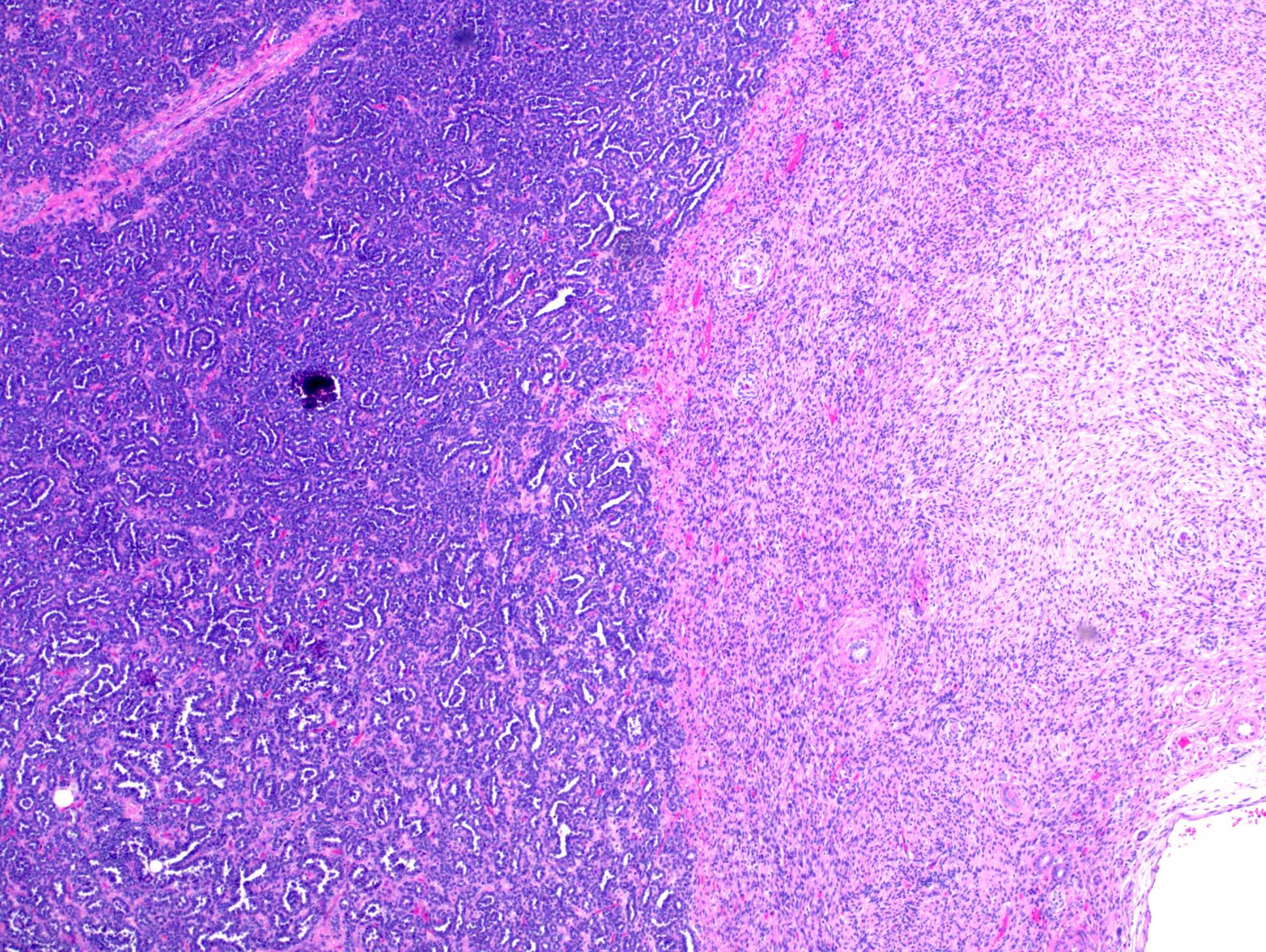
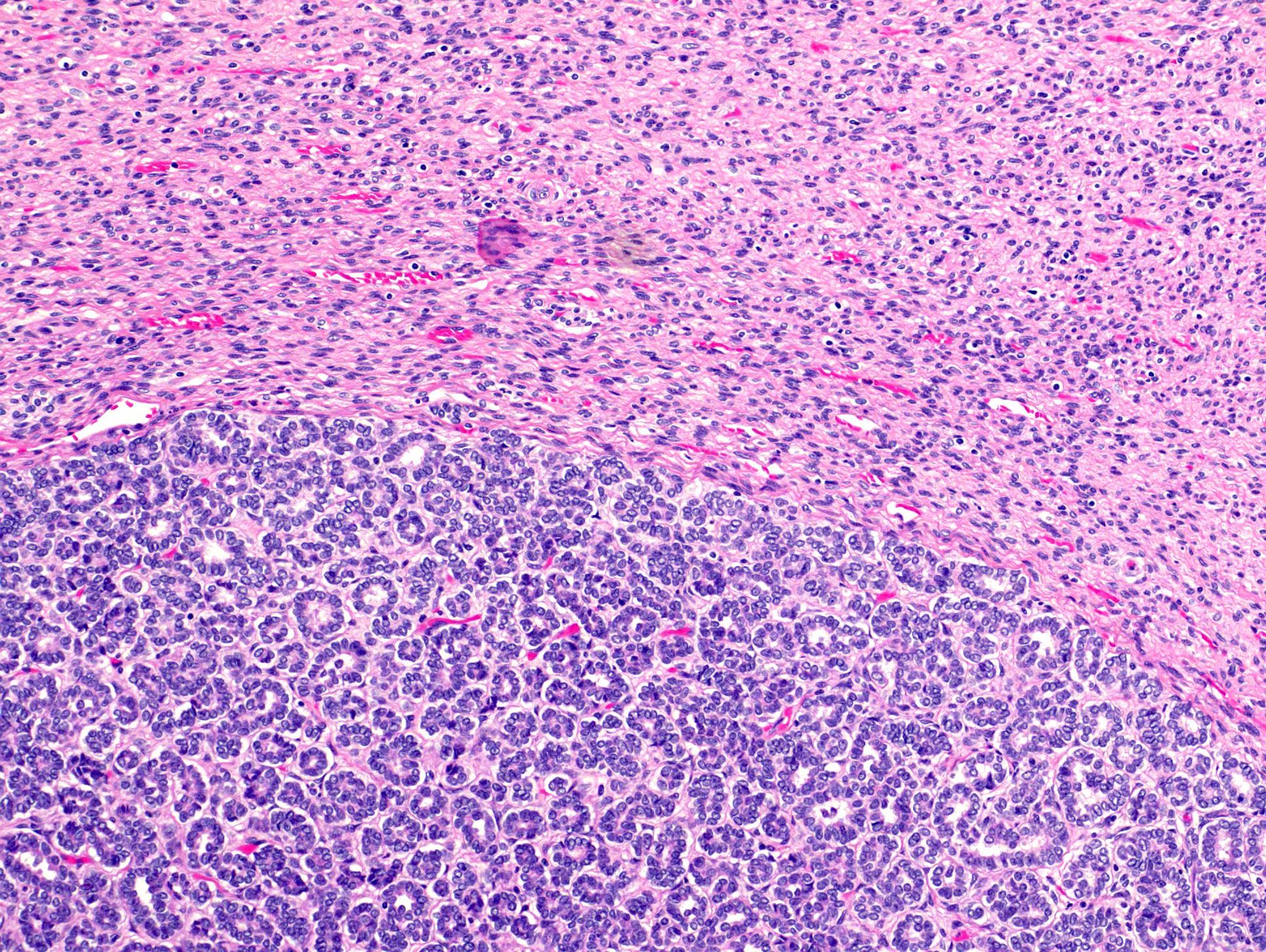
Microscopic (histologic) description
- Biphasic tumor composed of varying proportions of stromal and epithelial components
- Epithelial component identical to metanephric adenoma, tightly packed small tubules, papillary structures or glomeruloid structures (blunt short papillae resembling glomeruli)
- Epithelial cells have small round to oval nuclei with delicate chromatin and absent or inconspicuous nucleoli and slight pale cytoplasm; mitotic figures are absent or rare
- Psammoma bodies are common
- Mesenchymal component identical to metanephric stromal tumor, bland spindle and epithelioid / plump cells with thin hyperchromatic nuclei and indistinct cell borders
- Areas of intratumoral angiodysplasia, concentric cuffing of entrapped tubules ("onion skinning") and heterologous differentiation (cartilage, adipose tissue or glia) (Am J Surg Pathol 2001;25:433)
- No vascular invasion
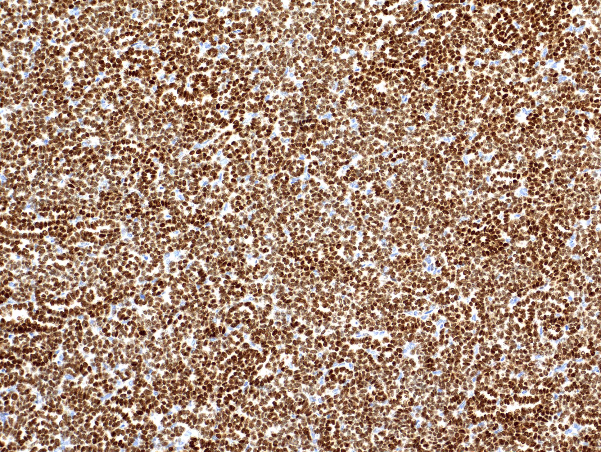
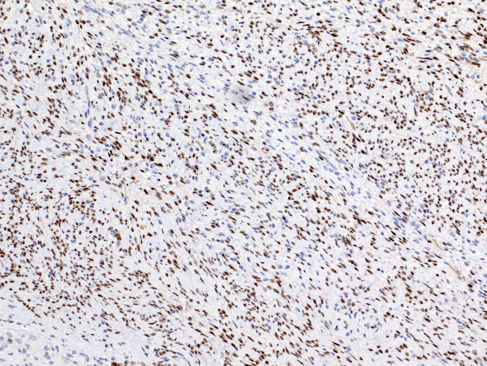
Microscopic (histologic) images
Contributed by Rose Chami, M.D.
Biphasic tumor with epithelial and stromal components
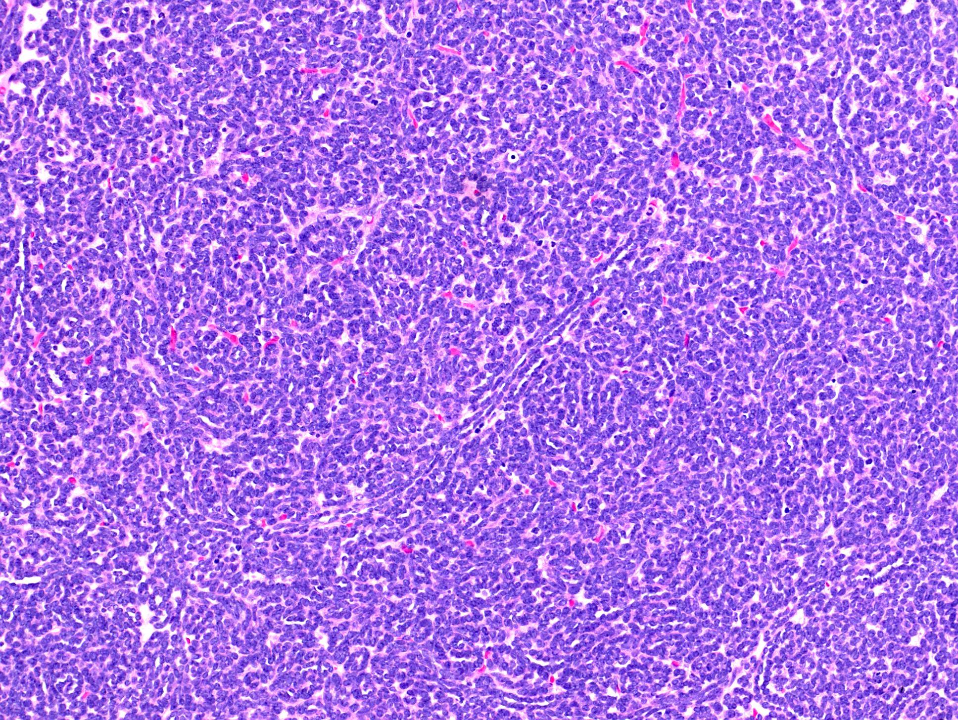
Highly cellular tumor
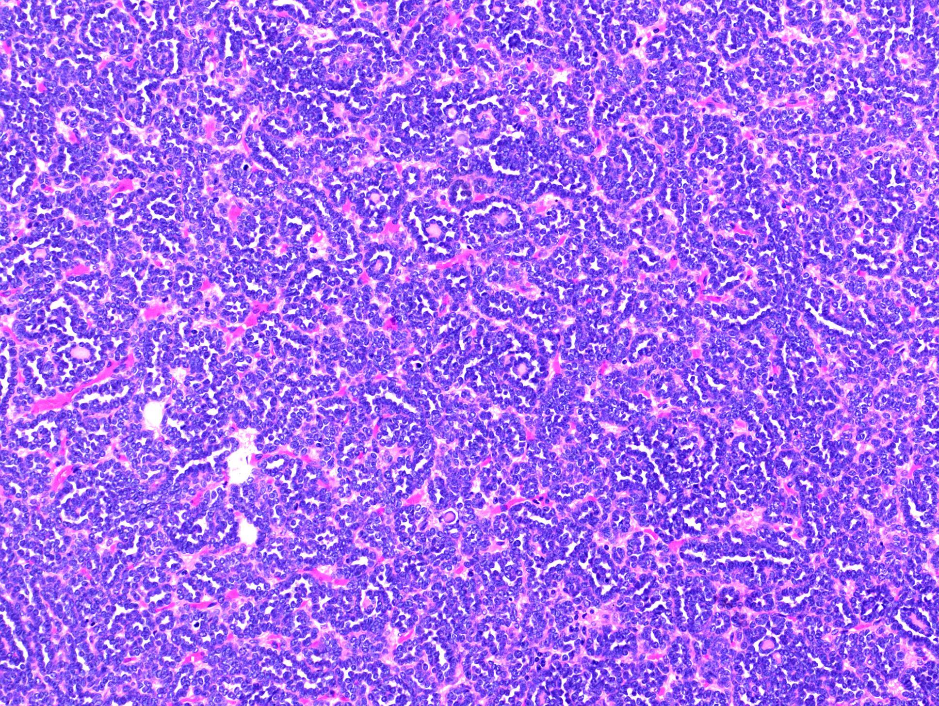
Tubular and glomeruloid structures
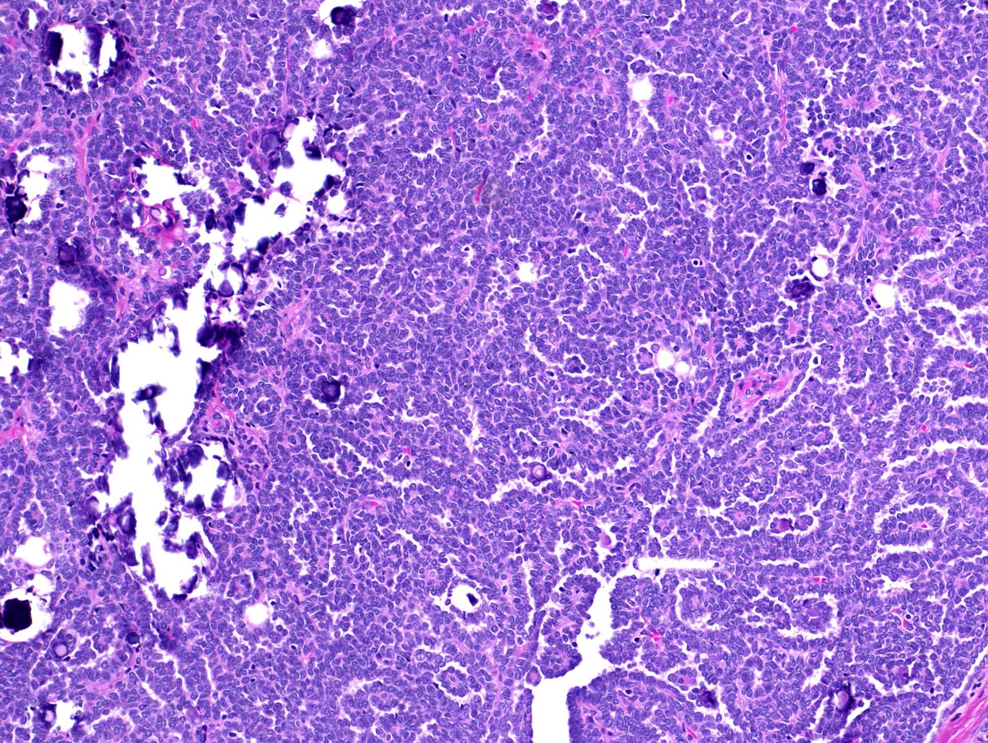
Psammoma bodies
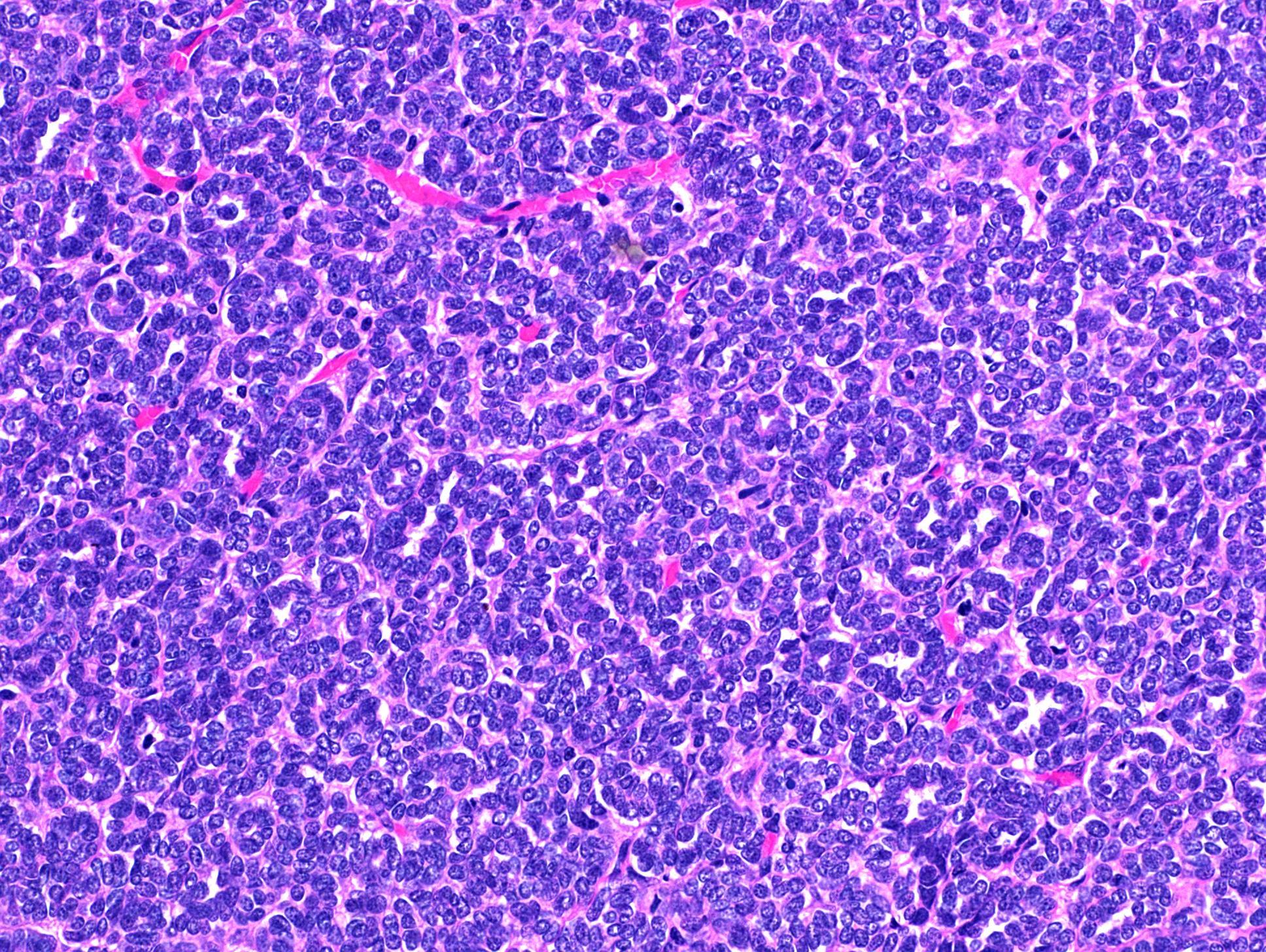
Cellular solid appearance
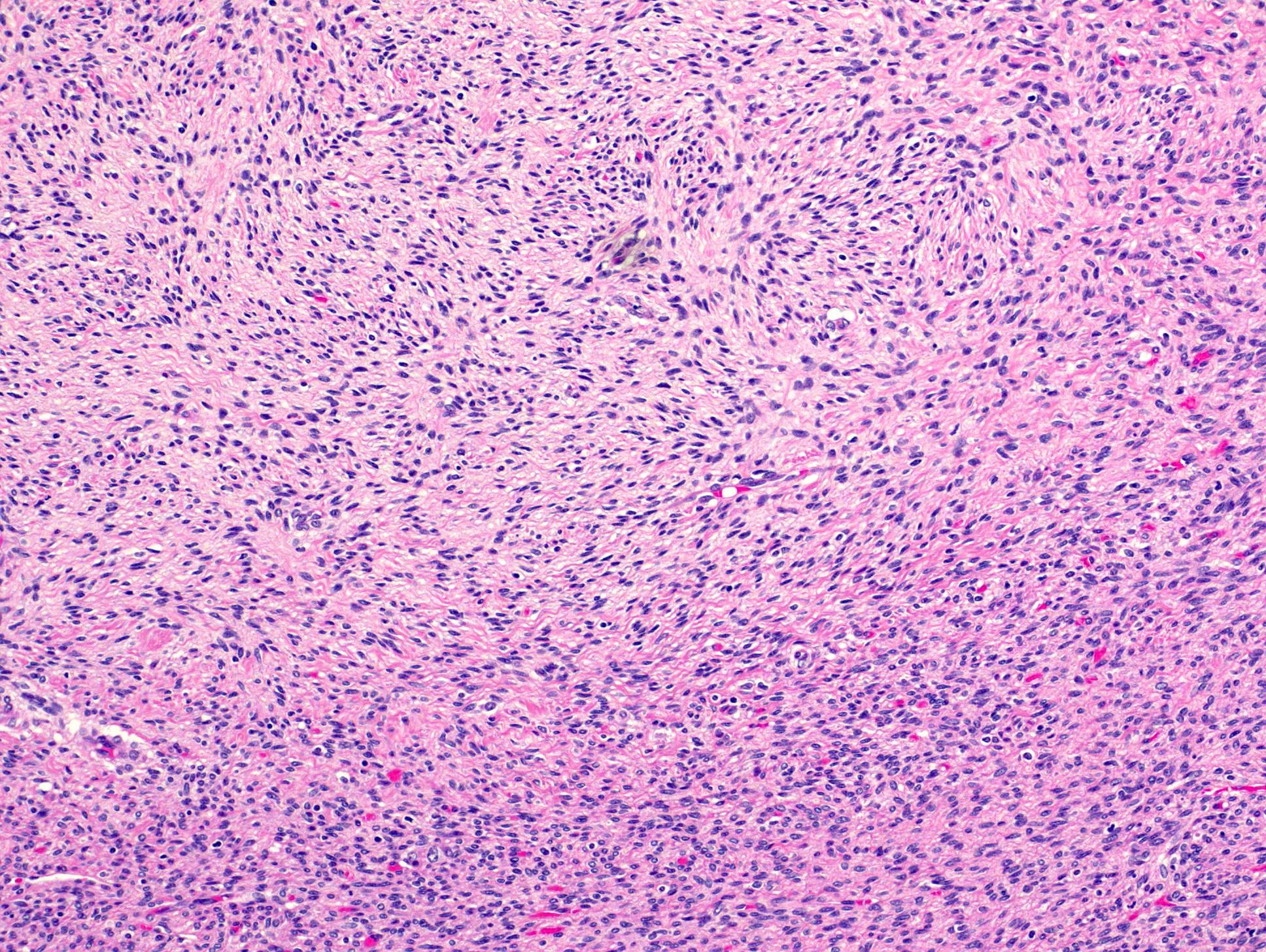
Spindle cell component
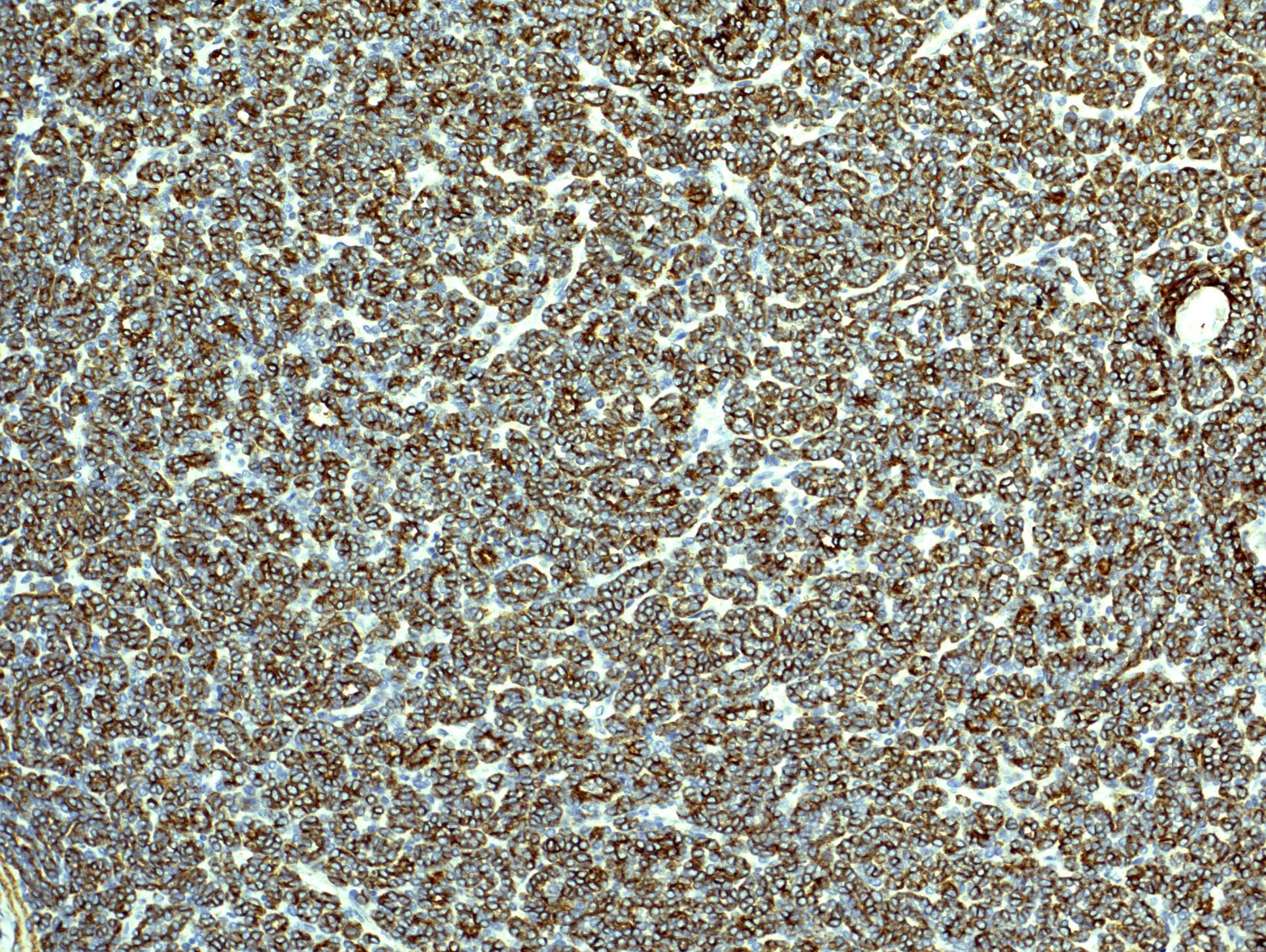
AE1/AE3
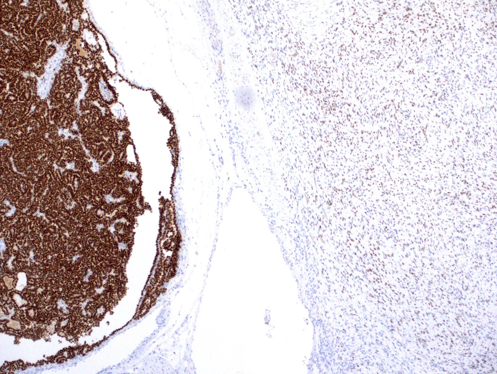
WT1, both components
WT1, epithelial component
WT1, stromal component
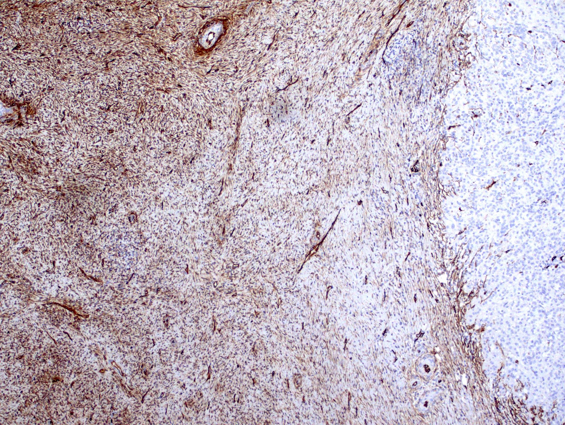
CD34, both components
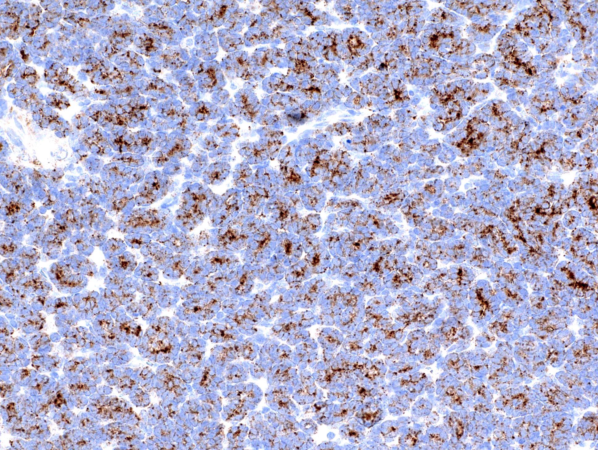
BRAF V600E, epithelial component
Positive stains
- Epithelial component (stains like metanephric adenoma): cytokeratin AE1/AE3, WT1 (nuclear, diffuse), CD57, BRAF V600E
- Stromal component: vimentin, CD34 (commonly), WT1, BRAF V600E (variable)
- References: Hum Pathol 2015;46:1153, Am J Surg Pathol 2015;39:1301
Negative stains
- Epithelial component: EMA, AMACR
- Stromal component: smooth muscle actin, desmin, keratin, S100 protein
- Reference: Am J Surg Pathol 1992;16:325
Molecular / cytogenetics description
- BRAF V600E mutation (Hum Pathol 2015;46:1153, Am J Surg Pathol 2015;39:1301)
Sample pathology report
- Right kidney, partial nephrectomy:
- Metanephric adenofibroma, 3.2 cm, resection margins negative for tumor (see comment)
- Comment: There is a nonencapsulated biphasic renal tumor composed of epithelial and stromal components. There is no nuclear atypia in both components and mitotic figures are rare or absent in the epithelial component. Immunopositive for BRAF V600E.
Differential diagnosis
- Metanephric adenoma:
- Lacks stromal component
- Metanephric stromal tumor:
- Lacks epithelial component
- Congenital mesoblastic nephroma:
- Wilms tumor:
- Particularly epithelial predominant histology, usually encapsulated and mitotically active
- Papillary renal cell carcinoma:
Board review style question #1
-
A pediatric renal tumor is shown. Which of the following is true about this entity?
- Biphasic neoplasm with varying proportions of stromal and epithelial components
- Epithelial component is mitotically active
- Malignant and frequently metastasizing
- Multifocal (typically)
Board review style answer #1
A. This is metanephric adenofibroma. It is a biphasic neoplasm with varying proportions of stromal and epithelial components.
Comment Here
Reference: Metanephric adenofibroma
Comment Here
Reference: Metanephric adenofibroma
Board review style question #2
-
Which of the following is true about the expression of metanephric adenofibroma?
- CD57-
- CK7+
- Desmin+
- WT1+
Board review style answer #2
