
Lymphoma & related disorders
Mature B cell neoplasms
Large B cell lymphomas-special subtypes
Primary effusion lymphoma
Author: Nikhil Sangle, M.D.

Last author update: 1 March 2011
Last staff update: 19 April 2024 (update in progress)
Copyright: 2001-2024, PathologyOutlines.com, Inc.
PubMed Search: Primary effusion lymphoma
Table of Contents
Definition / general | Terminology | Clinical features | Case reports | Microscopic (histologic) description | Microscopic (histologic) images | Cytology images | Positive stains | Negative stains | Molecular / cytogenetics description | Differential diagnosis | Additional references | Board review style question #1 | Board review style answer #1 | Board review style question #2 | Board review style answer #2Cite this page: Sangle N. Primary effusion lymphoma. PathologyOutlines.com website. https://www.pathologyoutlines.com/topic/lymphomaeffusion.html. Accessed May 13th, 2024.
Definition / general
- Rare type of diffuse large B cell lymphoma with lymphomatous effusions in pleural, pericardial and abdominal cavities but no tumor mass
Terminology
- Also called body cavity lymphoma
Clinical features
- Only one body cavity typically involved
- Strongly associated with HHV8 and advanced HIV; usually EBV+
- Also occurs in nonimmunosuppressed patients, often elderly
- Rarely, extracavitary tumors with features of PEL are seen in GI tract, skin, lung, CNS and lymph nodes
- Survival usually only months
- Solid variant: very rare; affects GI tract, skin, lung, cerebrum; rarely nodal (Hum Pathol 2002;33:846); may represent a heterogeneous group of disorders
Case reports
- 37 year old HIV+ man with coexisting small bowel mass (Am J Surg Pathol 2002;26:1363)
- 42 year old HIV+ man with pleural cavity and lingual tumor (Hum Pathol 2004;35:632)
- 51 year old man with HIV history, presenting with cough, dyspnea, chest pain and fever (Case of the Month #519)
- 68 year old HIV negative but HHV8+ patient (Arch Pathol Lab Med 2000;124:753)
- 70 year old man with HHV8+, EBV+ pleural effusions (J Med Case Rep 2011;5:60)
- 87 year old HIV- but HHV8+ man with T cell variant (Arch Pathol Lab Med 2001;125:1246)
- Two cases of HIV+, HHV8+ solid variant without primary effusion lymphoma (Hum Pathol 2002;33:846)
Microscopic (histologic) description
- Immunoblastic cells with abundant basophilic cytoplasm, round nucleus, single prominent nucleoli, perinuclear halo, variable nuclear pleomorphism with binucleated cells
- Occasional large Reed-Sternberg like and anaplastic cells
- Rarely is solid with plasmablastic features
- Solid variant: large pleomorphic cells; nodal case resembled anaplastic large cell lymphoma
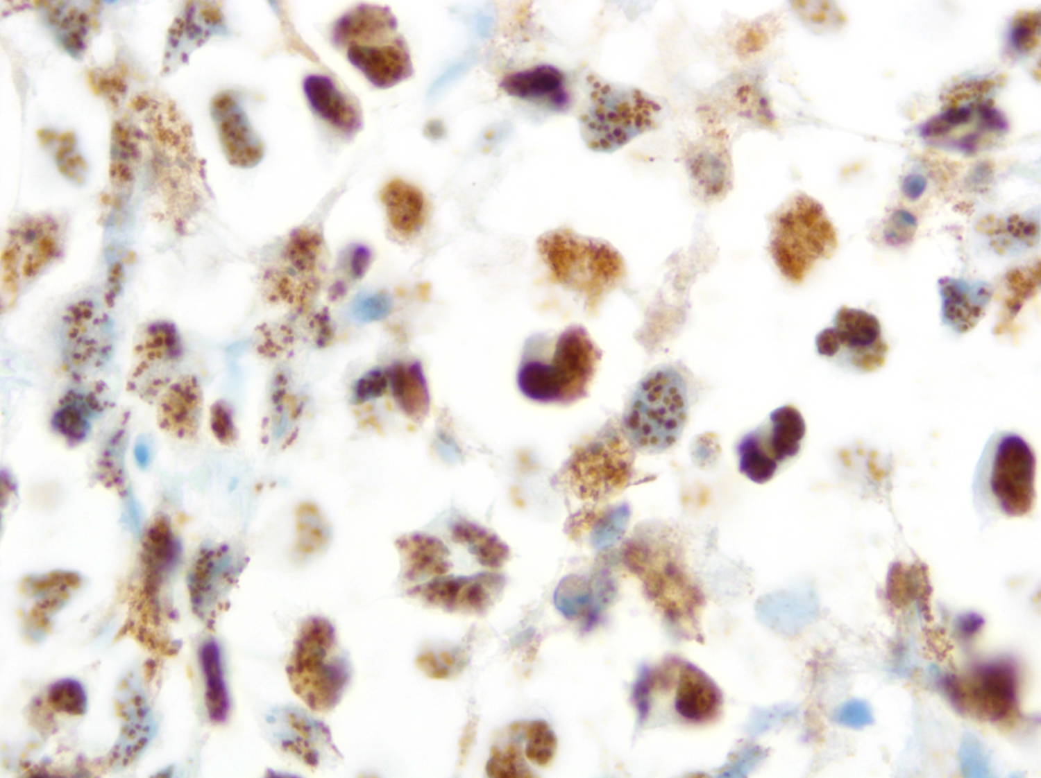
Microscopic (histologic) images
Contributed by Mario L. Marques-Piubelli, M.D. and Roberto N. Miranda, M.D. (Case #519)
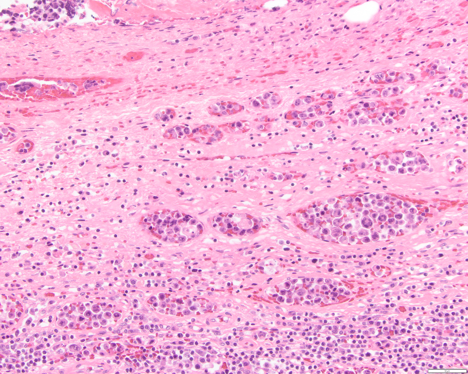
Extracavitary presentation of PEL
EBER
LANA-1
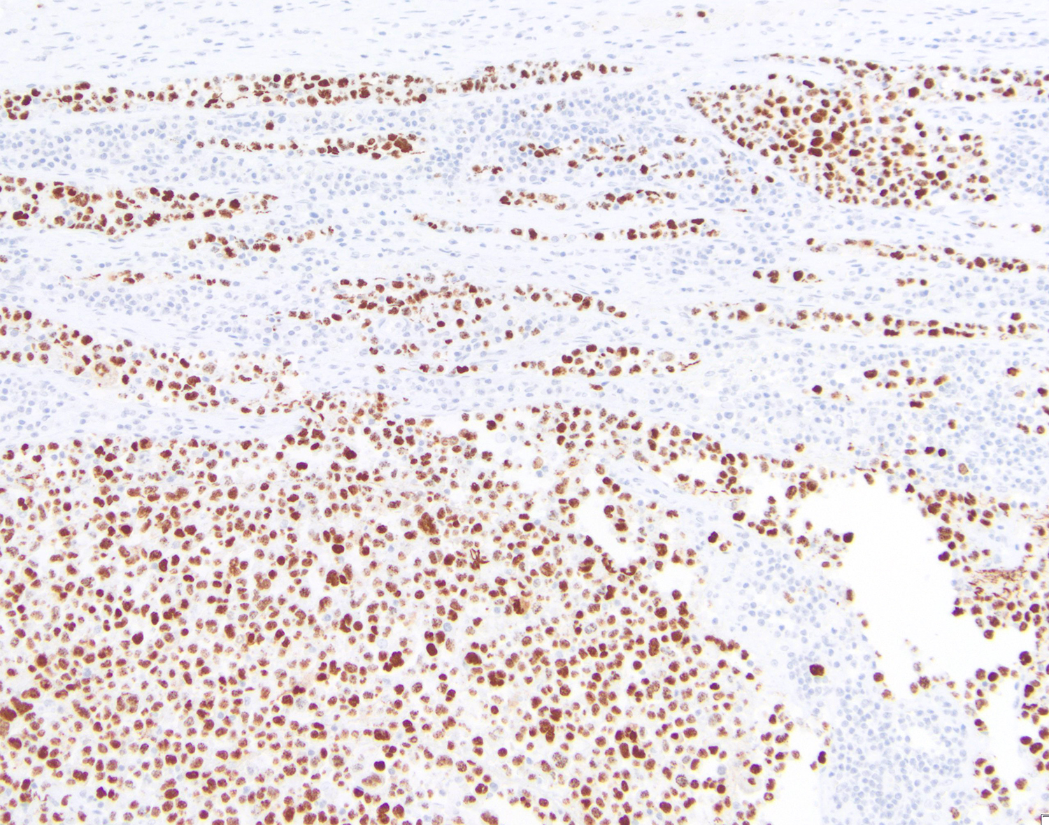
LANA-1 in an extracavitary presentation
Images hosted on other servers:

70 year old man with HHV8+, EBV+ pleural effusions
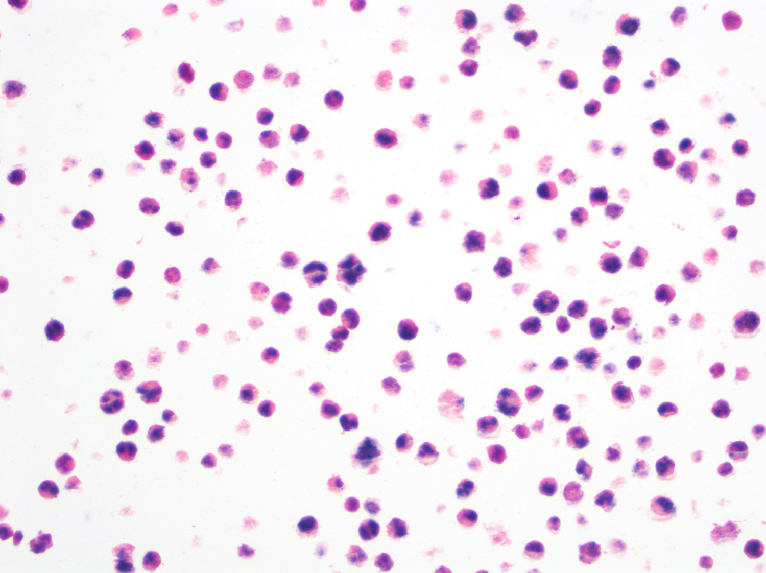
Cytology images
Contributed by Mario L. Marques-Piubelli, M.D. and Roberto N. Miranda, M.D. (Case #519)
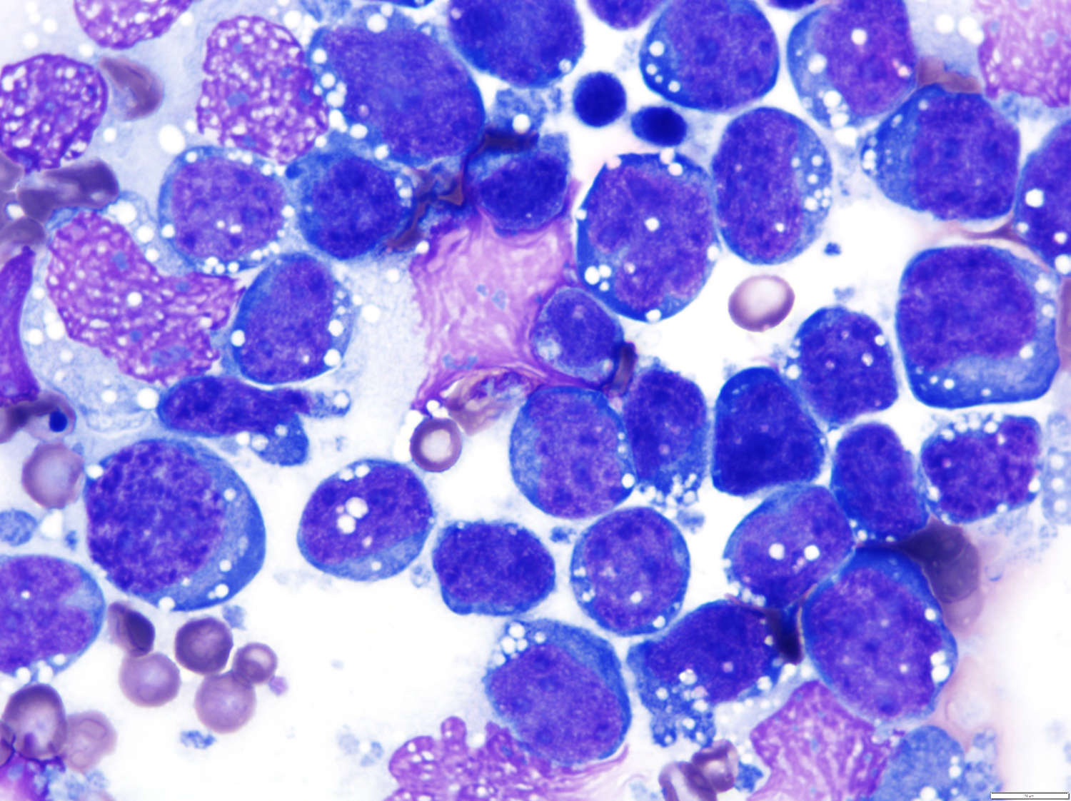
Diff-Quik
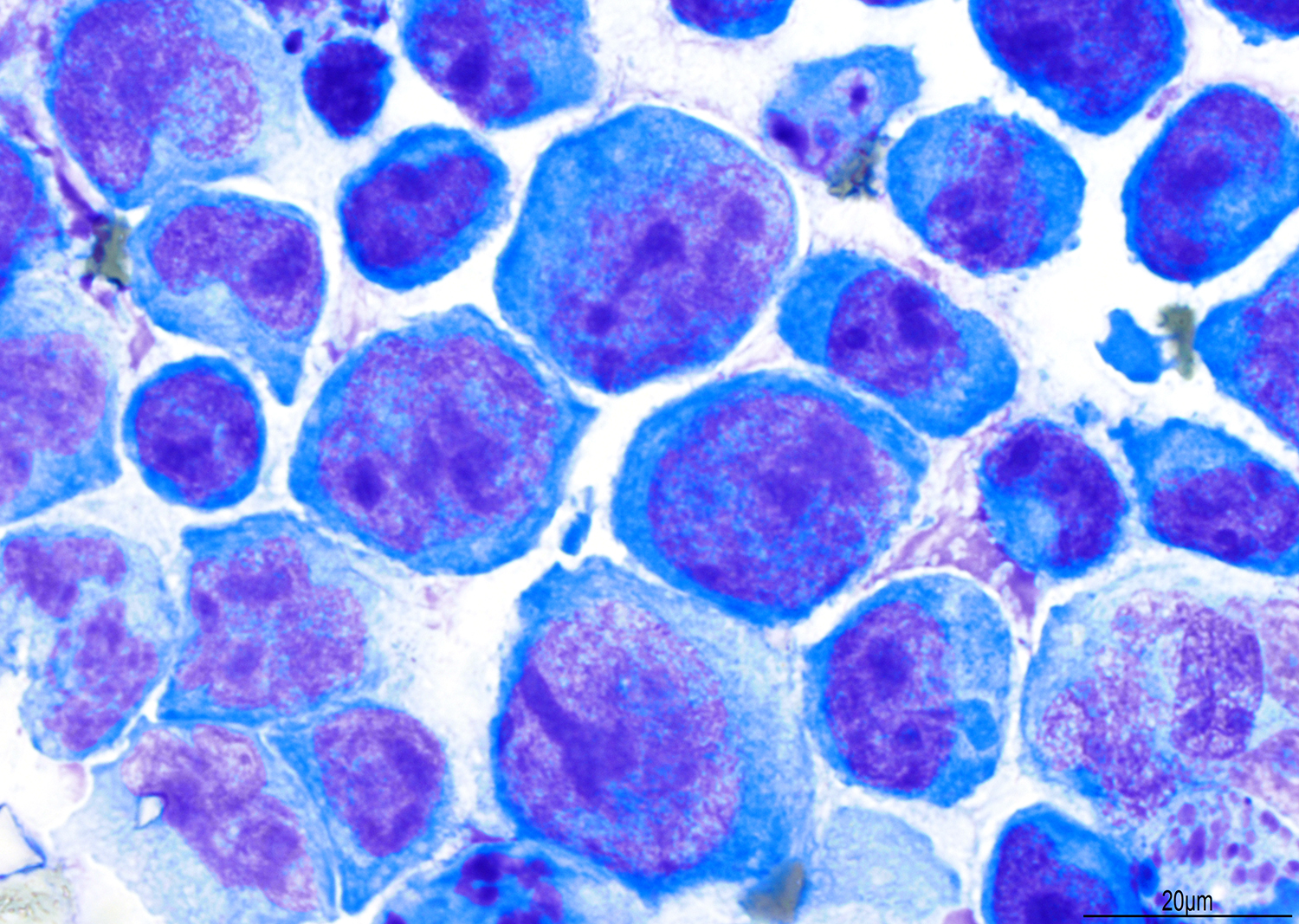
Wright-Giemsa
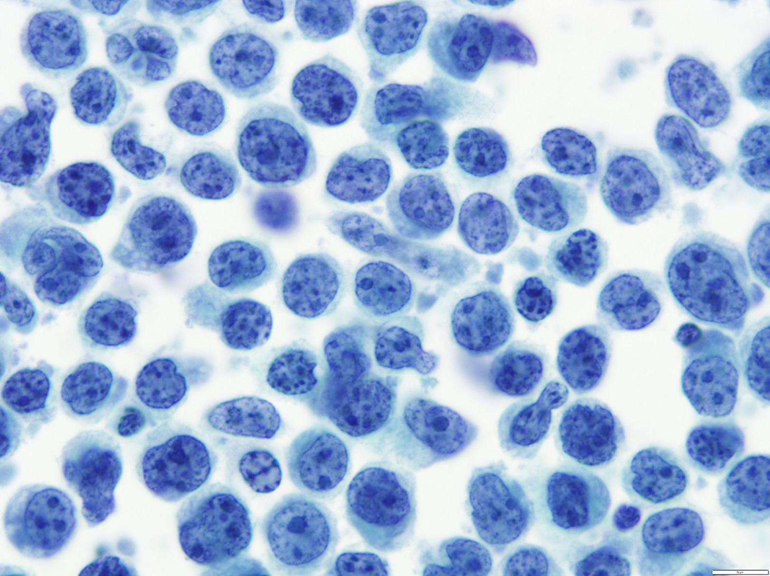
PEL Thinprep
Positive stains
Molecular / cytogenetics description
- Often required for diagnosis since negative for B cell stains
- Clonal rearrangements and somatic mutations of Ig heavy chain
- Clonal Epstein Barr virus and HHV8 genetic sequences
- Also gain of sequences in chromosomes 12 and X (Arch Pathol Lab Med 2000;124:824)
Differential diagnosis
- Plasmablastic lymphoma: not an effusion, positive for B cell markers
- Pyothorax associated lymphoma: different clinical history, positive for B cell markers, HHV8-
- Solid variant (HHV8+) resembles diffuse large B cell lymphoma with plasmablastic features (HHV8-)
Additional references
Board review style question #1
Which of the following pair of viruses are associated with primary effusion lymphoma (PEL)?
- EBV and HCV
- EBV and HHV-8
- HHV-8 and COVID-19
- HHV-8 and HPV
Board review style answer #1
Board review style question #2
Which is the latency pattern of the Epstein-Barr virus found associated with primary effusion lymphoma (PEL)?
- Latency Pattern 1: EBER(+), EBNA-1(+), LMP-1(-)
- Latency Pattern 1: EBER(+), EBNA-1(+), LMP-1(+)
- Latency Pattern 3: EBER(+), EBNA-2(-), LMP-1(+)
- Latency Pattern 2: EBER(+), EBNA-2(-), LMP-1(+)
Board review style answer #2
A. Latency Pattern 1: EBER(+), EBNA-1(+), LMP-1(-)
Comment Here
Reference: Primary effusion lymphoma
Comment Here
Reference: Primary effusion lymphoma
