
22 July 2005 - Case #13
All cases are archived on our website. To view them sorted by case number, diagnosis or category, visit our main Case of the Month page. To subscribe or unsubscribe to Case of the Month or our other email lists, click here.
This case was contributed by Dr. Volkan Adsay, Wayne State University, Department of Pathology, Detroit, Michigan, USA and his assistant Olca Batrk.
Case #13
Clinical history:
A 31 year old woman presented with a 14 cm pancreatic mass. Small nodules on the peritoneal surfaces were considered clinically to represent metastatic disease. Based on the frozen section diagnosis of poorly differentiated endocrine carcinoma of pancreatic origin, the patient underwent subtotal pancreatectomy.
Micro images:
What is your diagnosis?
Diagnosis: Desmoplastic round cell tumor of the pancreas
Immunohistochemistry images:
Discussion:
Desmoplastic round cell tumor is a rare and aggressive tumor of young males, usually of the abdomen or pelvis. It has also been reported in the pleura, thorax, paratesticular region, kidney and CNS but has not been previously reported (to our knowledge) in the pancreas (Urol Oncol 2005;23:132, Am J Surg Pathol 2004;28:1379). Patients typically present with large, symptomatic masses and may have widespread peritoneal tumor implants. The prognosis is usually poor.
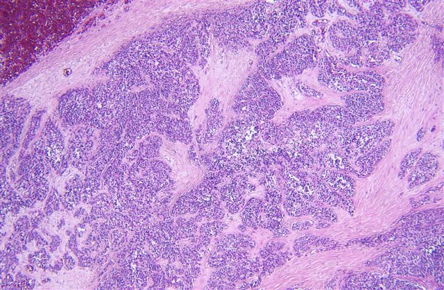
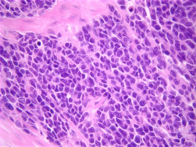
Histologically, these are small round blue cell tumors composed of solid nests of round / oval cells surrounded by cellular desmoplastic stroma. There is frequently necrosis, cystic degeneration and glandular arrangements and the tumors may contain signet ring-like cells, pseudorosettes, rhabdoid cells and spindled cells.
These tumors almost always have the translocation t(11;22)(p13;q12), which fuses the WT1 and EWS genes, leading to production of a chimeric protein with transcriptional activity that activates expression of target genes normally repressed by WT1, such as platelet derived growth factor A and transforming growth factor beta.
These tumors are typically immunoreactive for desmin, keratin (CAM 5.2 or AE1 / AE3), WT1 and often for PLAP and MIC2 / CD99, with variable immunostaining for calretinin, myoglobin, HER2 and KIT / CD117 (Am J Surg Pathol 2000;24:830, Mod Pathol 2003;16:229). The morphology and immunohistochemistry profile are usually sufficient for differentiating from other small blue cell tumors. If necessary, molecular identification of the characteristic translocation can be performed.
Reference: Am J Surg Pathol 2002;26:823
All cases are archived on our website. To view them sorted by case number, diagnosis or category, visit our main Case of the Month page. To subscribe or unsubscribe to Case of the Month or our other email lists, click here.
This case was contributed by Dr. Volkan Adsay, Wayne State University, Department of Pathology, Detroit, Michigan, USA and his assistant Olca Batrk.
Website news:
(1) This week's case is sponsored by Invitrogen. Invitrogen offers Zymed total system solution for pathology reagents and automation. From antibodies, kits and probes for IHC and CISH to immunostainers for automation, you can be confident that Zymed products will always deliver high quality staining results and offer the cutting edge pathology solutions you need. Note: sponsors do NOT have access in any manner to email addresses or other personal information in the possession of PathologyOutlines.com.
Visit and follow our Blog to see recent updates to the website.
Case #13
Clinical history:
A 31 year old woman presented with a 14 cm pancreatic mass. Small nodules on the peritoneal surfaces were considered clinically to represent metastatic disease. Based on the frozen section diagnosis of poorly differentiated endocrine carcinoma of pancreatic origin, the patient underwent subtotal pancreatectomy.
Micro images:
What is your diagnosis?
Click here for diagnosis and discussion:
Diagnosis: Desmoplastic round cell tumor of the pancreas
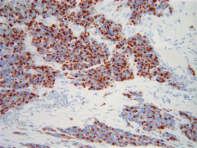
Immunohistochemistry images:
Desmin
Discussion:
Desmoplastic round cell tumor is a rare and aggressive tumor of young males, usually of the abdomen or pelvis. It has also been reported in the pleura, thorax, paratesticular region, kidney and CNS but has not been previously reported (to our knowledge) in the pancreas (Urol Oncol 2005;23:132, Am J Surg Pathol 2004;28:1379). Patients typically present with large, symptomatic masses and may have widespread peritoneal tumor implants. The prognosis is usually poor.
Histologically, these are small round blue cell tumors composed of solid nests of round / oval cells surrounded by cellular desmoplastic stroma. There is frequently necrosis, cystic degeneration and glandular arrangements and the tumors may contain signet ring-like cells, pseudorosettes, rhabdoid cells and spindled cells.
These tumors almost always have the translocation t(11;22)(p13;q12), which fuses the WT1 and EWS genes, leading to production of a chimeric protein with transcriptional activity that activates expression of target genes normally repressed by WT1, such as platelet derived growth factor A and transforming growth factor beta.
These tumors are typically immunoreactive for desmin, keratin (CAM 5.2 or AE1 / AE3), WT1 and often for PLAP and MIC2 / CD99, with variable immunostaining for calretinin, myoglobin, HER2 and KIT / CD117 (Am J Surg Pathol 2000;24:830, Mod Pathol 2003;16:229). The morphology and immunohistochemistry profile are usually sufficient for differentiating from other small blue cell tumors. If necessary, molecular identification of the characteristic translocation can be performed.
Reference: Am J Surg Pathol 2002;26:823
