Ovary
Mesenchymal tumors
Leiomyoma
Author: Nalini Gupta, M.D.

Last author update: 1 December 2014
Last staff update: 4 August 2025 (update in progress)
Copyright: 2003-2025, PathologyOutlines.com, Inc.
PubMed Search: Leiomyoma [title] ovary
Table of Contents
Definition / general | Epidemiology | Pathophysiology | Clinical features | Laboratory | Radiology description | Case reports | Treatment | Gross description | Gross images | Microscopic (histologic) description | Microscopic (histologic) images | Positive stains | Negative stains | Differential diagnosisCite this page: Gupta N. Leiomyoma. PathologyOutlines.com website. https://www.pathologyoutlines.com/topic/ovarytumorleiomyoma.html. Accessed September 15th, 2025.
Definition / general
- Accounts for 0.5 - 1% of all benign ovarian tumors
- Associated with synchronous leiomyomas of uterus
- Do not recur locally or metastasize, even if mitotically active
Epidemiology
- Age varies between 20 and 65 years
- About 16% occur after menopause (J Obstet Gynaecol Res 2005;31:257)
Pathophysiology
- Arises from smooth muscle cells in ovarian hilar blood vessels
- Other possible origins include cells in ovarian ligament, smooth muscle cells or multipotential cells in ovarian stroma, undifferentiated germ cells, or cortical smooth muscle metaplasia (Am J Surg Pathol 2004;28:1436)
Clinical features
- Abdominal pain, mass, vaginal discharge
Laboratory
- CA125, CA19-9 and CEA are within normal range
Radiology description
- Transvaginal ultrasonography: solid mass
- MRI T1 and T2 weighted images: well circumscribed low signal intensity mass (Eur Radiol 1998;8:1444)
Case reports
- 21 year old woman with bilateral massive ovarian leiomyomata (Mod Pathol 1992;5:586)
- 31 year old woman with primary ovarian leiomyoma associated with endometriotic cyst presenting with symptoms of acute appendicitis (Diagn Pathol 2009;4:25)
- 79 year old woman with leiomyoma of the ovary presenting with Meigs' syndrome (J Obstet Gynaecol Res 2005;31:257)
Treatment
- Surgical resection
Gross description
- Solid lobulated mass, median 3 cm (range 0.3 - 20 cm), often in hilar region
- Cut surface is gray-white, whorled
Gross images
AFIP images
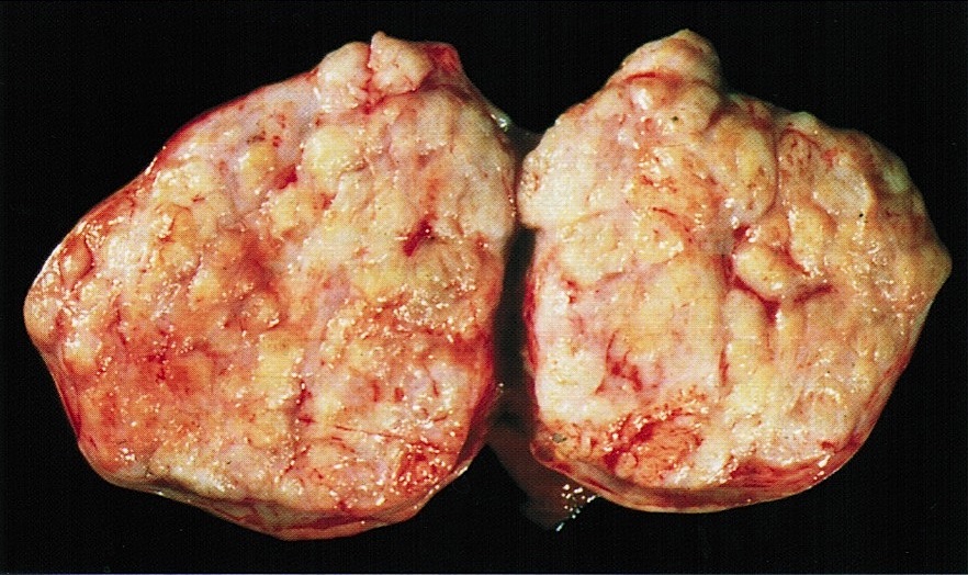
Lobulated sectioned surface
Images hosted on other servers:

Gray-white with whorled pattern

Ovary replaced with creamy mass
Microscopic (histologic) description
- Irregular bundles and whorling of spindle shaped cells with no atypia or pleomorphism
- Degenerative changes like hyaline degeneration and myxomatous changes can be seen
- May be cellular, have prominent mitotic activity or occasionally have bizarre nuclei or myxoid stroma
Microscopic (histologic) images
AFIP images
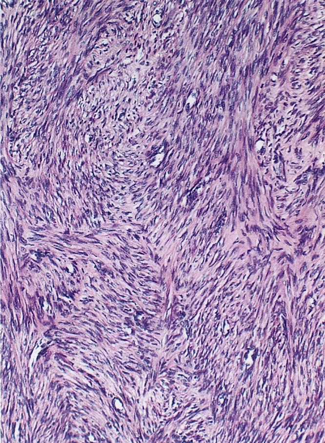
Spindle cells arranged in intersecting fascicles
Images hosted on other servers:

Long fascicle of spindle shaped cells

H&E, desmin, α smooth muscle actin

Positive for SMA
Positive stains
Negative stains
Differential diagnosis
