

Skin nonmelanocytic tumor
Other tumors of skin
CRTC1:TRIM11 cutaneous tumor
Deputy Editor-in-Chief: Jonathan D. Ho, M.B.B.S., D.Sc.


Last author update: 15 February 2024
Last staff update: 15 February 2024
Copyright: 2023-2024, PathologyOutlines.com, Inc.
PubMed Search: CRTC1:TRIM11 cutaneous tumor
Table of Contents
Definition / general | Essential features | Terminology | ICD coding | Epidemiology | Sites | Pathophysiology | Etiology | Clinical features | Diagnosis | Radiology description | Radiology images | Prognostic factors | Case reports | Treatment | Clinical images | Gross description | Gross images | Microscopic (histologic) description | Microscopic (histologic) images | Positive stains | Negative stains | Molecular / cytogenetics description | Molecular / cytogenetics images | Videos | Sample pathology report | Differential diagnosis | Additional references | Board review style question #1 | Board review style answer #1 | Board review style question #2 | Board review style answer #2Cite this page: Tseng C, Saeed Kamil Z. CRTC1:TRIM11 cutaneous tumor. PathologyOutlines.com website. https://www.pathologyoutlines.com/topic/skinmelanocyticCRTC1TRIM11.html. Accessed May 7th, 2024.
Definition / general
- Malignant dermal tumor with epithelioid and spindled cells of uncertain origin with partial melanocytic differentiation and CRTC1::TRIM11 translocation
Essential features
- Histological features include a dermal / subcutaneous circumscribed tumor composed of fascicles and nests of monomorphic epithelioid and spindle cells with prominent nucleoli divided by delicate collagen septa, no / very rare pigment
- Immunohistochemical features include consistent diffuse SOX10 and MITF staining; S100, MelanA and HMB45 stains are variable
- Defined by identifying CRTC1::TRIM11 translocation
- No evidence of EWSR1 rearrangement
- Appears to have at least some malignant potential, although cases and follow up are limited
Terminology
- Cutaneous melanocytic tumor with CRTC1::TRIM11 fusion
- Not recommended: cutaneous melanocytoma with CRTC1::TRIM11 fusion
Epidemiology
- Age: broad range (5 - 87); median age of 42 (Histopathology 2023;82:368)
- No clear gender bias (Am J Surg Pathol 2022;46:1457)
Sites
- Broad anatomic distribution
- Limbs (64%) appear to be involved more often than trunk (20%) and head (2%) based on current case reports (Am J Surg Pathol 2022;46:1457)
- 2 cases of oral and nasal mucosal involvement (Am J Surg Pathol 2022;46:1457)
Pathophysiology
- Exact mechanism of how the CRTC1::TRIM11 fusion transcript causes neoplasia is not known; hypotheses include
- CRTC1::TRIM11 fusion deletes a protein - protein interaction site on TRIM11 that is involved in directing misfolded proteins towards proteasomes for degradation (Cureus 2022;14:e33179)
- CRTC1::TRIM11 fusion retains an interaction site with CREB1, which in turn may drive expression of MITF; MITF acts like an oncogene and can drive MITF synthesis (Am J Surg Pathol 2022;46:1457)
Etiology
- Unknown
Clinical features
- Discrete, slowly growing, skin colored nodule without pigmentation (Am J Surg Pathol 2022;46:1457)
Diagnosis
- Biopsy
- Defined by molecular findings
Radiology description
- Lobulated lesion on imaging
- May be positron emission tomography (PET) avid
Radiology images
Contributed by Aaron Lloyd Hendler, M.D.
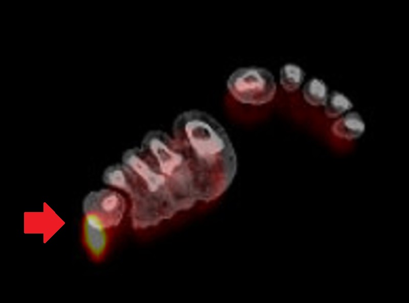
PET avid
Prognostic factors
- Limited case reports and follow up available but appears more indolent than melanoma and clear cell sarcoma at the time of writing
- 4 of 48 published cases reported have shown regional or distant metastasis (Am J Surg Pathol 2022;46:1457, Histopathology 2023;82:368, Am J Surg Pathol 2019;43:861, J Cutan Pathol 2024;51:181)
- A recent additional case of CRTC1:TRIM11 cutaneous tumor (CTCT) with regional lymph node metastasis was presented at the American Society of Dermatopathology in 2023 (Tseng, et al.: CRTC1::TRIM11 Cutaneous Tumor With Ulceration and Sentinel Lymph Node Metastasis, oral presentation at American Society of Dermatopathology, Oct 2 - 8, 2023)
Case reports
- 5 year old girl with right upper arm lesion (J Cutan Pathol 2022;49:1025)
- 27 year old man with fourth finger mass (Cureus 2022;14:e33179)
- 77 year old man with right thigh lesion (Pathol Int 2019;69:496)
Treatment
- Other than complete surgical excision, optimal treatment is unknown
Clinical images
Contributed by Alexandra Easson, M.D.
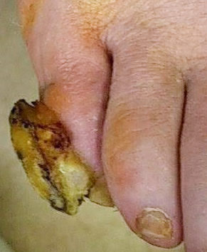
Ulcerated toe CTCT
Images hosted on other servers:

Arm CTCT

Finger CTCT
Gross description
- Circumscribed tumors with pale, firm cut surface without pigmentation
- Mean size: 1.4 cm (0.4 - 5.1 cm) (Histopathology 2023;82:368)
Gross images
Contributed by Zaid Saeed Kamil, M.B.Ch.B. and Calvin Tseng, M.D.
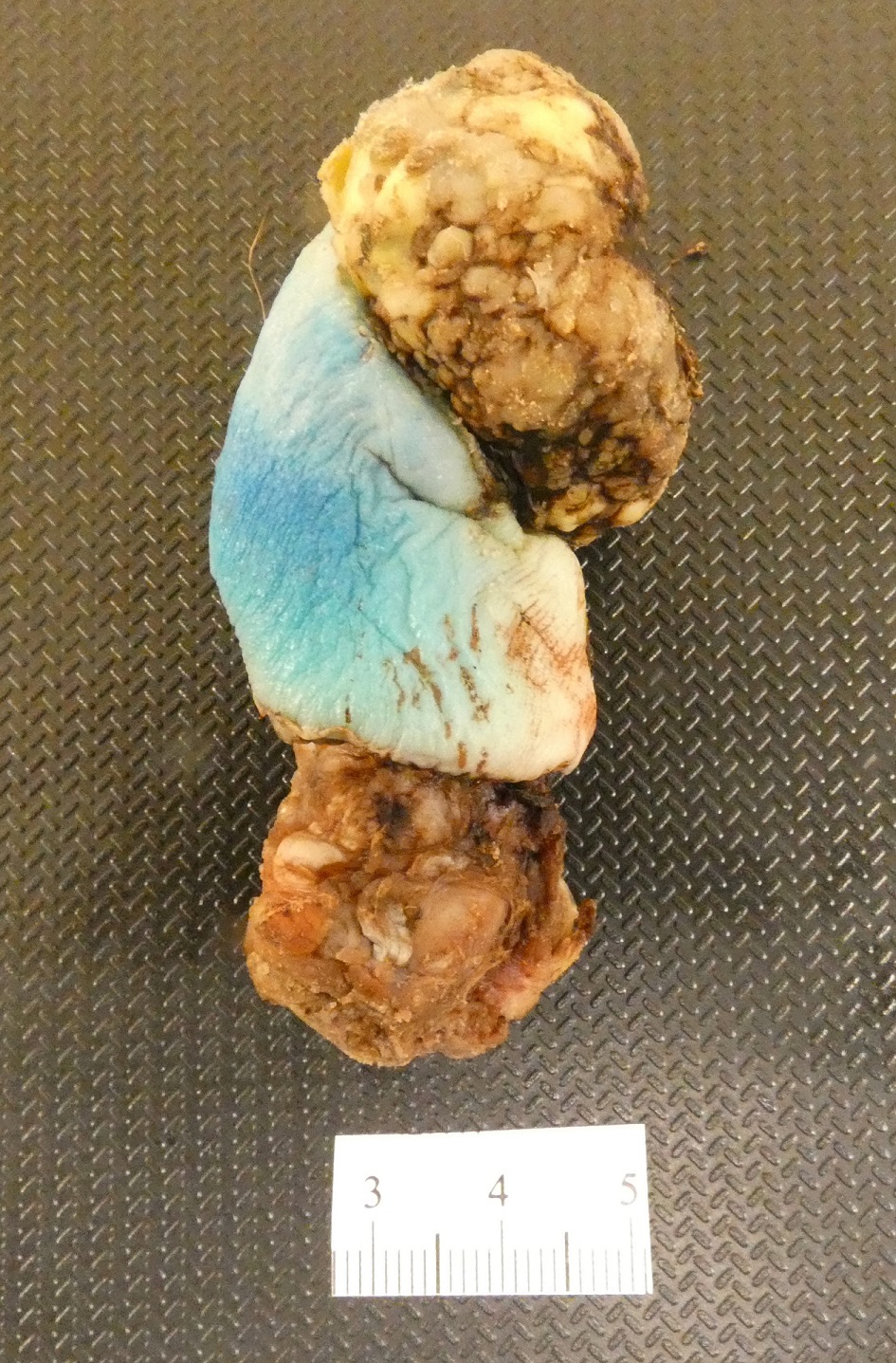
Amputation specimen
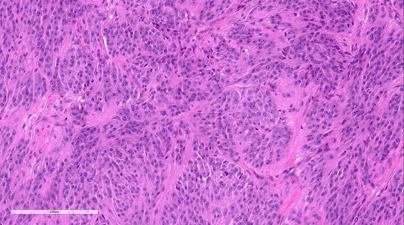
Microscopic (histologic) description
- Morphology is uniform and resembles clear cell sarcoma
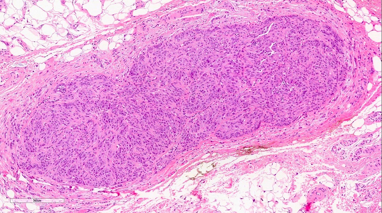
- Circumscribed, unencapsulated dermal nodule, no / very rare pigment, with only rare cases showing epidermal involvement (J Cutan Pathol 2022;49:1025)
- Some cases show extension into subcutis (Am J Surg Pathol 2019;43:861)
- Characteristic pattern of nests and intersecting (occasionally herringbone to vaguely palisaded) fascicles of uniform epithelioid to spindled cells with intervening delicate collagen bands (Am J Surg Pathol 2022;46:1457)
- Abundant pale eosinophilic cytoplasm, monomorphic nuclei with vesicular chromatin and prominent nucleoli
- Mitotic figures present (Am J Surg Pathol 2022;46:1457)
- Necrosis reported but uncommon (Am J Surg Pathol 2022;46:1457)
- Lymphovascular and perineural invasion uncommon (Am J Surg Pathol 2022;46:1457)
- Pigment and multinucleate giant cells may very rarely be seen
- A recent case of CTCT with ulceration was reported (Tseng, et al.: CRTC1::TRIM11 Cutaneous Tumor With Ulceration and Sentinel Lymph Node Metastasis, oral presentation at American Society of Dermatopathology, Oct 2 - 8, 2023)
Microscopic (histologic) images
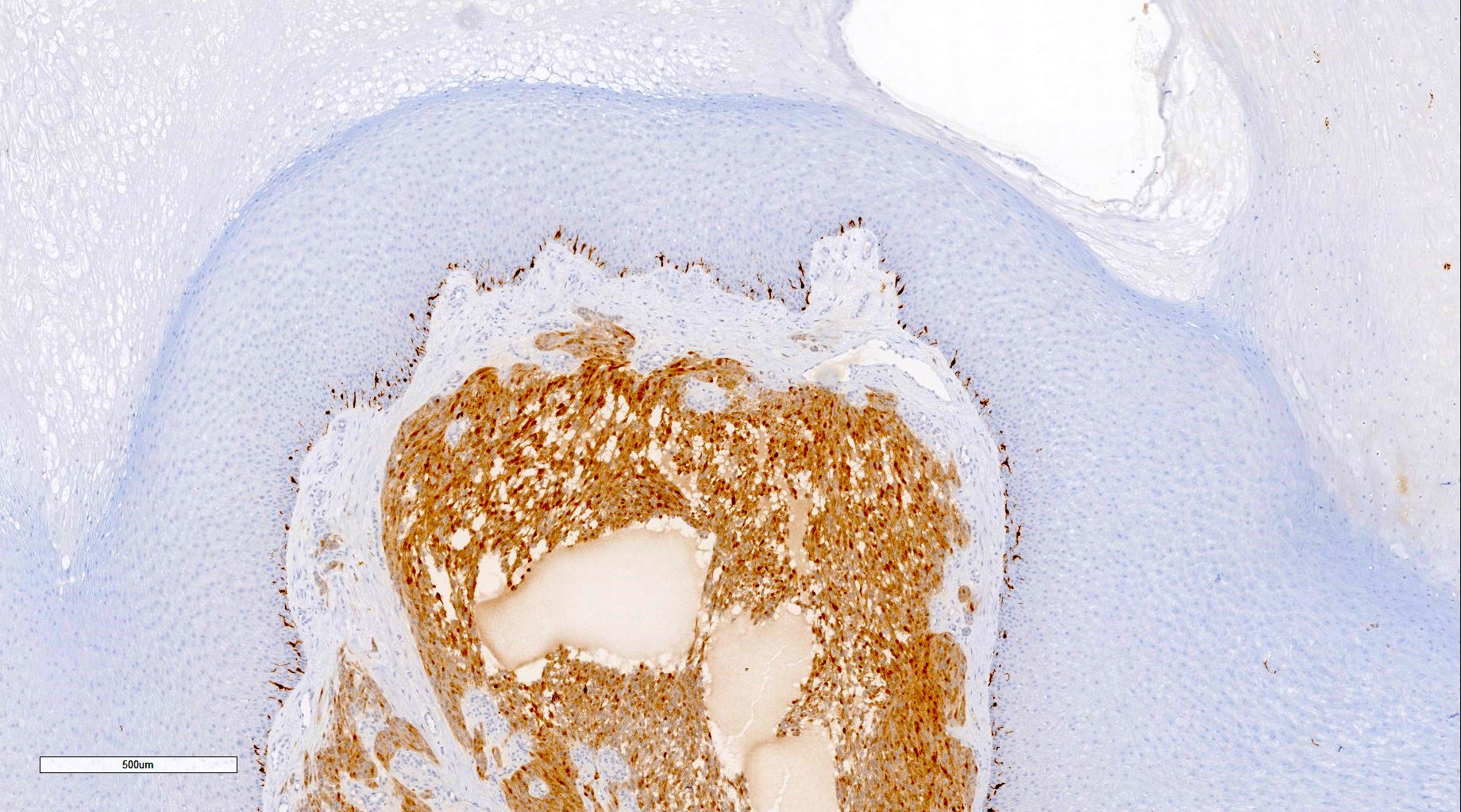
Contributed by Zaid Saeed Kamil, M.B.Ch.B., Stephen M. Smith, M.D. and Calvin Tseng, M.D.
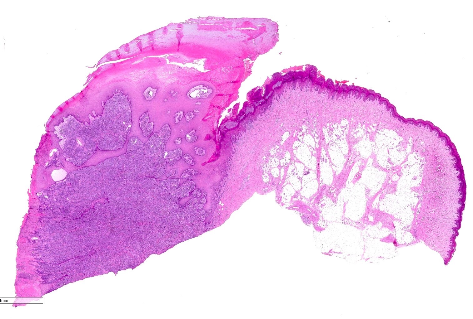
Dermal lesion
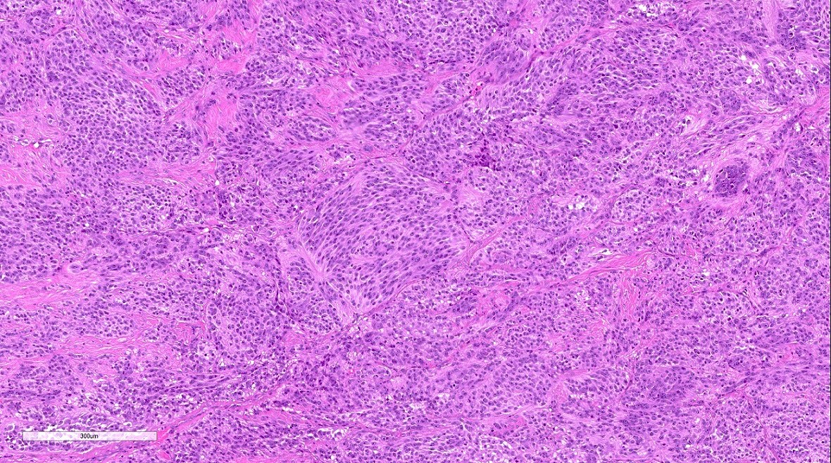
Fascicular growth pattern
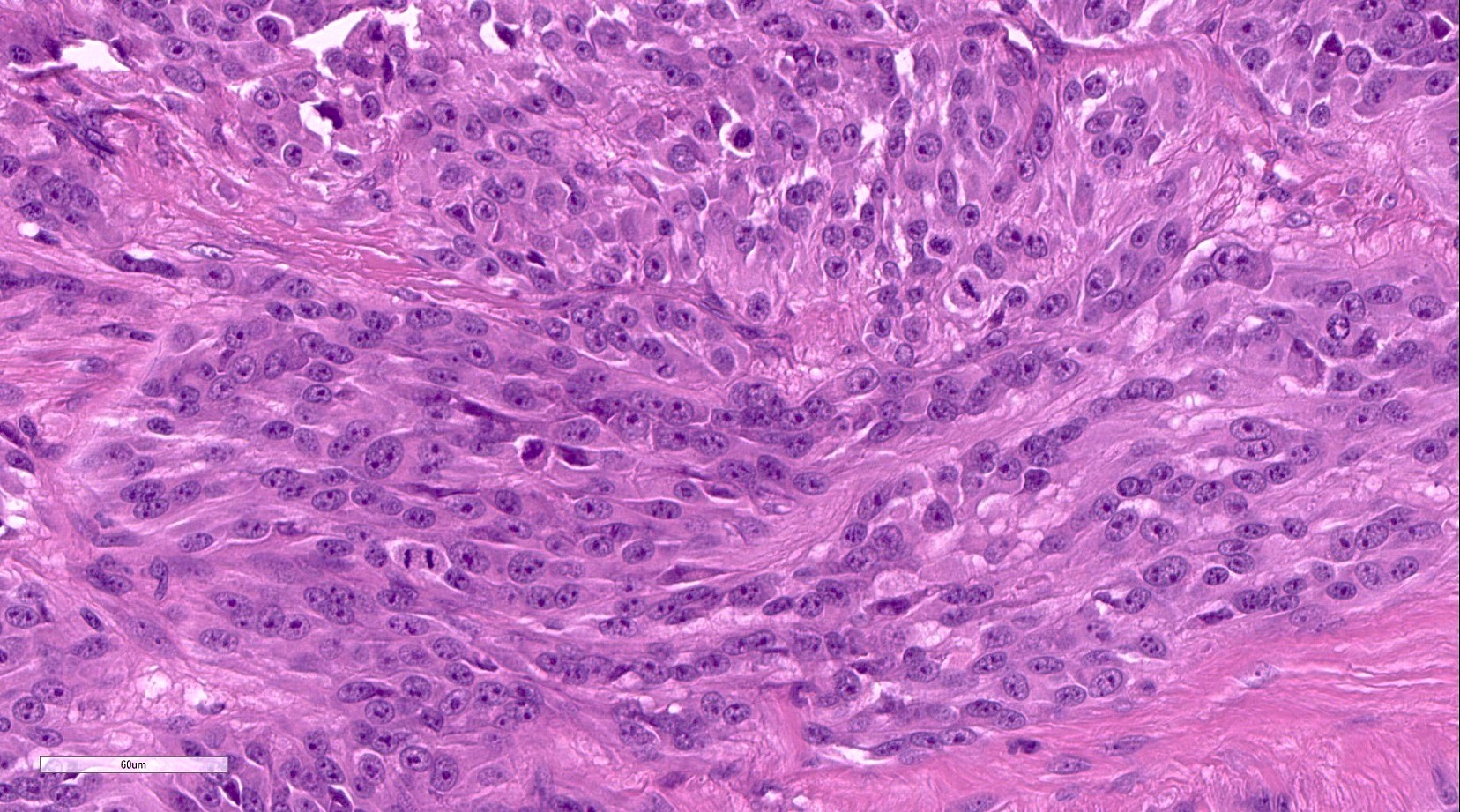
Monomorphic cells
Vascular invasion
SOX10 diffuse positive
S100 positive
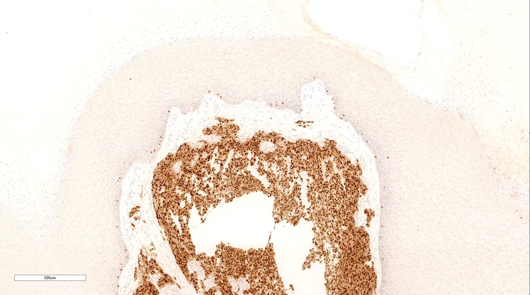
MITF diffuse positive
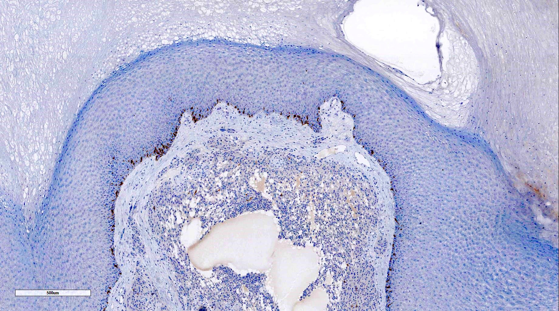
MelanA negative
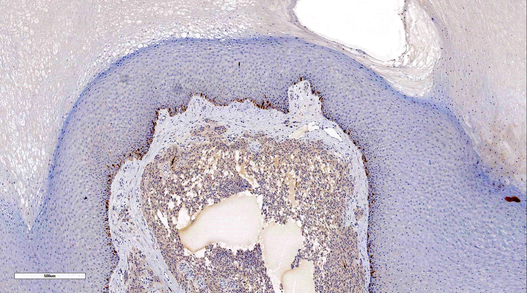
HMB45 negative
PRAME negative

Pan-TRK positive
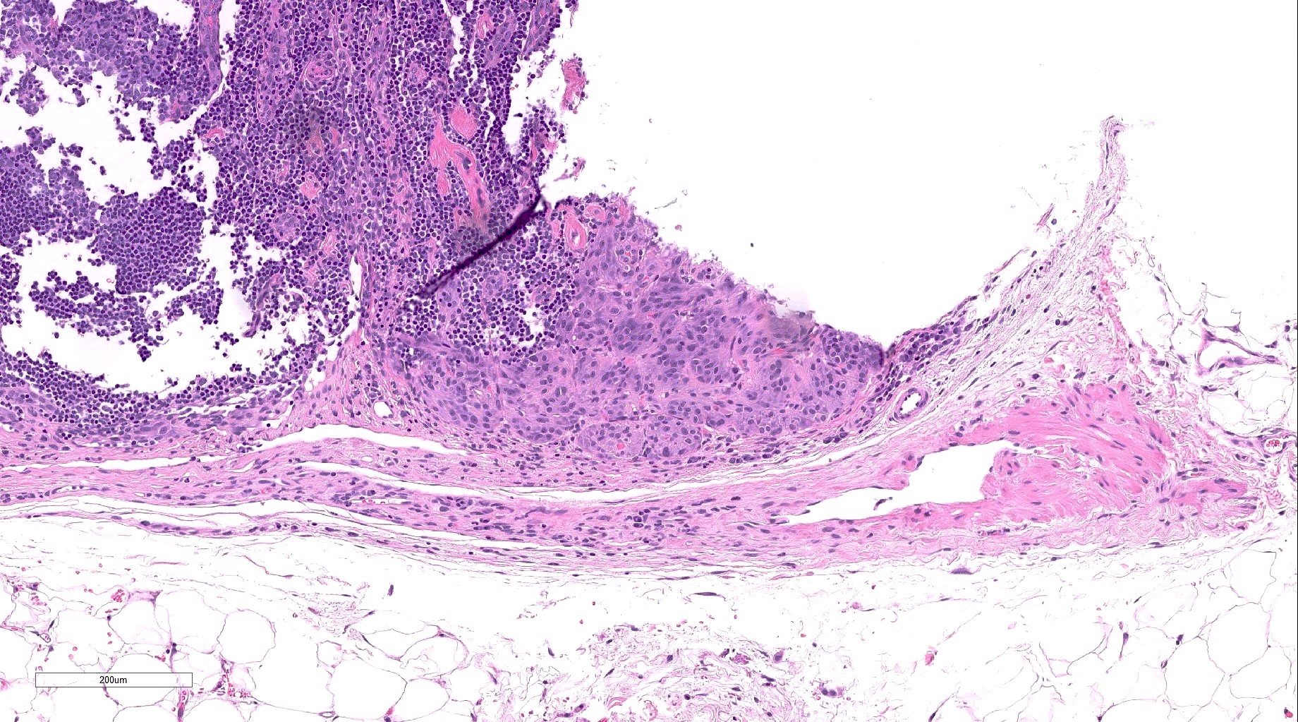
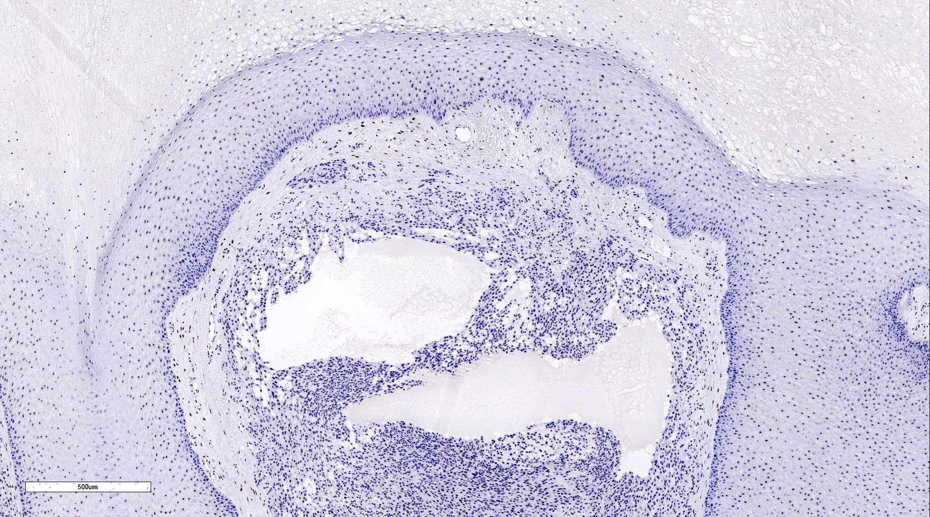
Lymph node metastasis
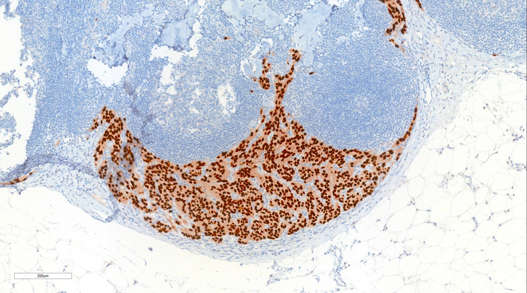
Lymph node metastasis SOX10
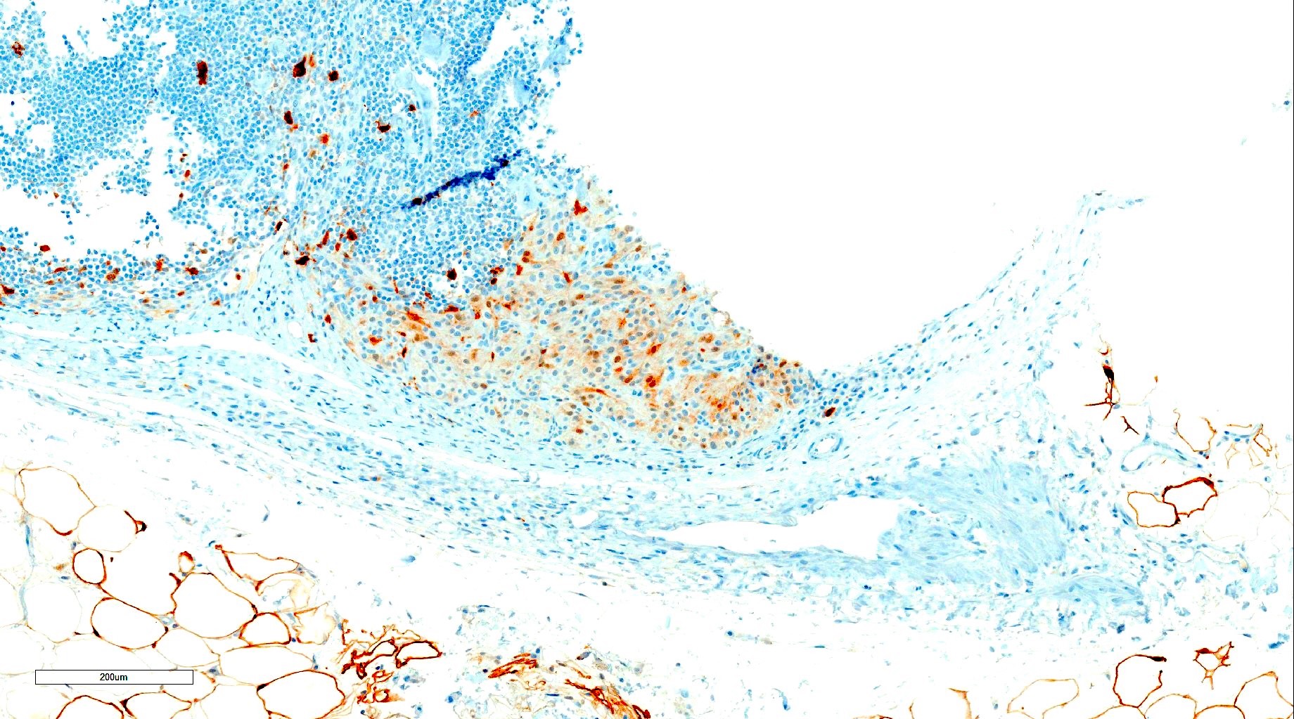
Lymph node weak S100
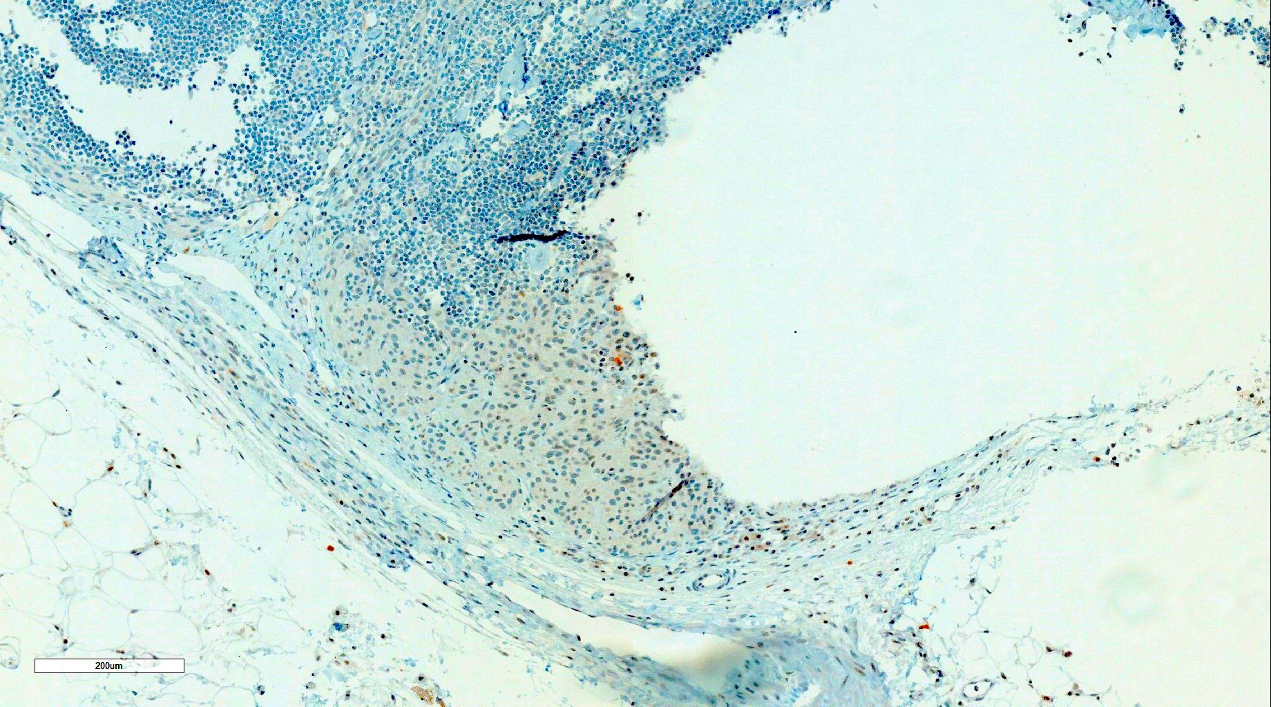
Negative MelanA and HMB45
Positive stains
- SOX10 diffuse positive in all cases (Am J Surg Pathol 2022;46:1457)
- MITF diffuse positive in majority of cases (J Cutan Pathol 2022;49:1025)
- S100 diffuse positive in 44%, partial positive in 36% (Am J Surg Pathol 2022;46:1457)
- MelanA diffuse positive in 9%, partial positive in 52% (Am J Surg Pathol 2022;46:1457)
- HMB45 diffuse positive in 4%, partial positive in 52% (Am J Surg Pathol 2022;46:1457)
- Ki67 5 - 20% (Am J Surg Pathol 2018;42:382)
- Pan-TRK diffuse cytoplasmic positive in 57%, partial cytoplasmic positive in 36% despite lack of TRK mutations (Am J Surg Pathol 2022;46:1457)
- TRIM11 diffuse nuclear positive in 94% (Am J Surg Pathol 2022;46:1457)
Negative stains
Molecular / cytogenetics description
- CRTC1::TRIM11 fusion (Am J Surg Pathol 2022;46:1457)
- No EWRS1 rearrangement (Am J Surg Pathol 2022;46:1457)
- No melanoma driver mutations (Am J Surg Pathol 2022;46:1457)
- No NTRK fusions despite positivity for pan-TRK immunohistochemistry (Am J Surg Pathol 2018;42:382)
Molecular / cytogenetics images
Contributed by Ian King, Ph.D.
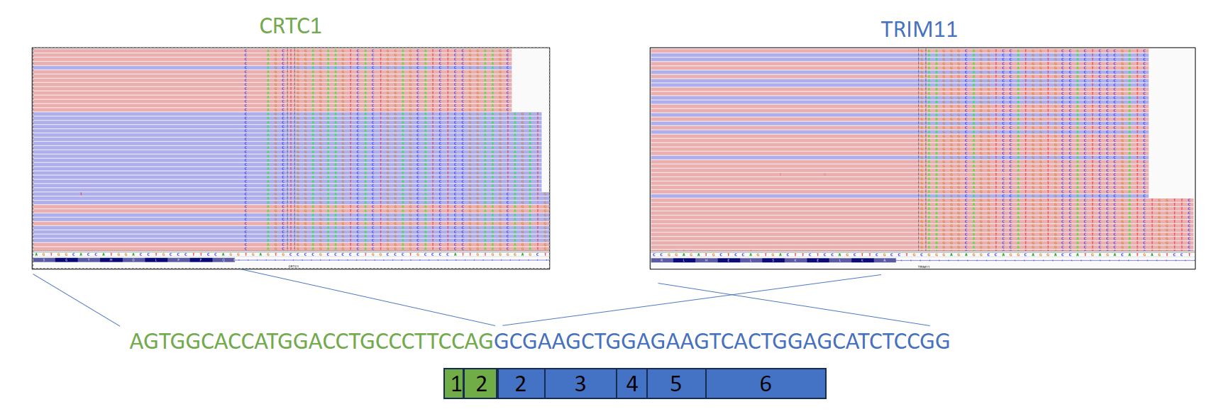
RNA CRTC1::TRIM11 fusion
Images hosted on other servers:

CRTC1::TRIM11 FISH

CRTC1::TRIM11 fusion
Videos
CRTC::TRIM11 cutaneous tumor
Sample pathology report
- Skin, amputation, right fifth toe:
- CRTC1::TRIM11 cutaneous tumor (see comment)
- Comment: The neoplasm is composed of intersecting fascicles and nests of relatively uniform epithelioid and spindled cells with intervening bands of fibrocollagenous stroma. The neoplastic cells have abundant amounts of amphophilic to eosinophilic cytoplasm and moderately pleomorphic nuclei with prominent nucleoli. Mitotic figures including deep mitoses are identified. There is no evidence of necrosis, lymphovascular invasion or perineural invasion. The neoplastic cells are diffusely positive for SOX10, patchy positive for S100 and negative for MelanA and HMB45. Molecular analysis demonstrated the presence of CRTC1::TRIM11 fusion. Overall, the features are compatible with the recently described CRTC1::TRIM11 cutaneous tumor. Based on the limited reported cases and limited follow up data, the clinical course was indolent in most of the reported cases. However, 4 of the reported cases presented with regional nodal metastasis or distant metastasis as of January 2024. The available data at the time of this report suggest that these tumors have at least some malignant potential.
Differential diagnosis
- Clear cell sarcoma:
- Predominantly affects adults ages 30 - 40s
- Often deeper location (associated with tendons or aponeuroses)
- Infiltrative growth
- May have clear cells and melanin pigment
- Wreath-like multinucleated tumor giant cells
- More consistently expresses S100, MelanA and HMB45
- Has ESWR1 translocations, most commonly EWSR1::ATF1 or EWSR1::CREB1
- Lacks CRTC1::TRIM11 fusion
- More aggressive behavior
- Melanoma:
- In situ component (may not be present for previously excised melanoma, metastatic melanoma or primary dermal melanoma)
- Infiltrative growth
- Pleomorphic cytology
- Pigment production
- More consistently expresses S100, MelanA and HMB45 (other than in desmoplastic melanoma)
- PRAME positive
- Has melanoma driver mutations
- Lacks CRTC1::TRIM11 fusion
- More aggressive behavior
- Spitz tumor:
- Cellular blue nevus:
- Myoepithelial tumors:
- Epithelioid schwannoma:
- MITF negative
- 40% show loss of INI1 (Am J Surg Pathol 2017;41:1013)
- Lacks CRTC1::TRIM11 fusion
- Epithelioid MPNST:
- PEComa:
- MITF pathway activated melanocytic tumors:
Additional references
Board review style question #1
A superficial dermal tumor with the above histology is identified. IHC shows diffuse SOX10 positivity, diffuse MITF positivity, partial S100 positivity, partial MelanA positivity and partial HMB45 positivity. PRAME IHC is negative. Molecular investigations identify a CRTC1::TRIM11 fusion. Is it important to differentiate this entity from melanoma and clear cell sarcoma and why?
- No, as this entity currently appears to behave similarly to melanoma and clear cell sarcoma
- No, as this entity is currently thought to be a subtype of melanoma
- Yes, as this entity currently appears to behave less aggressively than melanoma and clear cell sarcoma
- Yes, as this entity currently has specific targeted treatments available
Board review style answer #1
C. Yes, as this entity currently appears to behave less aggressively than melanoma and clear cell sarcoma. Based on follow up data available as of October 2023, CRTC1:TRIM11 cutaneous tumor (CTCT) appears to be a low grade malignancy that behaves less aggressively than typical melanomas and clear cell sarcomas. Answer B is incorrect because CTCT has not had melanoma driver mutations identified as of October 2023. Answer A is incorrect because CTCT appears to behave less aggressively than melanoma and clear cell sarcoma based on follow up data available as of October 2023. Answer D is incorrect because no targeted treatment has been identified for CTCT as of October 2023.
Comment Here
Reference: CRTC1:TRIM11 cutaneous tumor
Comment Here
Reference: CRTC1:TRIM11 cutaneous tumor
Board review style question #2
Of the following features, which is typical of CRTC1:TRIM11 cutaneous tumor (CTCT)?
- Absence of mitotic activity
- Presence of an epidermal component
- Presence of CRTC1::TRIM11 fusion
- Presence of infiltrative growth
Board review style answer #2
C. Presence of CRTC1::TRIM11 fusion. CRTC1:TRIM11 cutaneous tumor (CTCT) is defined by the CRTC1:TRIM11 fusion, which has not yet been identified in other entities. Answer B is incorrect because the majority of CTCT cases do not show an epidermal component. Answer D is incorrect because the majority of CTCT are well circumscribed. Answer A is incorrect because CTCT show mitotic activity.
Comment Here
Reference: CRTC1:TRIM11 cutaneous tumor
Comment Here
Reference: CRTC1:TRIM11 cutaneous tumor
