Skin nonmelanocytic tumor
Adnexal tumors
Sweat gland derived (apocrine & eccrine glands)
Tubular apocrine adenoma
Editorial Board Member: Bethany R. Rohr, M.D.


Last author update: 5 August 2024
Last staff update: 8 August 2024
Copyright: 2002-2025, PathologyOutlines.com, Inc.
PubMed Search: Tubular apocrine adenoma
Table of Contents
Definition / general | Essential features | Terminology | ICD coding | Epidemiology | Sites | Pathophysiology | Clinical features | Diagnosis | Prognostic factors | Case reports | Treatment | Clinical images | Gross description | Microscopic (histologic) description | Microscopic (histologic) images | Positive stains | Negative stains | Electron microscopy description | Molecular / cytogenetics description | Sample pathology report | Differential diagnosis | Practice question #1 | Practice answer #1 | Practice question #2 | Practice answer #2Cite this page: Hamza M, Shalin SC. Tubular apocrine adenoma. PathologyOutlines.com website. https://www.pathologyoutlines.com/topic/skintumornonmelanocyticapocrinetubularadenoma.html. Accessed August 18th, 2025.
Definition / general
- Benign dermal adnexal neoplasm of apocrine derivation
- May be associated with organoid nevus, nevus sebaceus of Jadassohn and syringocystadenoma papilliferum (SCAP)
- Most cases show apocrine differentiation but eccrine differentiation may be present as well
- Reference: Int J Mol Sci 2021;22:5077
Essential features
- Rare, benign adnexal neoplasm
- Most common location is the scalp but can occur on other sites (Hum Pathol 2018;73:59)
- Microscopically, it is a well circumscribed intradermal tumor composed of tubules lined by 2 cell layers or more in a fibrous, sometimes hyalinized stroma
Terminology
- Also called apocrine adenoma, tubular adenoma, tubulopapillary hidradenoma, papillary tubular adenoma
- Considerable overlap with papillary eccrine adenoma; may be part of the same spectrum (Am J Dermatopathol 1992;14:149, Am J Dermatopathol 1993;15:482, Int J Mol Sci 2021;22:5077)
ICD coding
- ICD-10: D23.9 - other benign neoplasm of skin, unspecified
Epidemiology
- Age distribution of tubular apocrine adenoma is very wide, ranging from 28 to 85 years according to one study (Hum Pathol 2018;73:59)
Sites
- Most common location is scalp
- Rarely occurs in the nose, eyelid, leg, trunk, axilla, chest, external auditory meatus, cheek, vulva
Pathophysiology
- Associated with organoid nevus, nevus sebaceus of Jadassohn and syringocystadenoma papilliferum (SCAP) (J Cutan Pathol 1989;16:230)
- BRAF p.V600E mutations are detected in 50 - 64% of syringocystadenomas papilliferum and 66% of tubular adenomas, respectively (Cancers (Basel) 2022;14:476)
- BRAF and KRAS mutations may be present (Hum Pathol 2018;73:59)
Clinical features
- Clinically asymptomatic, well defined nodule
- Usually < 2 cm but reported up to 7 cm (Int J Mol Sci 2021;22:5077)
Diagnosis
- Skin biopsy
Prognostic factors
- Lesion is considered benign; complete excision is recommended to prevent recurrence (Medicine (Baltimore) 2021;100:e28002)
Case reports
- 34 year old woman with 10 year history of foot nodule consistent with tubular apocrine adenoma (J Dermatol 2019;46:e45)
- 36 year old woman with an asymptomatic tubular apocrine adenoma of the vulva (Indian Dermatol Online J 2018;9:346)
- 63 year old man with recurrence of a tubular apocrine adenoma of the left upper eyelid after incomplete excision (Saudi J Ophthalmol 2019;33:304)
Treatment
- Complete excision is curative (Int J Mol Sci 2021;22:5077)
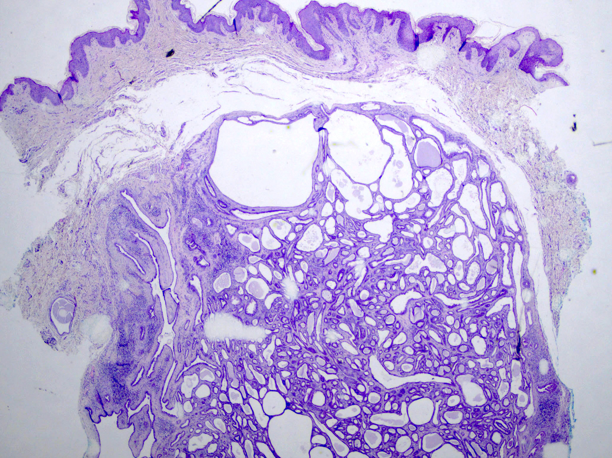
Clinical images
Images hosted on other servers:

Mimics basal cell carcinoma
Gross description
- Firm, slow growing, dermal, skin colored nodule
Microscopic (histologic) description
- Well circumscribed dermal neoplasm that may extend into subcutis
- Lobular pattern of dermal and subcutaneous tubular apocrine structures often encased by a fibrous, sometimes hyalinized stroma
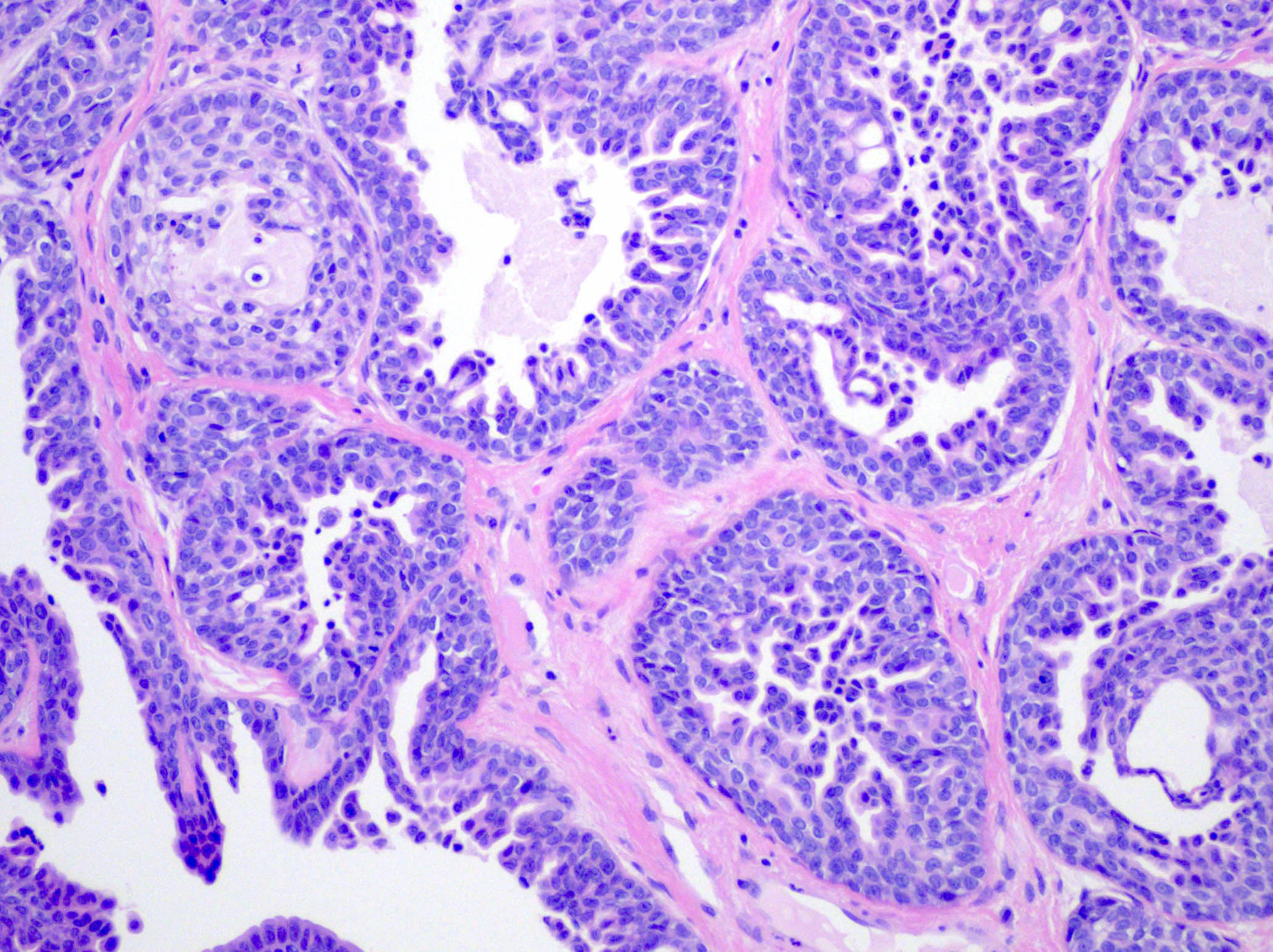
- Lobules have dilated, variably sized, well formed tubules lined by 2 layers of epithelial cells
- Pseudopapillae are common but true papillae are more often associated with SCAP
- Decapitation secretion by apical layer and flattened outer myoepithelial layer
- Cuboidal to columnar cells with eosinophilic cytoplasm and round bland nuclei
- Often hyaline and clear cell change
- May show cyst formation with papillae or pseudopapillae protruding into the lumen
- Variable overlying epidermal hyperplasia
- Rare connection with overlying epidermis
- References: J Cutan Pathol 1987;14:114, Saudi J Ophthalmol 2019;33:304, Int J Mol Sci 2021;22:5077
Microscopic (histologic) images
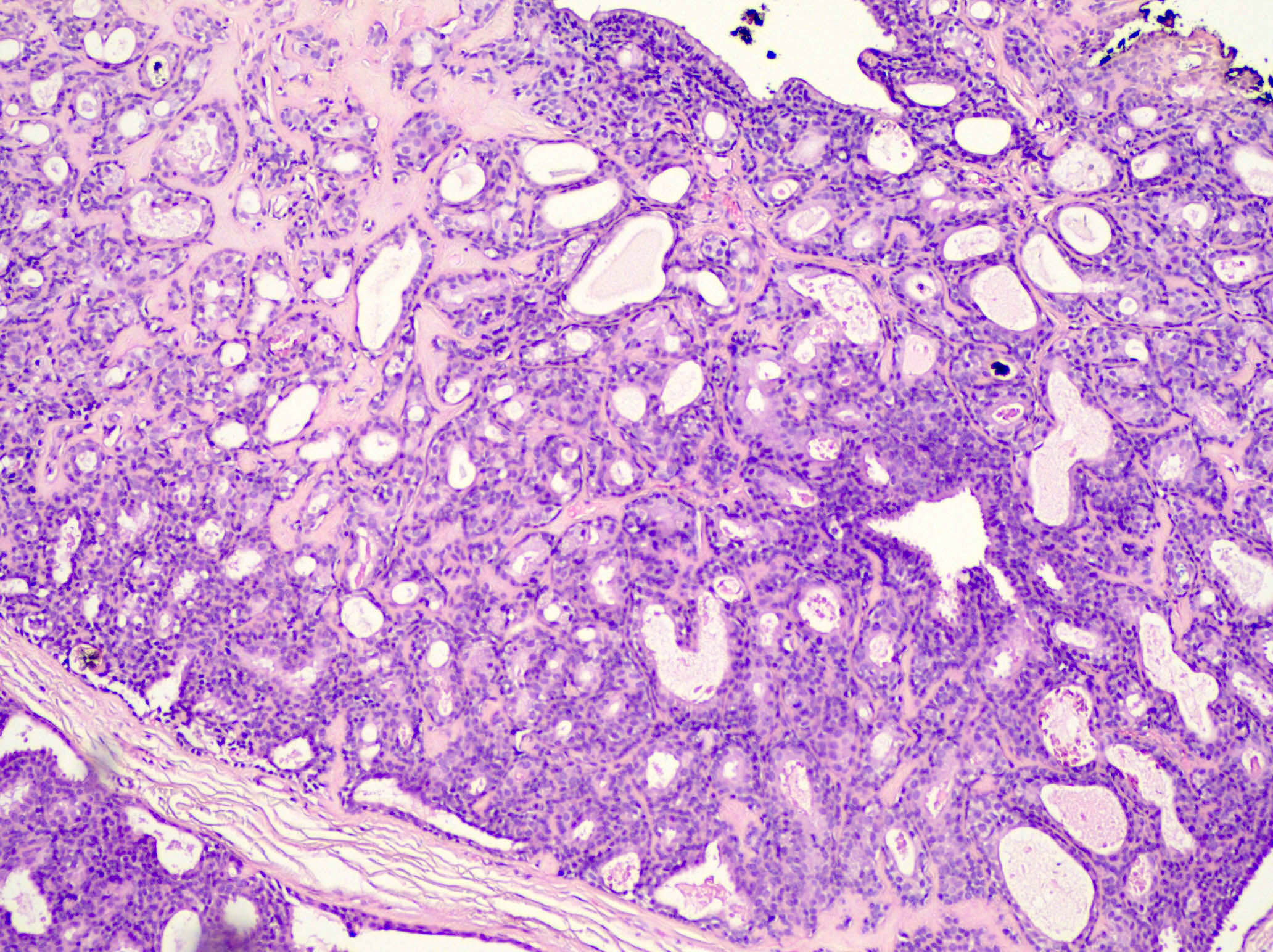
Contributed by Mugahed Hamza, M.B.B.S. and Sara C. Shalin, M.D., Ph.D.
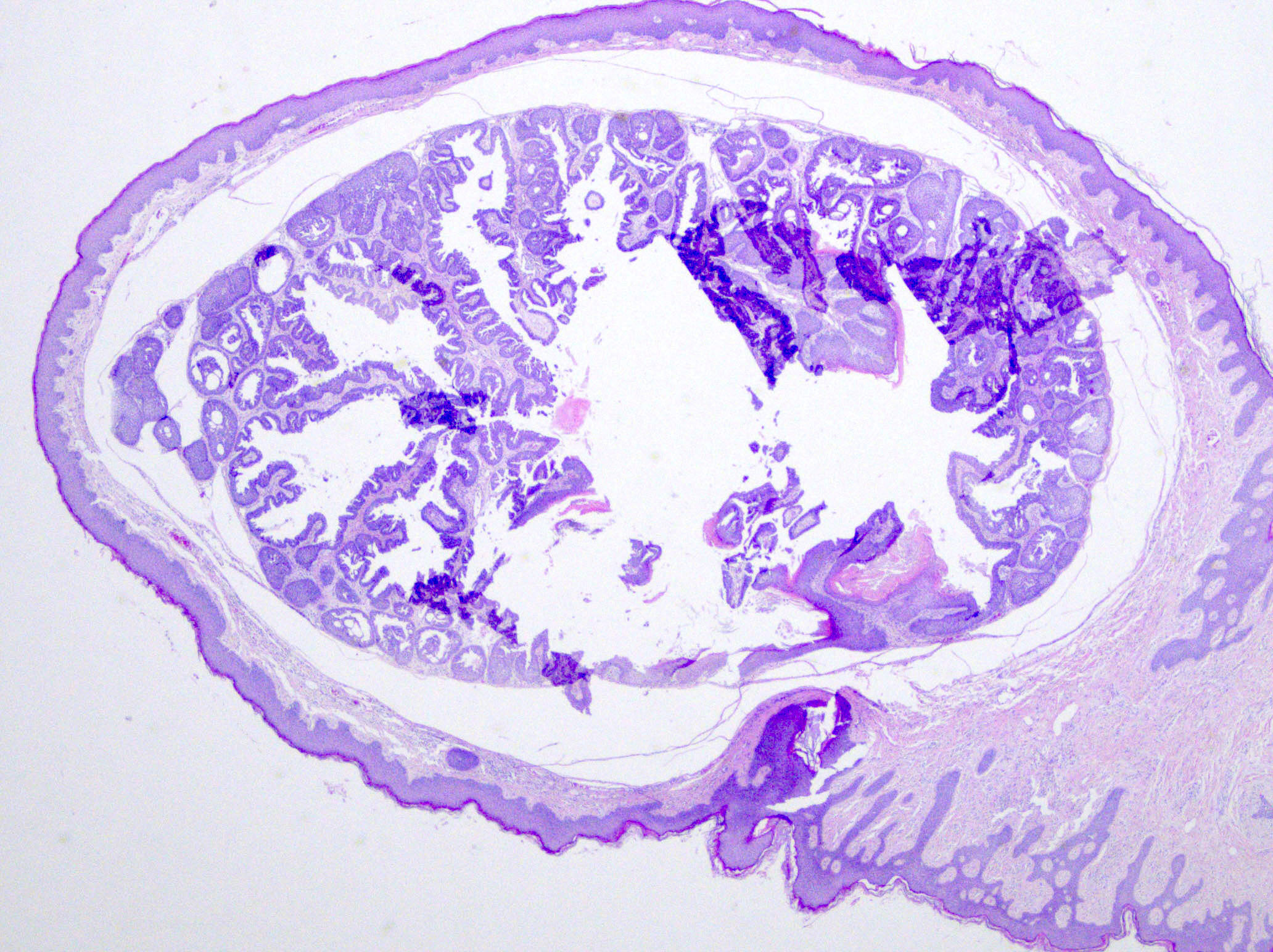
Well circumscribed dermal neoplasm
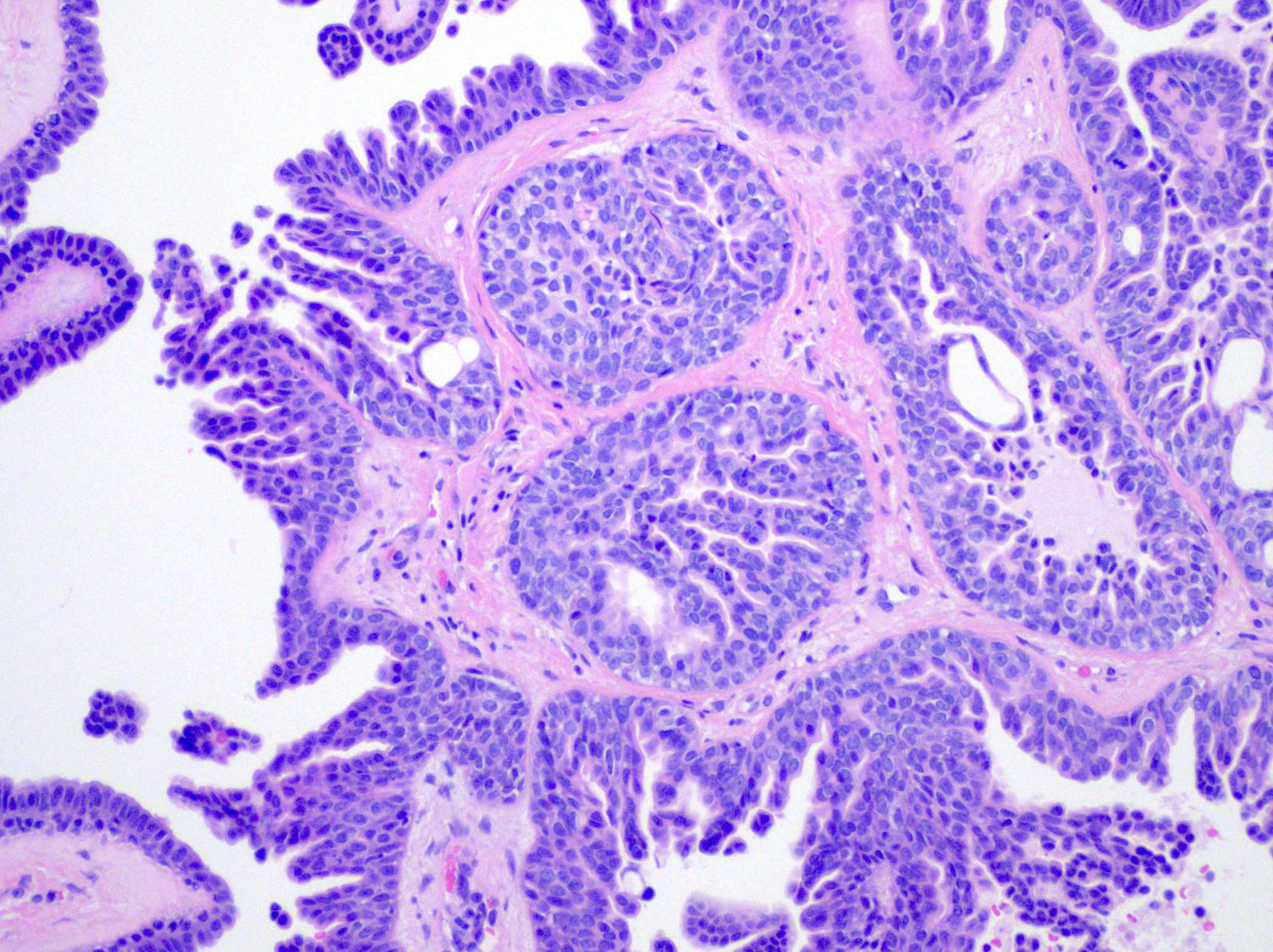
Pseudopapillae protruding into the lumen
Lobules have dilated, variably sized tubules
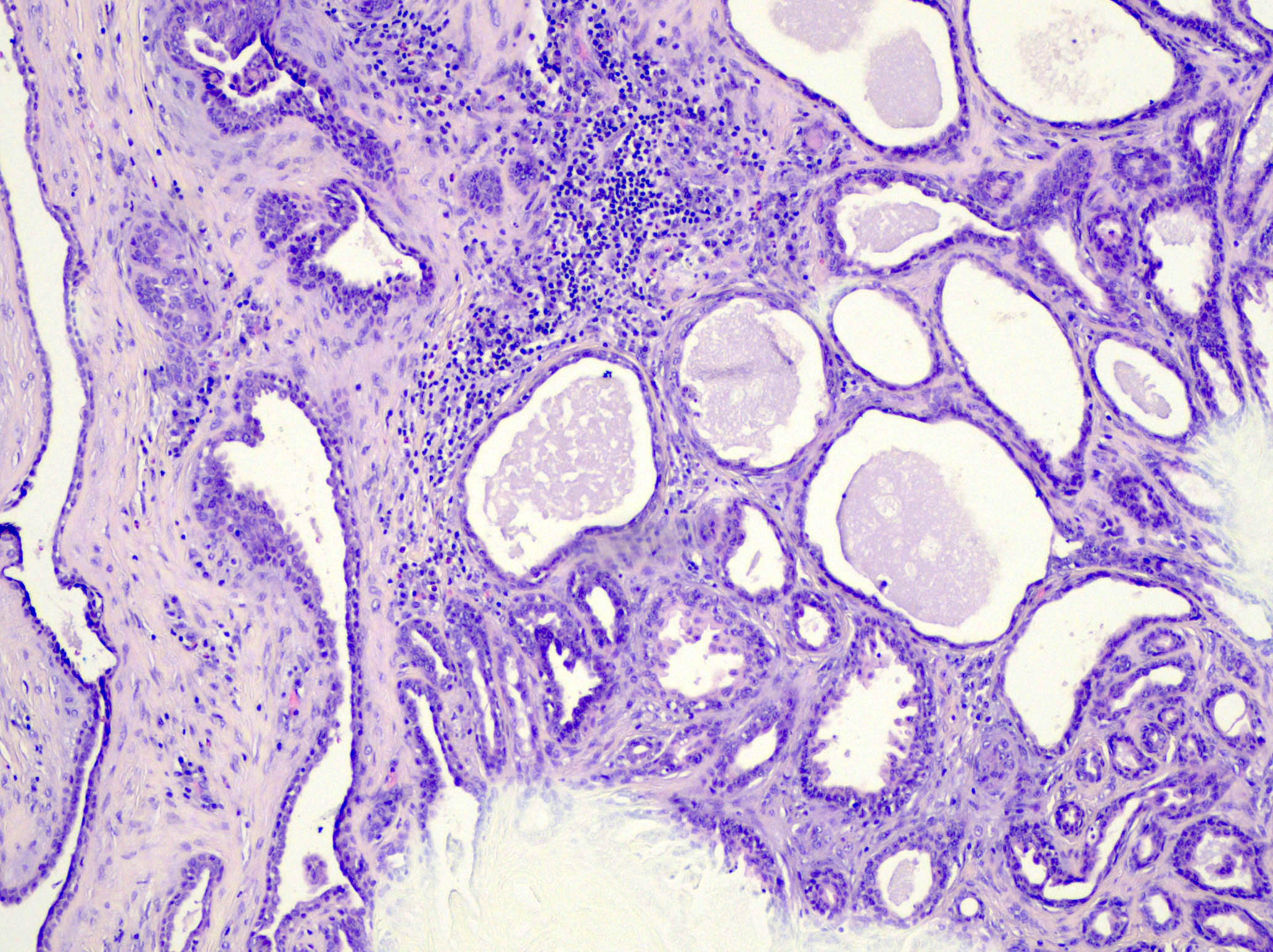
Variably sized tubules with 2 cell layer lining
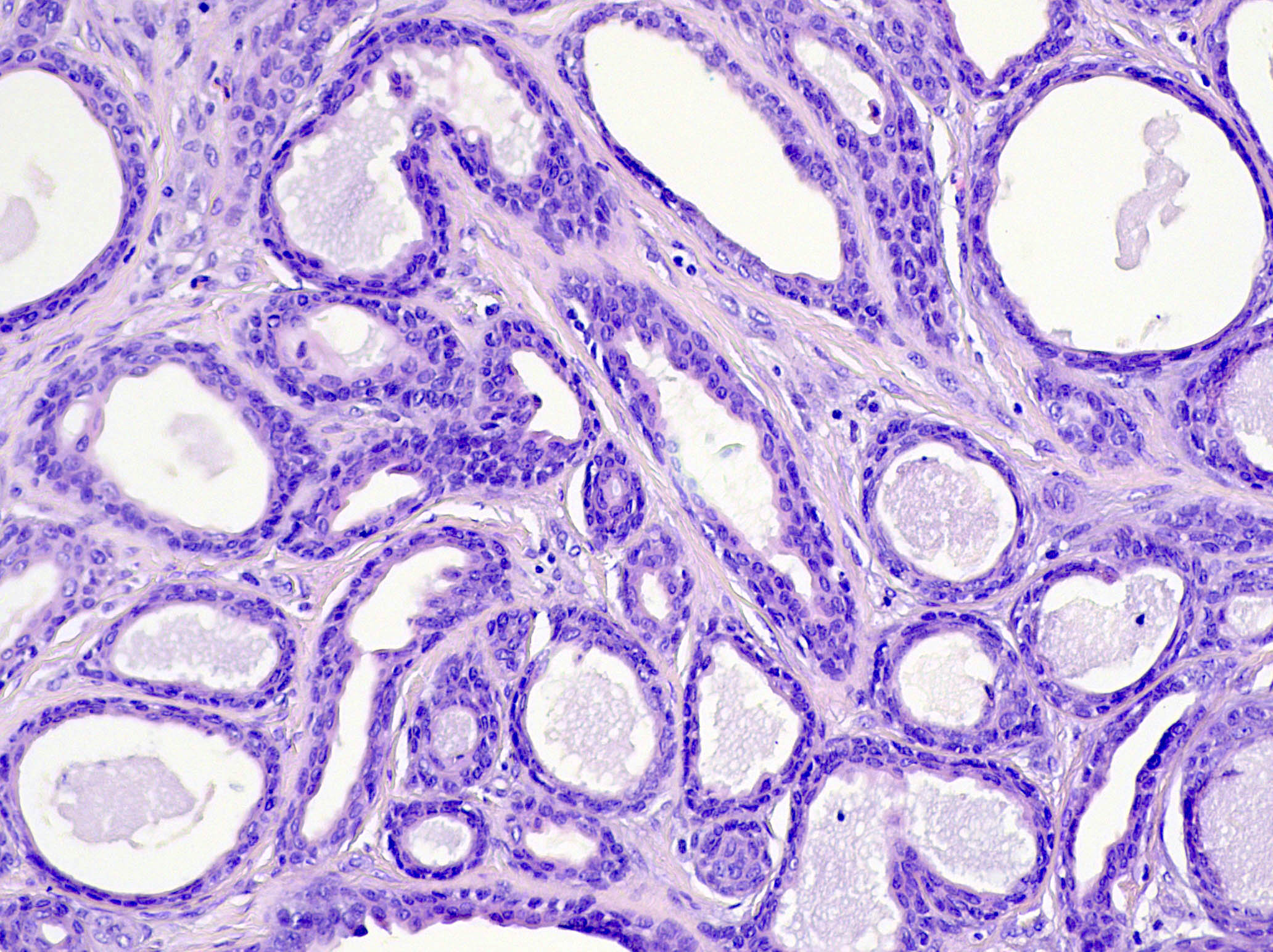
Well formed tubules
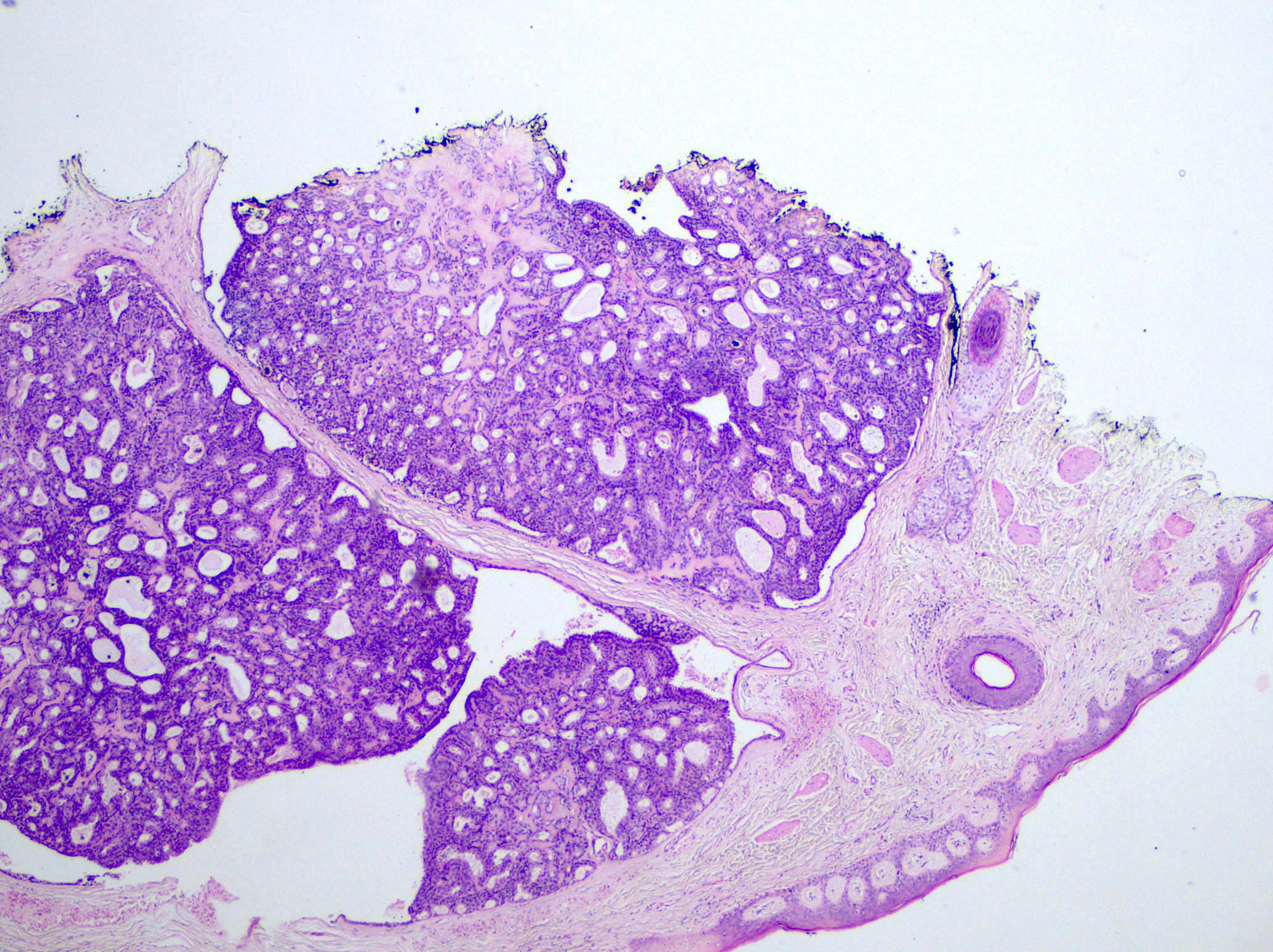
Dermal neoplasm with variably sized tubules
Tubules with multilayered epithelial cell layer
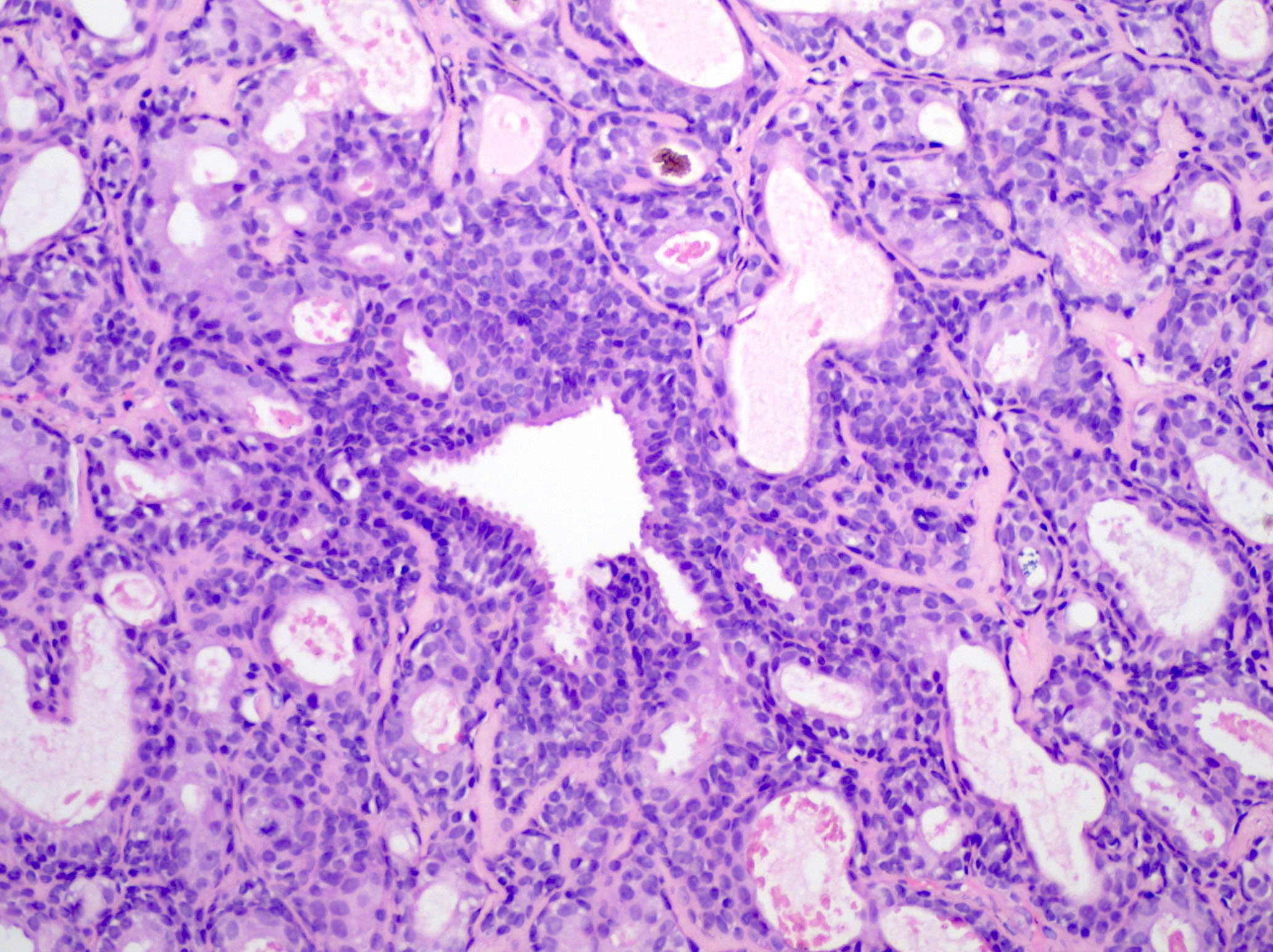
Focal decapitation secretion
Positive stains
Negative stains
Electron microscopy description
- Tall columnar cells on basal lamina forming acini
- Cells lining tubules have luminal microvilli and apical pinching
- Conspicuous mitochondria, prominent Golgi
- Lipid rich cytoplasmic secretory vacuoles
- Decapitation secretion (J Am Acad Dermatol 1984;11:639)
Molecular / cytogenetics description
- BRAF p.V600E mutations are detected in 50 - 64% of syringocystadenomas papilliferum and 66% of tubular adenomas, respectively (Cancers (Basel) 2022;14:476)
- BRAF and KRAS mutations may be present (Hum Pathol 2018;73:59)
Sample pathology report
- Skin, scalp, shave biopsy:
- Tubular apocrine adenoma
Differential diagnosis
- Apocrine cystadenoma:
- More dilated, cystic spaces rather than tubules
- Hidradenoma papilliferum:
- Often has complex arborizing papillae, with more closely arranged tumor cells and glands
- Limited to female genital region
- Papillary apocrine carcinoma:
- More cytologic atypia, irregular nuclear contours and a higher mitotic rate along with infiltrative growth
- Papillary eccrine adenoma:
- Classically has features of eccrine rather than apocrine derivation
- Lacks decapitation secretion
- Different clinical presentation and distribution
- Syringocystadenoma papilliferum:
- Usually connects to epidermis
- Fibrovascular cores within papillary structures
- Plasma cells within stroma
- Tubular apocrine adenoma may be a variant
Practice question #1
Which of the following is true regarding tubular apocrine adenoma?
- Associated with Cowden syndrome
- Associated with mucinous carcinoma
- Most common on the extremities
- Sometimes associated with syringocystadenoma papilliferum
Practice answer #1
D. Sometimes associated with syringocystadenoma papilliferum. Tubular apocrine adenoma most commonly presents on the scalp, often arising in a background of nevus sebaceus and is sometimes associated with syringocystadenoma papilliferum. Answer A is incorrect because while Cowden syndrome is associated with other cutaneous adnexal tumors, it is not associated with tubular apocrine adenoma. Answer B is incorrect because it is not associated with mucinous carcinoma. Answer C is incorrect because the most common location is on the scalp, not the extremities.
Comment Here
Reference: Tubular apocrine adenoma
Comment Here
Reference: Tubular apocrine adenoma
Practice question #2
A 30 year old man presents with a 1.2 cm nodule on the scalp. Sections from the tumor are shown in the photos above. This tumor can be associated with which of the following conditions?
- Cowden syndrome
- Mucinous carcinoma
- Muir-Torre syndrome
- Nevus sebaceus
Practice answer #2
D. Nevus sebaceus. The tumor is a tubular apocrine adenoma, which often arises in a background of nevus sebaceus and is sometimes associated with syringocystadenoma papilliferum. Answer A is incorrect because while Cowden syndrome is associated with other cutaneous adnexal tumors, it is not associated with tubular apocrine adenoma. Answer B is incorrect because it is also not associated with mucinous carcinoma. Answer C is incorrect because while Muir-Torre syndrome is associated with various sebaceous cutaneous neoplasms, it is not associated with tubular apocrine adenoma.
Comment Here
Reference: Tubular apocrine adenoma
Comment Here
Reference: Tubular apocrine adenoma
