Soft tissue
Adipose tissue tumors
Liposarcoma
Pleomorphic liposarcoma
Authors: Stacy D. Webb, M.D., David Suster, M.D.
Editorial Board Member: Farres Obeidin, M.D.
Deputy Editor-in-Chief: Borislav A. Alexiev, M.D.


Last author update: 11 May 2022
Last staff update: 22 January 2025
Copyright: 2002-2025, PathologyOutlines.com, Inc.
PubMed Search: Pleomorphic liposarcoma pathology
Table of Contents
Definition / general | Essential features | ICD coding | Epidemiology | Sites | Pathophysiology | Etiology | Clinical features | Diagnosis | Laboratory | Radiology description | Radiology images | Prognostic factors | Case reports | Treatment | Gross description | Gross images | Frozen section description | Microscopic (histologic) description | Microscopic (histologic) images | Virtual slides | Cytology description | Positive stains | Negative stains | Electron microscopy description | Electron microscopy images | Molecular / cytogenetics description | Molecular / cytogenetics images | Videos | Sample pathology report | Differential diagnosis | Practice question #1 | Practice answer #1 | Practice question #2 | Practice answer #2Cite this page: Webb SD, Suster D. Pleomorphic liposarcoma. PathologyOutlines.com website. https://www.pathologyoutlines.com/topic/softtissueadiposepleomorphiclipo.html. Accessed September 10th, 2025.
Definition / general
- Pleomorphic, high grade sarcoma with variable numbers of pleomorphic lipoblasts, without areas that resemble atypical lipomatous tumor / well differentiated liposarcoma (or other lines of differentiation) and absence of MDM2 gene alterations by cytogenetic and molecular studies
Essential features
- Pleomorphic spindle / epithelioid cell sarcoma with variable number of pleomorphic lipoblasts
ICD coding
- ICD-O: 8854/3 - pleomorphic liposarcoma
- ICD-11: 2B59.Y & XH25R1 - liposarcoma, other specified primary site & pleomorphic liposarcoma
Epidemiology
- < 5% of all liposarcomas (Ann Diagn Pathol 2018;37:118, Semin Diagn Pathol 2019;36:122)
- Peak incidence in the seventh decade of life; pediatric cases are rare (Am J Surg Pathol 2009;33:645)
- M > F
Sites
- 66% of cases occur on the extremities (lower limbs > upper)
- Trunk, retroperitoneum and spermatic cord are less frequently affected (Mod Pathol 2001;14:179, Am J Surg Pathol 2004;28:1257, Am J Surg Pathol 2002;26:601, Mod Pathol 1999;12:722)
- Rare sites include the mediastinum, heart, lung, pleura, breast, scalp, colon and orbit (Mod Pathol 2015;28:721, Am J Surg Pathol 2002;26:601, Am J Surg Pathol 2004;28:1257)
- Very rarely occur in bone as primary tumors (Clin Sarcoma Res 2018;8:2)
- Purely dermal cases rarely reported, primarily as case reports (Am J Dermatopathol 1998;20:332, Mod Pathol 2001;14:179, Am J Surg Pathol 2004;28:1257, Am J Surg Pathol 2002;26:601)
- Often arise in deep soft tissue or subcutaneous fat (Am J Surg Pathol 2002;26:601, Am J Surg Pathol 2004;28:1257)
Pathophysiology
Etiology
- Unknown
Clinical features
- Rapidly growing, painless mass
- Some report pain or other symptoms related to tumor location
Diagnosis
- Requires histological examination on core needle biopsy or excision
Laboratory
- No specific laboratory abnormality is known
Radiology description
- Soft tissue mass with heterogeneous areas of necrosis and hemorrhage, containing less / no fatty tissue (Radiographics 2005;25:1371)
Radiology images
Images hosted on other servers:

MRI of breast mass

CT heterogeneously enhanced mass
Prognostic factors
- Aggressive, often exhibiting local recurrence and metastatic rates of 30 - 50%
- 5 year overall survival of ~60%
- Most common site of metastasis are lungs; however, may occur in various other organs, including pleura, liver and bone
- Poor prognostic factors: central location, increased tumor depth, greater size and higher mitotic count (Am J Surg Pathol 2002;26:601, Am J Surg Pathol 2004;28:1257)
Case reports
- 38 year old woman with growing palpable mass in her left breast (J Breast Cancer 2020;23:567)
- 45 year old man complaining of a progressive painless swelling in the right inguinoscrotal region (Int J Surg Case Rep 2021;81:105725)
- 67 year old man with swelling over the left ring finger (Indian J Surg Oncol 2019;10:699)
- 72 year old man with a 10 cm back mass (Pathol Res Pract 2004;200:545)
- 80 year old woman with face mass involving the parotid gland (Am J Otolaryngol 2004;25:432)
Treatment
- Wide or radical resection with negative margins or amputation with postoperative radiotherapy; also chemotherapy (Am J Surg Pathol 2004;28:1257, Medicine (Baltimore) 2018;97:e9986)
Gross description
- White-yellow, relatively firm, often large (median 8 - 10 cm), multinodular tumors
- Tumors are usually well demarcated but not encapsulated
- Often demonstrate myxoid changes with foci of necrosis and hemorrhage
- Can show foci or scattered areas of cystic degeneration (Mod Pathol 2001;14:179)
Gross images
Contributed by David Suster, M.D.
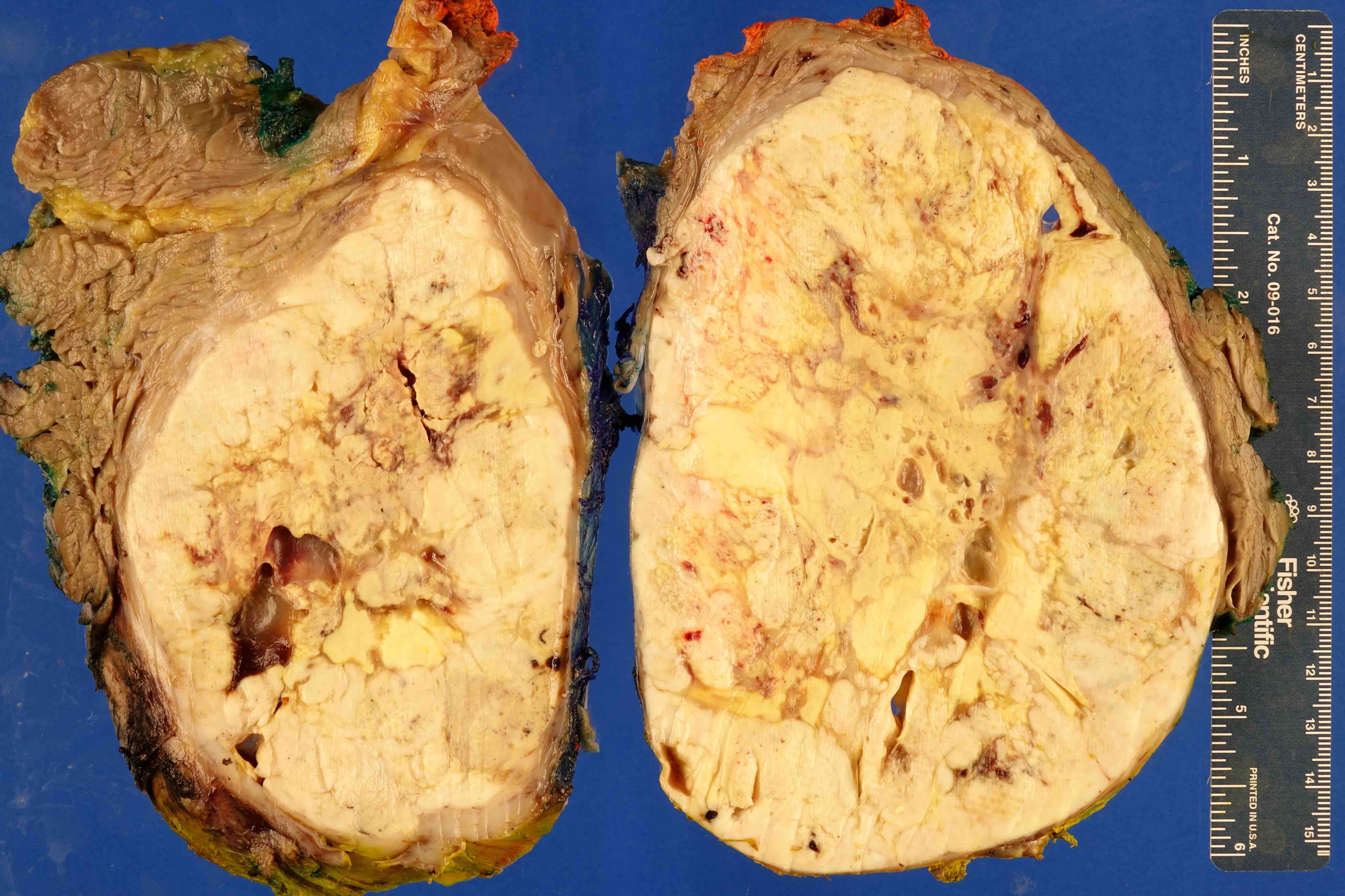
Well circumscribed tumor mass
Frozen section description
- Diagnosis typically not made on frozen section; however, generally a diagnosis of high grade sarcoma may be rendered based on degree of cytologic atypia, mitotic activity and whether necrosis is present
Microscopic (histologic) description
- Varying proportion of pleomorphic lipoblasts in a background of a high grade, usually pleomorphic, undifferentiated sarcoma (Surg Pathol Clin 2019;12:63, Histopathology 2014;64:38)
- Well circumscribed but nonencapsulated with infiltrative borders
- May display significant morphologic overlap with myxofibrosarcoma, undifferentiated pleomorphic sarcoma and high grade dedifferentiated liposarcoma
- Cytologically, tumors are composed of high grade cells with varying numbers of pleomorphic and often bizarre, multinucleated tumor cells
- These tumor cells will often show subtle to prominent cytoplasmic vacuolization with some forms appearing as classic lipoblasts with scalloped nuclei
- Signet ring lipoblasts may also be scattered throughout tumor in association with malignant cells
- Epithelioid morphology is seen in about 25% of cases
- Cases with epithelioid morphology may be difficult to distinguish from other high grade tumors that display epithelioid morphology, including vascular tumors, melanoma, carcinoma and other epithelioid subtypes of sarcomas (Am J Surg Pathol 2002;26:601, Am J Surg Pathol 2004;28:1257, Mod Pathol 1999;12:722, Pathology 2014;46:126, Ultrastruct Pathol 2002;26:299)
- Tumor necrosis common
- Median 25 mitotic figures/10 high power fields
- May have neutrophils within giant cells (emperipolesis-like finding), extra and intracellular eosinophilic hyaline droplets (not specific)
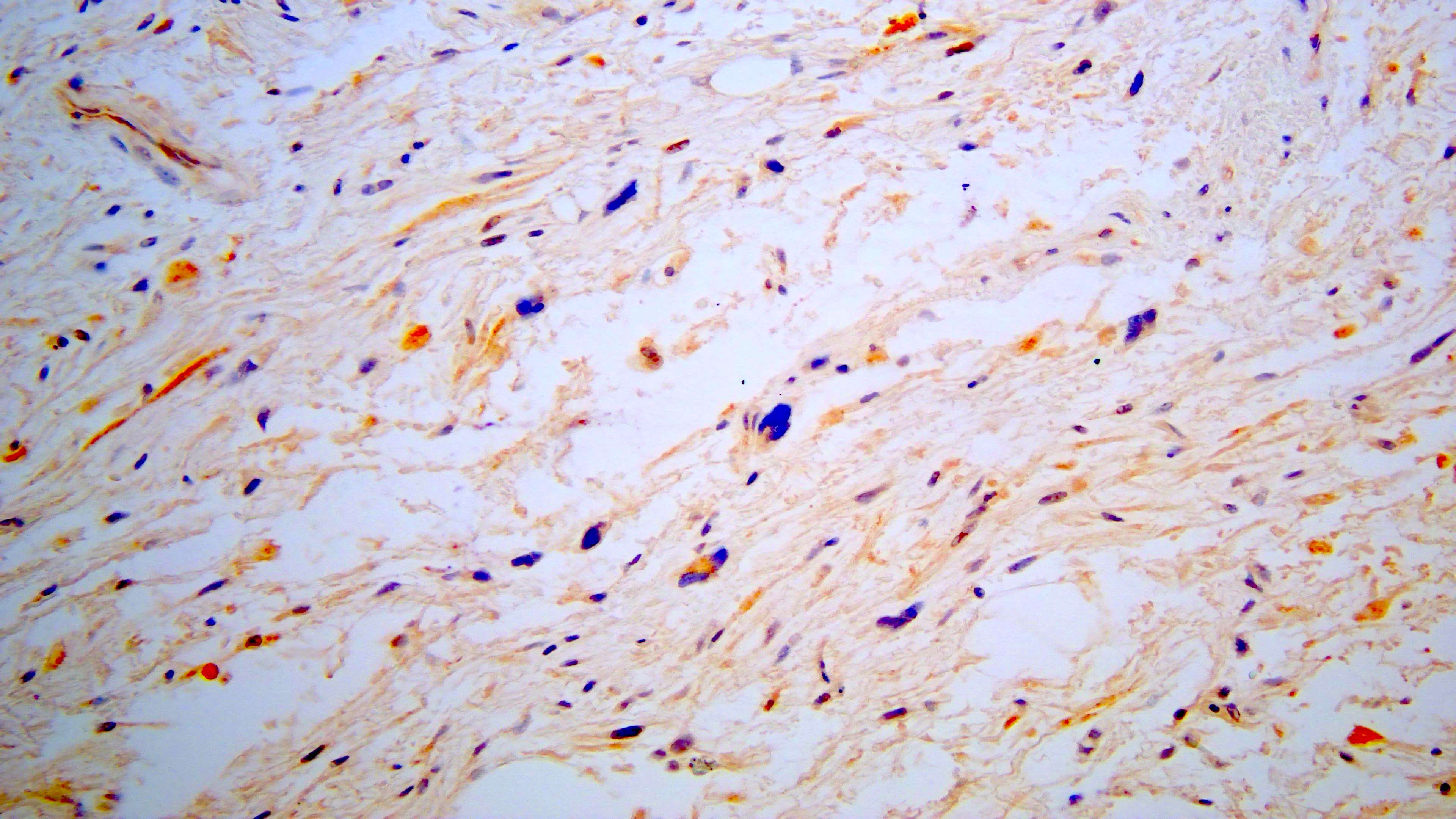
Microscopic (histologic) images
Contributed by David Suster, M.D.

Sheets of atypical cells
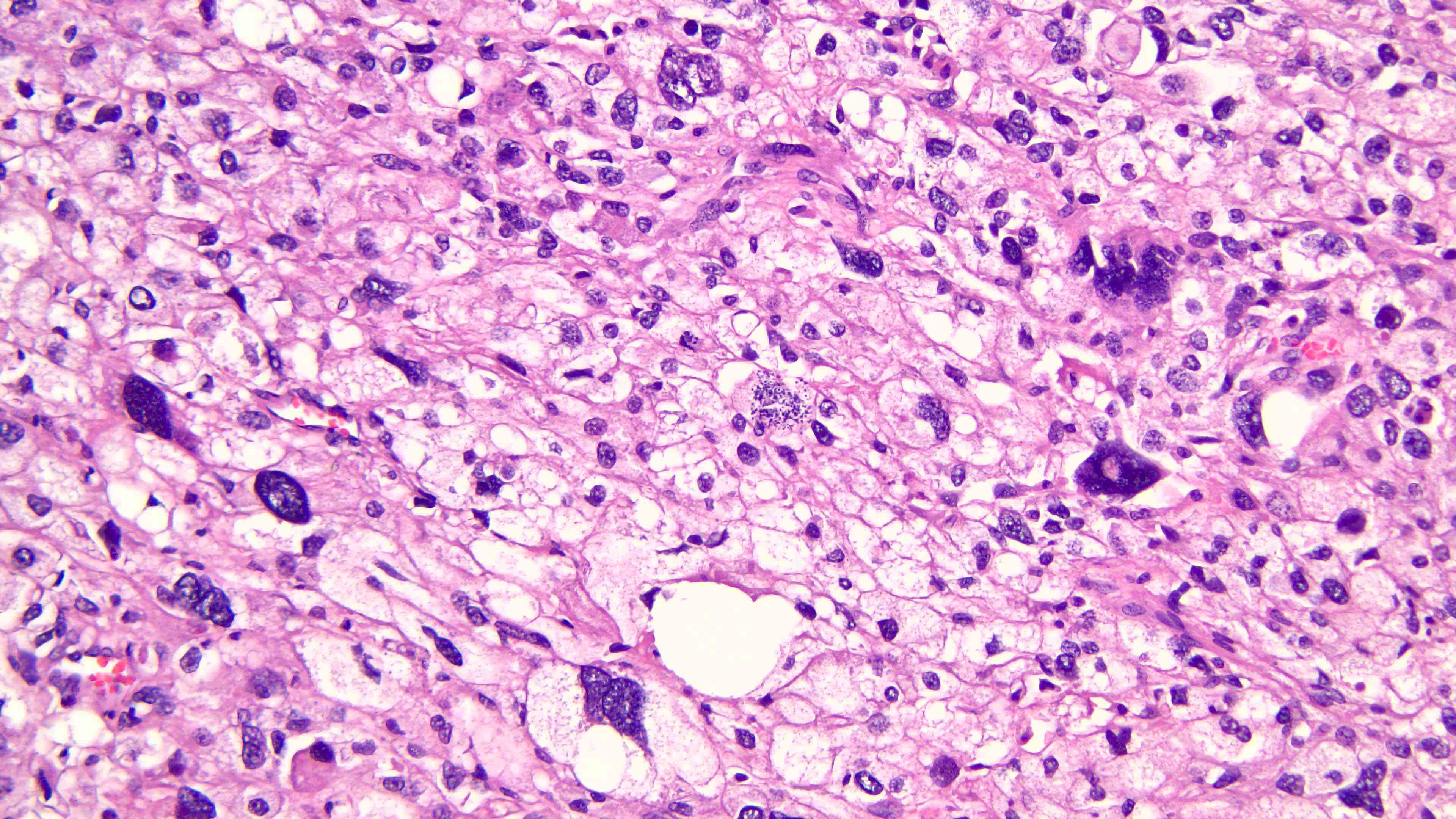
Extensive necrosis

Bizarre cell shapes
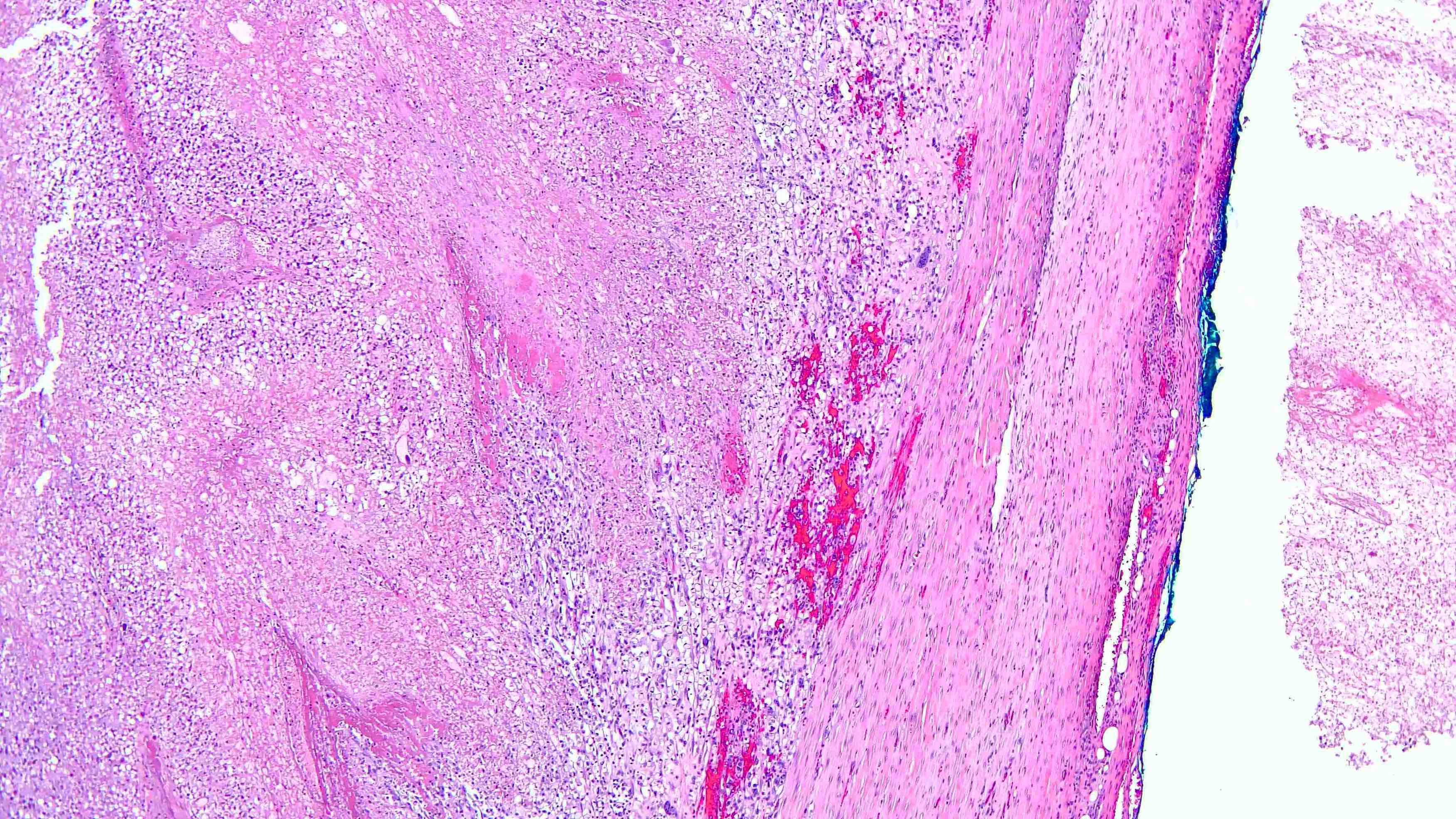
Pleomorphic lipoblast

Bone metastasis
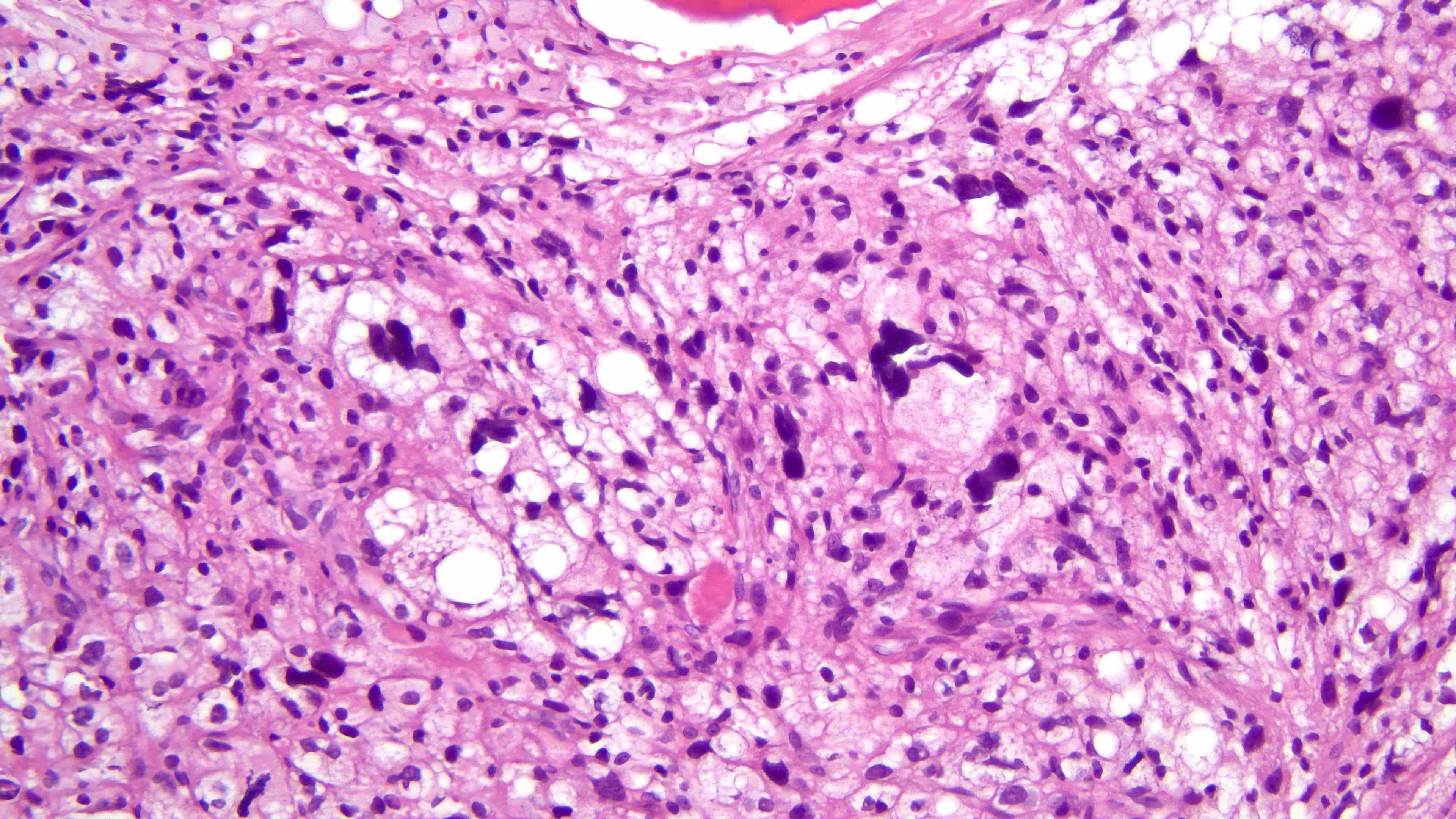
Pleomorphic lipoblasts

Epithelioid morphology

S100 positivity
Negative MDM2
Virtual slides
Images hosted on other servers:

PLPS: soft tissue, lower leg, excision
Cytology description
- Pleomorphic spindle cells of high grade sarcoma with occasional lipoblasts
Positive stains
- Vimentin, S100 (lipogenic areas in 33%), smooth muscle actin (45%)
- CD34, keratin (21%), desmin (13%)
- Epithelioid subtype may be positive for keratins and MelanA (Semin Diagn Pathol 2019;36:122)
Negative stains
- MDM2 and CDK4 are typically negative (Mod Pathol 2010;23:1657, Am J Surg Pathol 2002;26:601, Am J Surg Pathol 2004;28:1257, Am J Surg Pathol 2010;34:1122)
Electron microscopy description
- Abundant coalescing lipid droplets, numerous cytoplasmic organelles
Electron microscopy images
Images hosted on other servers:

Lipid vacuoles

Ultrastructure
Molecular / cytogenetics description
- No pathognomonic recurrent molecular alterations have been identified
- Most important negative finding is identification of the absence of supernumerary ring chromosomes with amplification of MDM2 / CDK4, as this excludes a high grade dedifferentiated liposarcoma with pleomorphic features
- Complex karyotypes (Cancer Res 2002;62:2993, Lab Invest 2005;85:176, Int J Cancer 2002;99:68, Mod Pathol 2005;18:638, PLoS One 2008;3:e3179)
- Molecular profiles of pleomorphic liposarcomas more closely resemble those of other pleomorphic sarcomas (such as high grade myxofibrosarcoma and undifferentiated pleomorphic sarcoma) than those of well differentiated and dedifferentiated liposarcoma, indicating a separate underlying molecular pathway (Cell 2017;171:950, Cancer Res 2002;62:2993, Lab Invest 2005;85:176, Nat Genet 2010;42:715)
- Somatic mutations in TP53 and NF1 (Nat Genet 2010;42:715, J Cancer Res Clin Oncol 1998;124:532, PLoS One 2008;3:e3179)
- Myxoid liposarcoma may demonstrate epithelioid morphology and pleomorphic features but is characterized by a recurrent characteristic t(12;16)(q13;p11) FUS-DDIT3 rearrangement (Histopathology 2005;46:334)
Molecular / cytogenetics images
Contributed by Bei You, Ph.D.

Complex karyotype
Videos
Pleomorphic liposarcoma: 5 minute pathology pearls
Pleomorphic liposarcoma (epithelioid variant)
Sample pathology report
- Thigh, left mass, wide resection:
- Pleomorphic liposarcoma (19 x 12 x 13 cm), high grade (see synoptic report)
- Tumor necrosis is present, ~30%
- No lymphovascular or perineural invasion seen
- Margins of resection, no tumor seen
- Pathologic stage: pT3 N0 (no lymph nodes submitted or found)
Differential diagnosis
- Dedifferentiated liposarcoma:
- May show a high grade component with pleomorphic features
- Defined by amplification of 12q13-15 region (MDM2 / CDK4 genes) and will sometimes show an adjacent well differentiated component
- Metastatic carcinoma:
- May grow as sheets of poorly differentiated pleomorphic cells
- Usually lacks adipocytic differentiation and stains positive with cytokeratins
- Undifferentiated pleomorphic sarcoma and other high grade pleomorphic sarcomas:
- May show prominent tumor cell pleomorphism but lacks evidence of adipocytic differentiation / lipoblastic cells
- Pleomorphic lipoma / spindle cell lipoma:
- May show scattered pleomorphic cells; however, degree of pleomorphism is significantly less than in pleomorphic liposarcoma
- No overt features of malignancy, such as necrosis, high mitotic activity
- Characterized by loss of RB1
- Pleomorphic rhabdomyosarcoma:
Practice question #1
A 77 year old woman presents with a 19 cm deep soft tissue mass in the left lower extremity. A core needle biopsy shows the high grade sarcoma shown in the image above. Which is the most likely diagnosis?
- Dedifferentiated liposarcoma
- Pleomorphic liposarcoma
- Pleomorphic rhabdomyosarcoma
- Undifferentiated pleomorphic sarcoma
- Well differentiated liposarcoma
Practice answer #1
B. Pleomorphic liposarcoma. The image shows a high grade sarcoma with bizarre pleomorphic tumor cells that show evidence of adipocytic differentiation in the form of cytoplasmic vacuolization.
Comment Here
Reference: Pleomorphic liposarcoma
Comment Here
Reference: Pleomorphic liposarcoma
Practice question #2
What is pleomorphic liposarcoma molecularly characterized by?
- Amplification of 12q13-15
- Amplification of MDM2 and CDK4
- Complex cytogenetic abnormalities without specific, recurrent molecular alterations
- Deletions of SMARCB1 gene and loss of INI1 expression
- Gene fusion between FUS and DDIT3
Practice answer #2
C. Complex cytogenetic abnormalities without specific, recurrent molecular alterations. Pleomorphic sarcoma is a high grade sarcoma characterized by complex molecular alterations. The tumors show complex karyotypes with numerous cytogenetic abnormalities. Specific, recurrent alterations have not been identified. The tumors are negative for amplification of MDM2 / CDK4 (12q-13-15 region).
Comment Here
Reference: Pleomorphic liposarcoma
Comment Here
Reference: Pleomorphic liposarcoma
