

Soft tissue
Uncertain differentiation
Clear cell sarcoma
Author: Kemal Kösemehmetoğlu, M.D.
Editorial Board Member: Jose G. Mantilla, M.D.
Deputy Editor-in-Chief: Borislav A. Alexiev, M.D.

Last author update: 30 July 2021
Last staff update: 30 December 2021
Copyright: 2002-2024, PathologyOutlines.com, Inc.
PubMed Search: Clear cell sarcoma[title] soft tissue[title]
Table of Contents
Definition / general | Essential features | Terminology | ICD coding | Epidemiology | Sites | Pathophysiology | Etiology | Clinical features | Diagnosis | Radiology description | Radiology images | Prognostic factors | Case reports | Treatment | Clinical images | Gross description | Gross images | Frozen section description | Microscopic (histologic) description | Microscopic (histologic) images | Cytology description | Cytology images | Positive stains | Negative stains | Electron microscopy description | Molecular / cytogenetics description | Molecular / cytogenetics images | Videos | Sample pathology report | Differential diagnosis | Additional references | Board review style question #1 | Board review style answer #1 | Board review style question #2 | Board review style answer #2Cite this page: Kösemehmetoğlu K. Clear cell sarcoma. PathologyOutlines.com website. https://www.pathologyoutlines.com/topic/softtissueclearcellsarcoma.html. Accessed May 10th, 2024.
Definition / general
- Clear cell sarcoma (CCS) is a malignant soft tissue sarcoma composed of monotonous epithelioid and spindle cells with clear to eosinophilic cytoplasm characterized by melanocytic differentiation and EWSR1-ATF1 / CREB1 rearrangement
Essential features
- Young adult, M:F = 1, distal extremity, slow growing
- Not related to epidermis (hence, tendon and aponeuroses)
- Do not confuse with clear cell sarcoma of kidney
- Sheets and nests of monotonous epithelioid spindle cells with clear to eosinophilic cytoplasm separated by a delicate framework of fibrocollageneous stroma
- Scattered Touton type or wreath-like multinucleated giant cells are relatively common
- S100 / SOX10+, Melan A+, HMB45+
- EWSR1-ATF1 or EWSR1-CREB1
Terminology
- Melanoma of soft parts (not recommended)
- Clear cell sarcoma of tendon and aponeuroses
- Clear cell sarcoma of soft parts
ICD coding
- ICD-O: 9044/3 - clear cell sarcoma, NOS (except of kidney M-8964/3)
- ICD-10: C49.9 - malignant neoplasm of connective and soft tissue, unspecified
- ICD-11: 2B5K & XH77N6 - unspecified malignant soft tissue tumors or sarcomas of bone or articular cartilage of other or unspecified sites & clear cell sarcoma of soft tissue
Epidemiology
- < 1% of all soft tissue tumors
- Young adults (third decade)
- No gender predilection
Sites
- Foot and ankle > hand > thigh > trunk > head & neck
- 75% distal extremities
- Rarely located in penis, mediastinum, lung, retroperitoneum, abdomen and bone
- References: J Clin Pathol 2010;63:416, Dermatol Res Pract 2012;2012:984096, Eur J Surg Oncol 2014;40:505
Pathophysiology
- Microphthalmia associated transcription factor (MITF) seems to be critical oncogenic target of EWSR1-ATF1 in CCS leading to melanocytic phenotype but not in angiomatoid fibrous histiocytoma with the same EWSR1-ATF1 rearrangement (Cancer Cell 2006;9:473, Am J Surg Pathol 2012;36:e1)
- Regarding gene expression profile, CCSs cluster with melanomas, not soft tissue sarcomas (J Clin Oncol 2003;21:1775)
- Classified as tumor of uncertain differentiation in WHO 2020
Etiology
- Unknown
Clinical features
- Not uncommon to get a long history (prebiopsy duration)
- Pain and mass are common presenting symptoms
Diagnosis
- Magnetic resonance imaging (MRI)
- Biopsy and molecular pathology
Radiology description
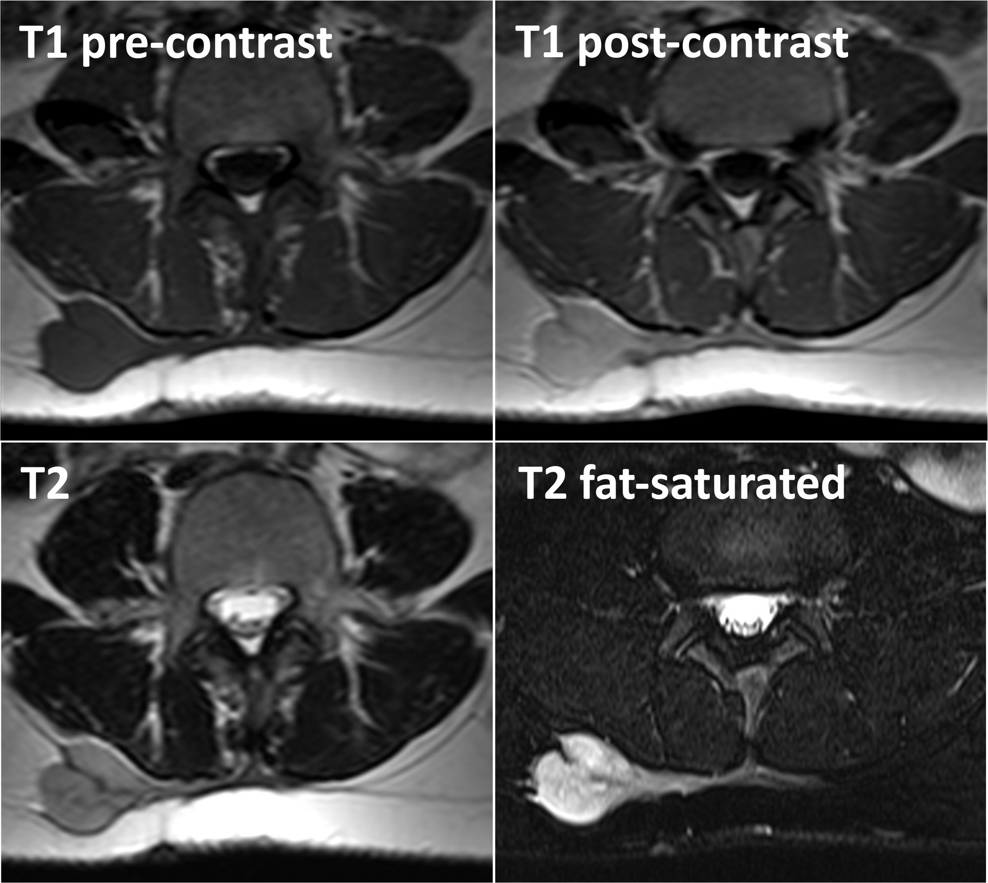
- Well defined, homogeneous and benign looking mass
- Magnetic resonance imaging
- Homogeneous iso hyperintense on T1 and heterogeneous on T2 with variable contrast enhancement
- Reference: Skeletal Radiol 2000;29:187
Radiology images
Contributed by Ustun Aydingoz, M.D.
Magnetic resonance imaging
Images hosted on other servers:
MRI of CCS
Prognostic factors
- High recurrence rate (up to 40%)
- Metastasis to lung and lymph nodes in 30 - 50% of patients (1999 Sep;86:969, J Cancer Res Clin Oncol 2018;144:1711, Cancer 1983;52:1482, Am J Surg Pathol 2008;32:452, Am J Surg Pathol 1992;16:1197)
- 5 year survival 60%, 10 year survival 40%, 20 year survival 10% (J Clin Pathol 2010;63416, Am J Surg Pathol 2008;32:452, Dermatol Res Pract 2012;2012:984096, J Cancer Res Clin Oncol 2018;144:1711)
- Poor prognostic factors: Tumor size > 5 cm, necrosis, recurrence and lymph node metastases (Dermatol Res Pract 2012;2012:984096, 1999 Sep;86:969, Cancer 1983;52:1482, Am J Surg Pathol 1992;16:1197, Cancer 1990;65:367)
Case reports
- 32 year old man with penile mass metastatic to the lymph nodes and bone (Am J Clin Exp Urol 2015;3:43)
- 33 year old man and 37 year old woman with clear cell sarcoma in unusual sites mimicking metastatic melanoma (World J Clin Oncol 2019;10:213)
- 36 year old man with distal femoral clear cell sarcoma (BMC Musculoskelet Disord 2021;22:99)
- 43 year old woman with a neck mass (Balkan Med J 2018;35:203)
- 57 year old man with primary mediastinal CCS (Diagn Pathol 2017;12:5)
- 80 year old woman with mass in large toe (Cureus 2020;12:e11719)
Treatment
- Complete surgical excision with or without radiotherapy
- Chemotherapy / immunotherapy has no effect
Clinical images
Images hosted on other servers:
Clear cell sarcoma of large toe
Gross description
- 1 - 15 cm, median of 3 - 4 cm (usually < 4 cm)
- Solid, homogeneous, white-cream colored, well demarcated
- Usually sits on tendon and aponeuroses
- References: Cancer 1965;18:1163, Eur J Surg Oncol 2014;40:505
Gross images
Contributed by Kemal Kösemehmetoğlu, M.D. and Mark R. Wick, M.D.
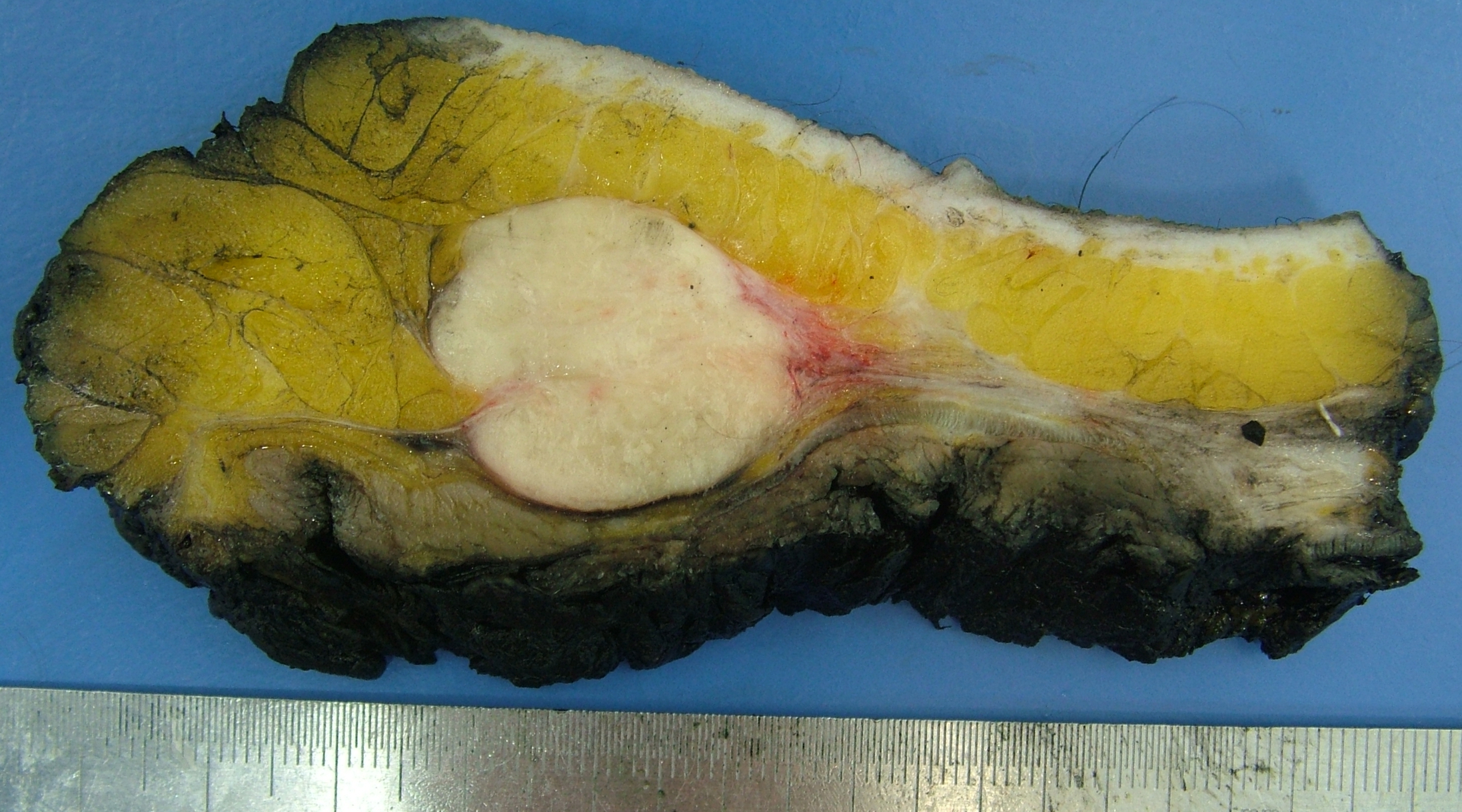
Clear cell sarcoma of soft tissue
Frozen section description
- Sentinel lymph node can be encountered
- Melanoma is the main differential diagnosis and may be impossible to separate in this setting
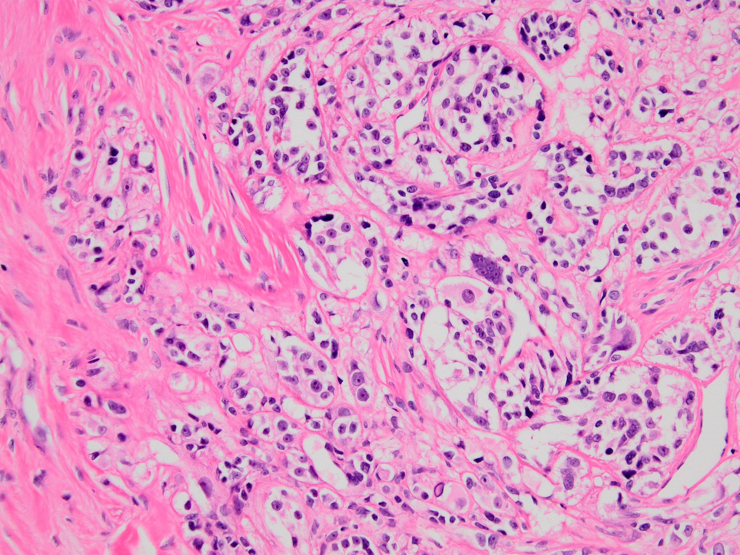
Microscopic (histologic) description
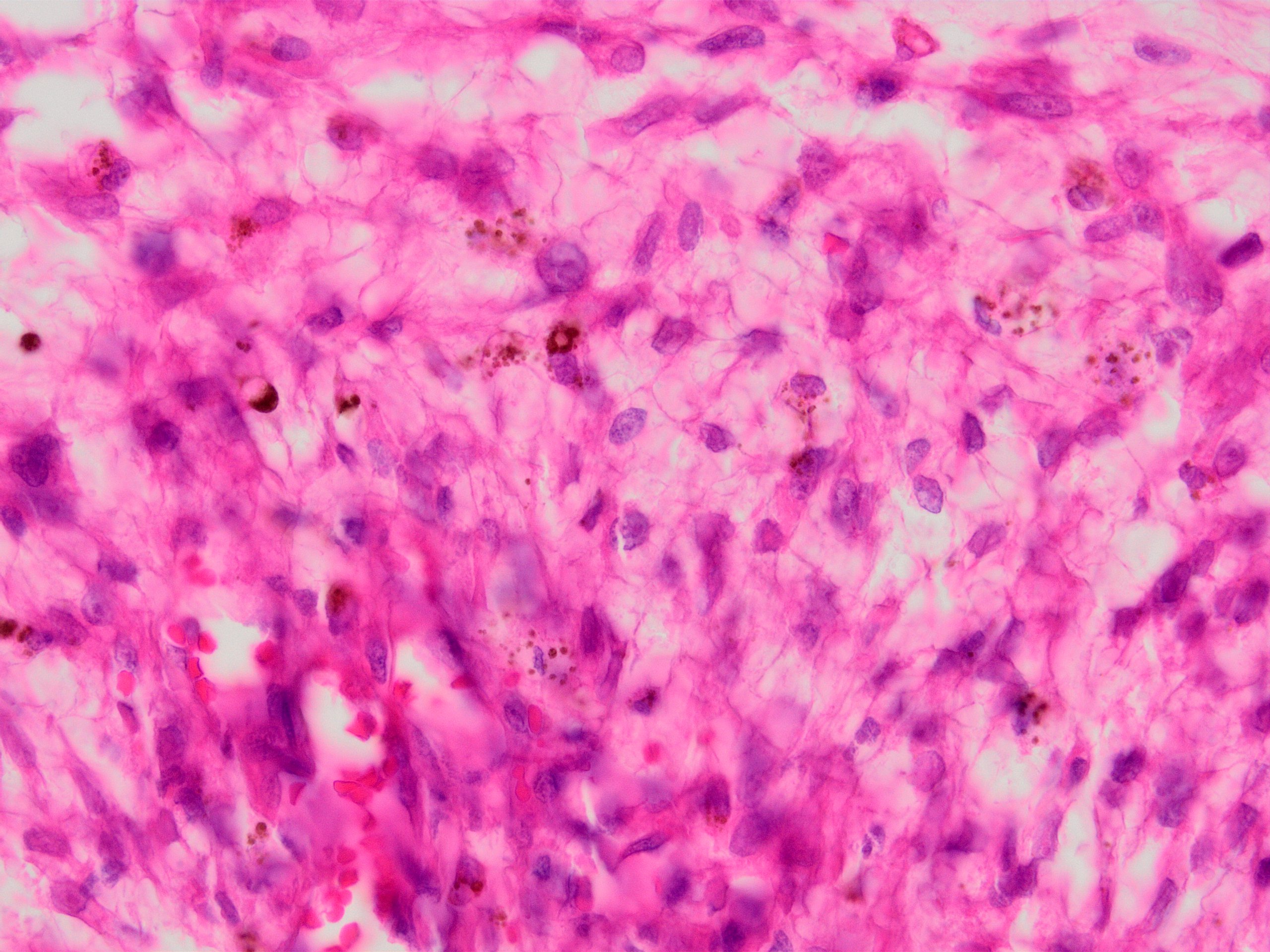
- Essential: nests of epithelioid spindle cells with clear eosinophilic cytoplasm and prominent nucleoli plus melanocytic differentiation
- Morphological patterns (Am J Surg Pathol 2008;32:452, J Clin Pathol 2010;63:416):
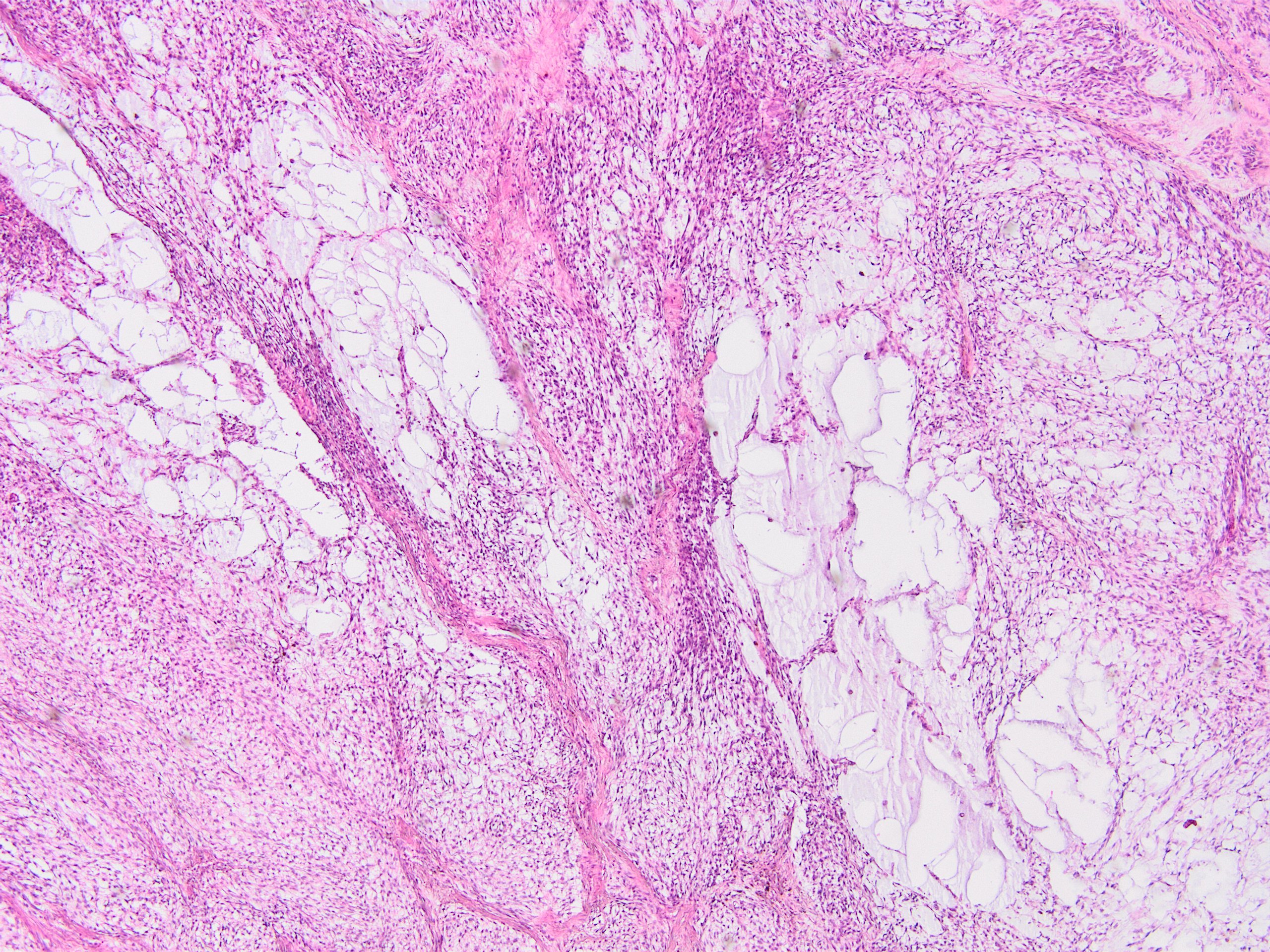
- Vaguely organoid (neuroendocrine-like) pattern: nests and short fascicles of epithelioid or spindle cells, surrounded by a delicate framework of fibrocollagenous tissue contiguous with the adjacent tendons and aponeurosis
- Diffuse pattern: solid sheets of epithelioid to spindle cells
- Pseudoalveolar pattern: reminiscent of alveolar soft part sarcoma
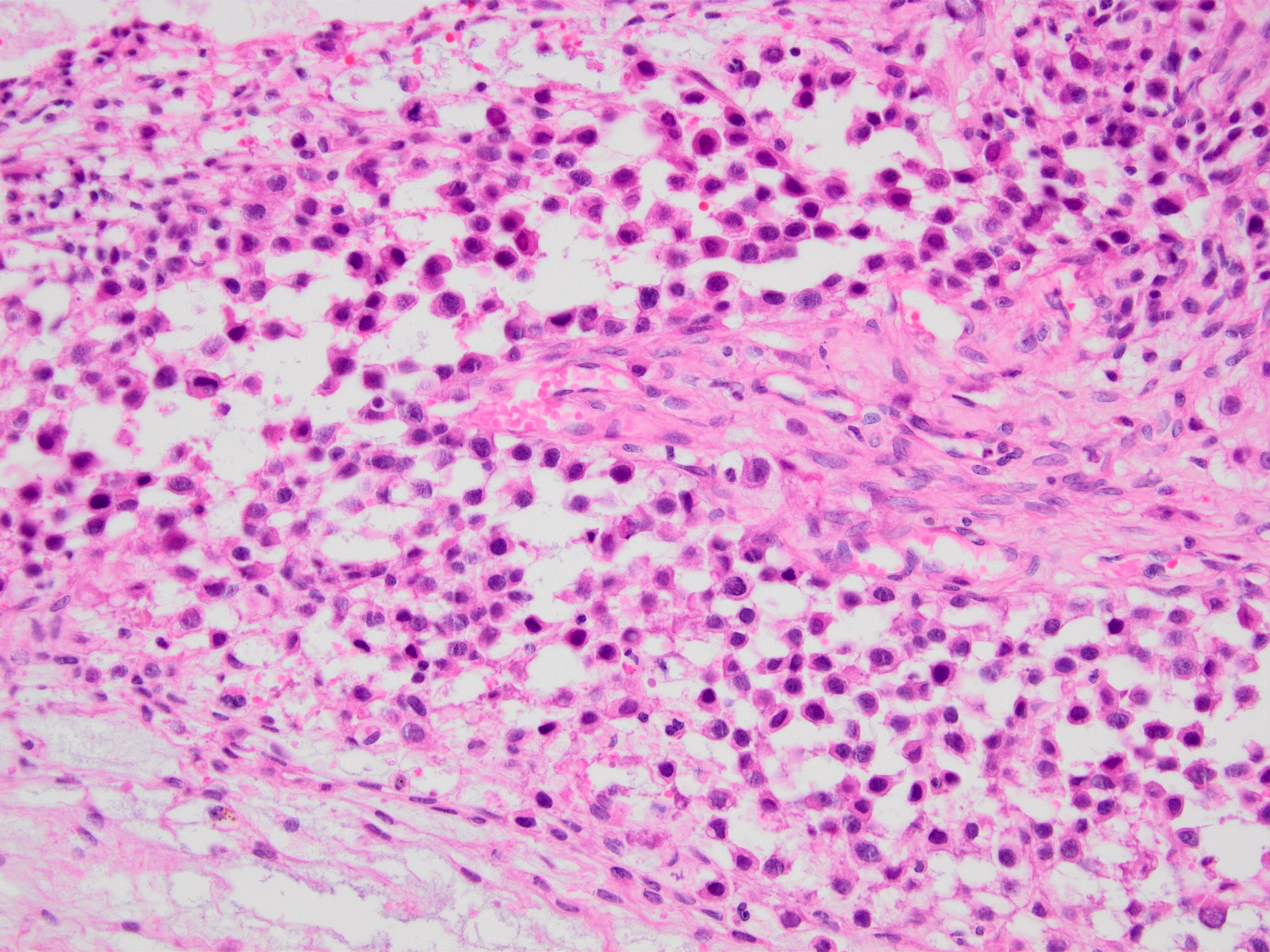
- Myxoid / microcystic pattern
- Inflammatory pattern: reminiscent of seminoma
- Cells:
- Epithelioid to spindled
- Monotonous round to oval nuclei with prominent nucleoli
- Lightly eosinophilic to amphophilic cytoplasm
- Clear cells typically comprise only a subset of the tumor
- Scattered rhabdoid and pleomorphic cells
- Scattered Touton type or wreath-like multinucleated giant cells are relatively common
- Rare mitosis
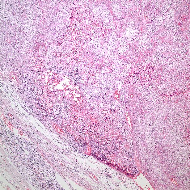
- Necrosis usually focally in 33%
- Melanin pigment in scattered cells in half of the cases
- No involvement of epidermis or no epidermal melanocytic proliferation but longstanding lesions (especially at fingers) may enlarge and therefore tumors may ulcerate
Microscopic (histologic) images
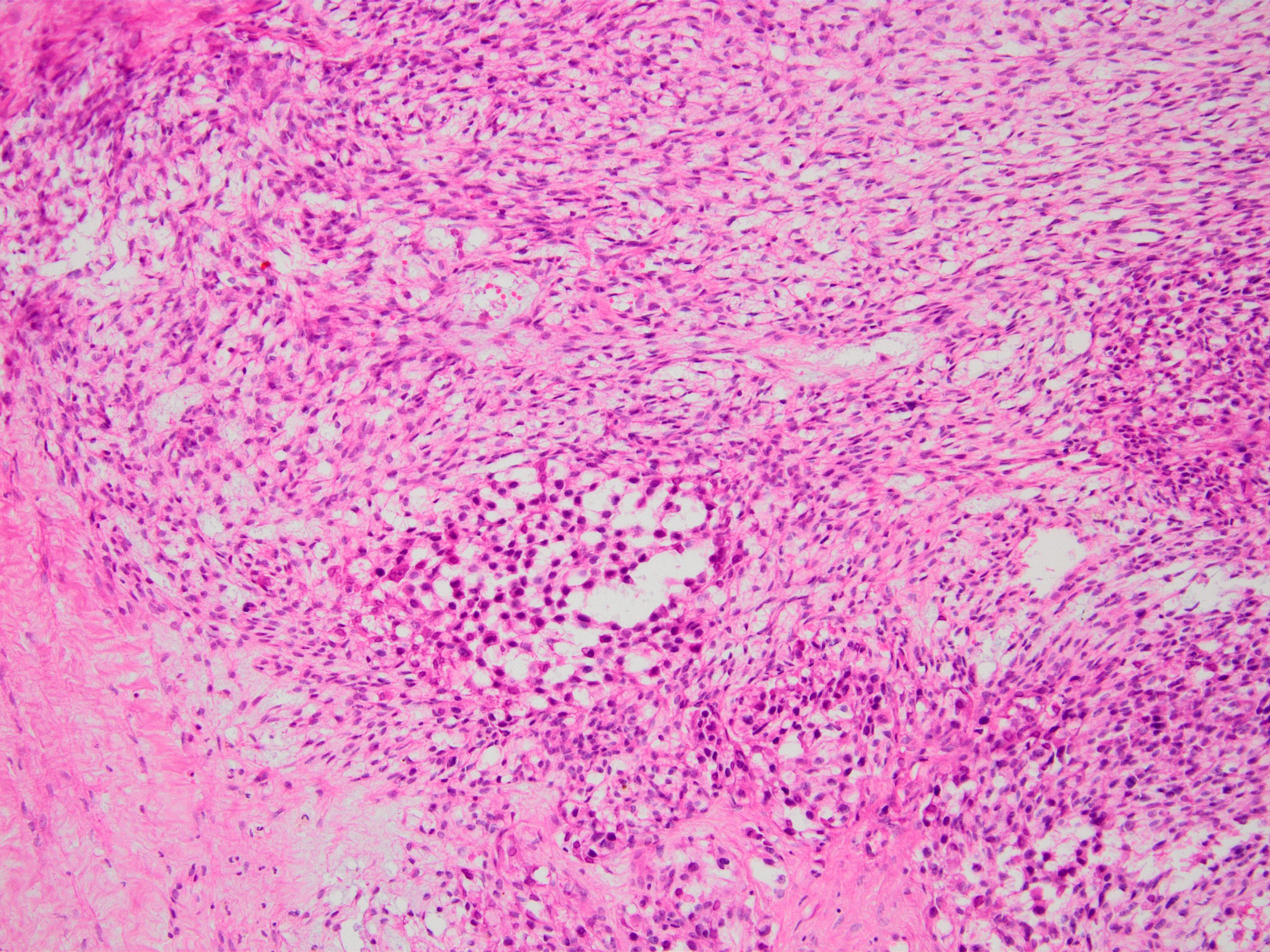
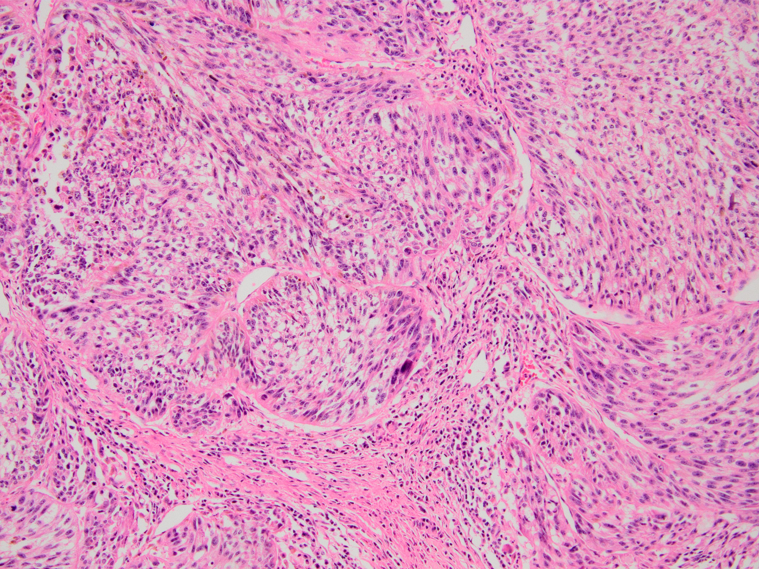
Contributed by Kemal Kösemehmetoğlu, M.D.
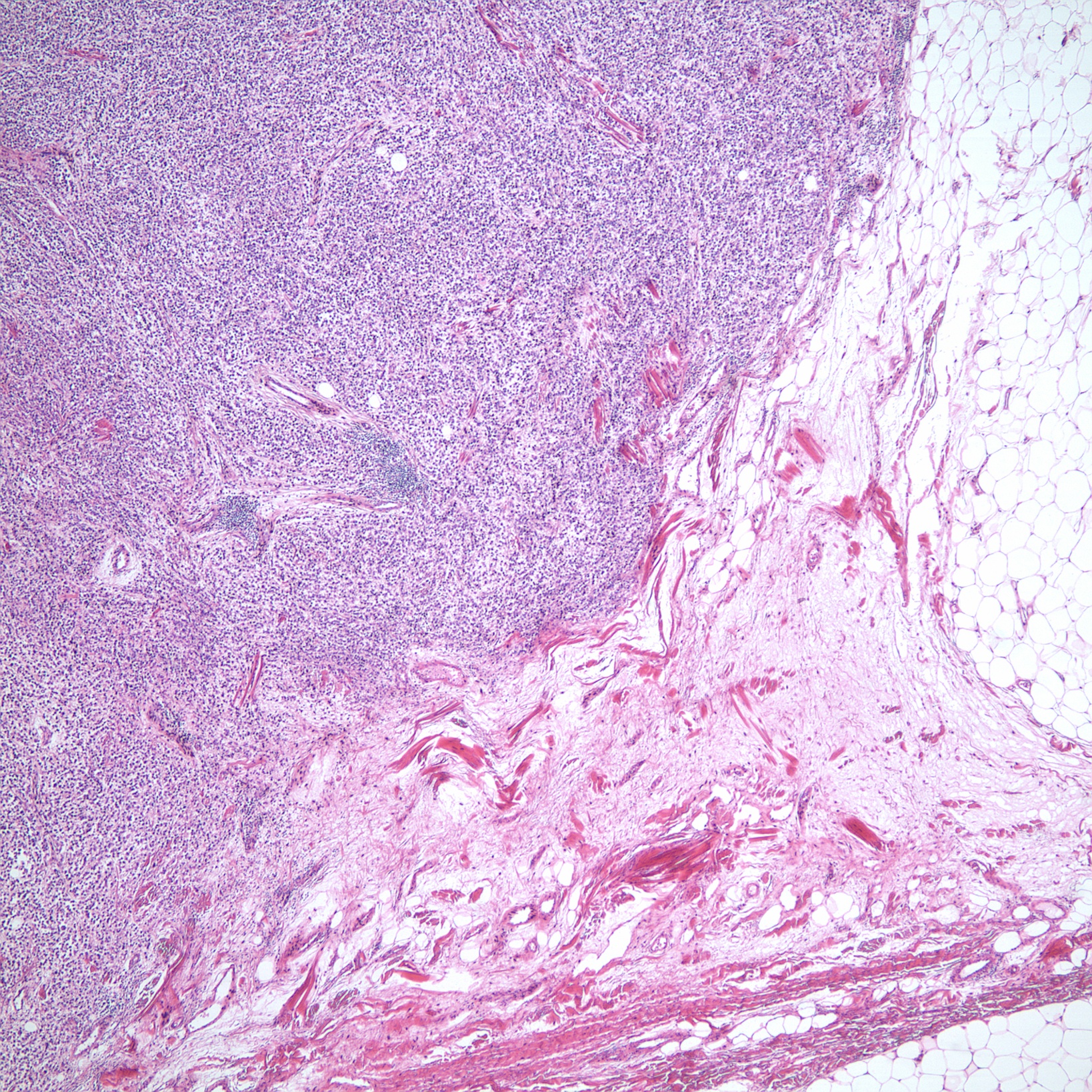
CCS attached to aponeurosis
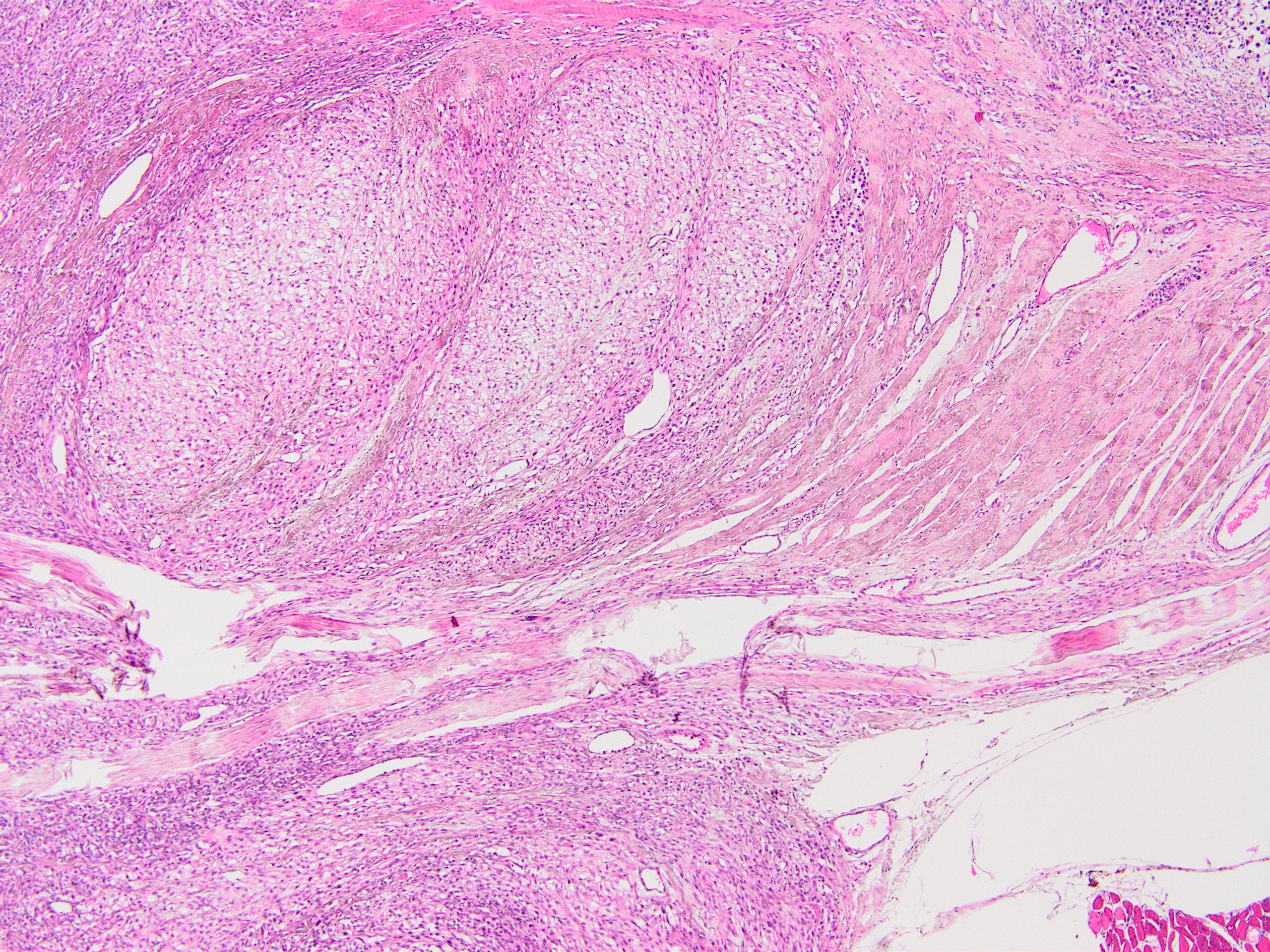
Tumor adjacent to tendon and aponeurosis
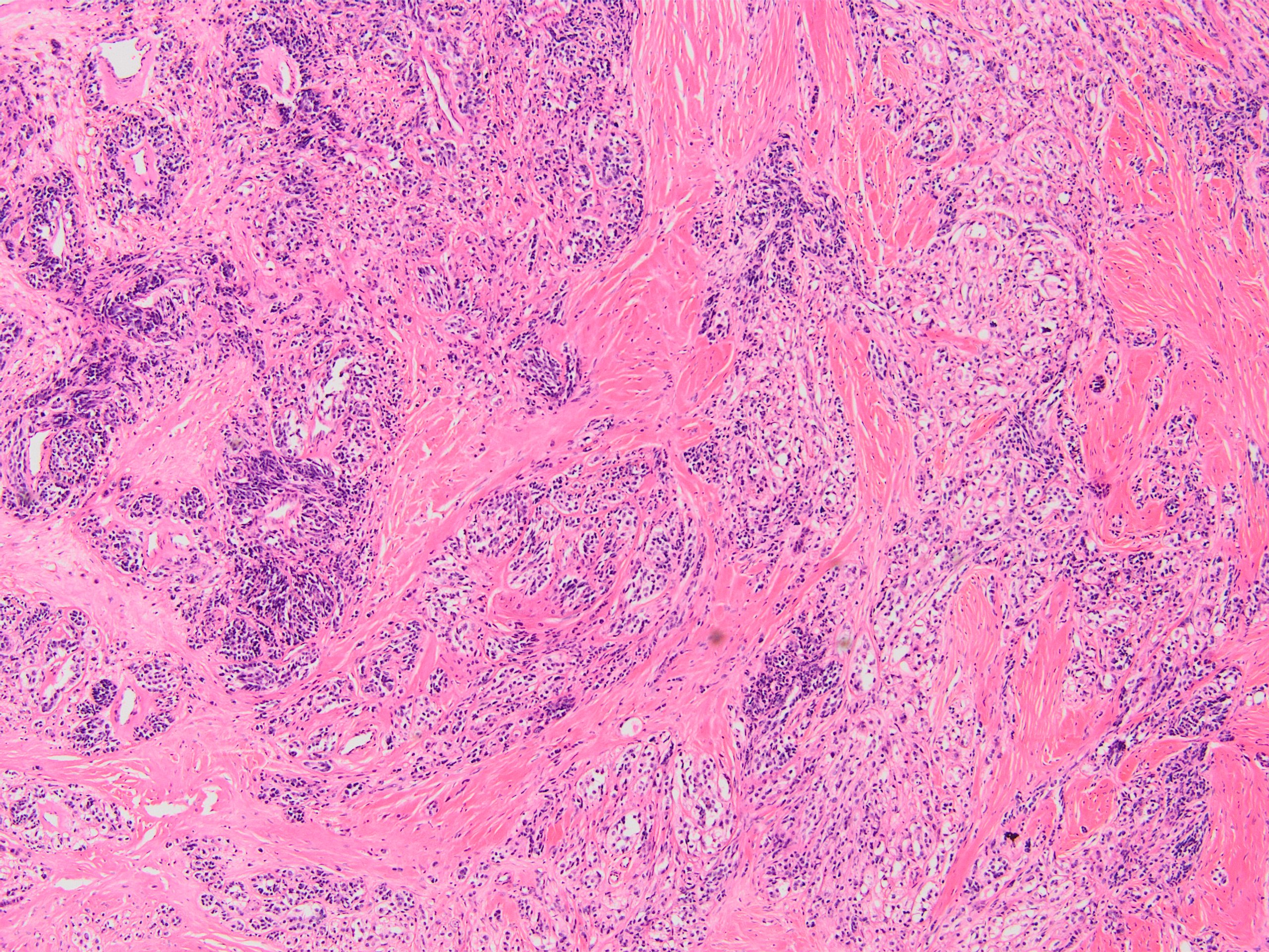
Classical (organoid / neuroendocrine-like) morphology
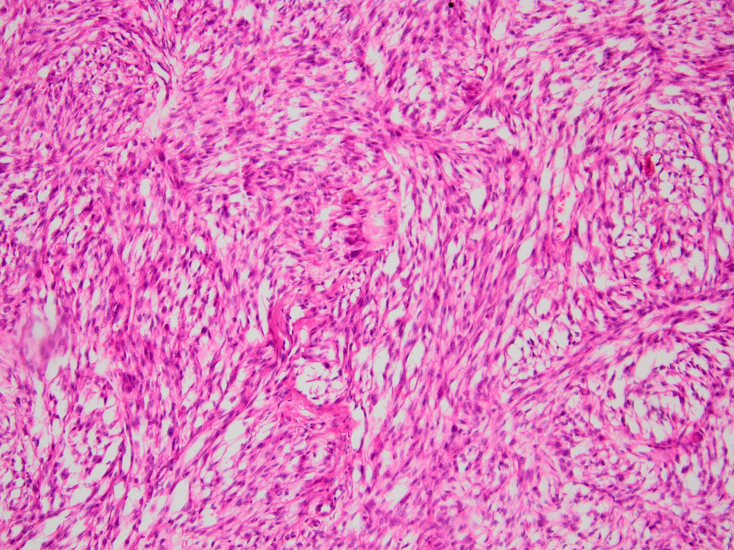
Short fascicles of spindle cells
Mixed spindle and epithelioid cells
Intermixed spindle and epithelioid cells
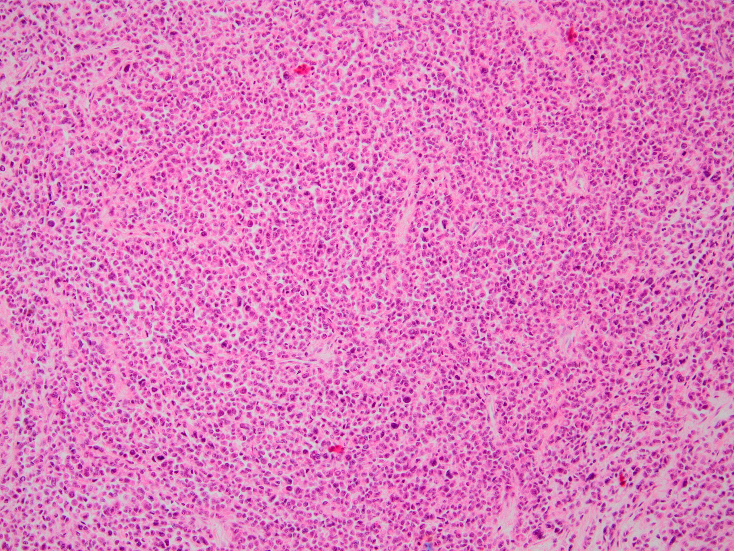
Solid sheets of epithelioid cells
Classical CCS
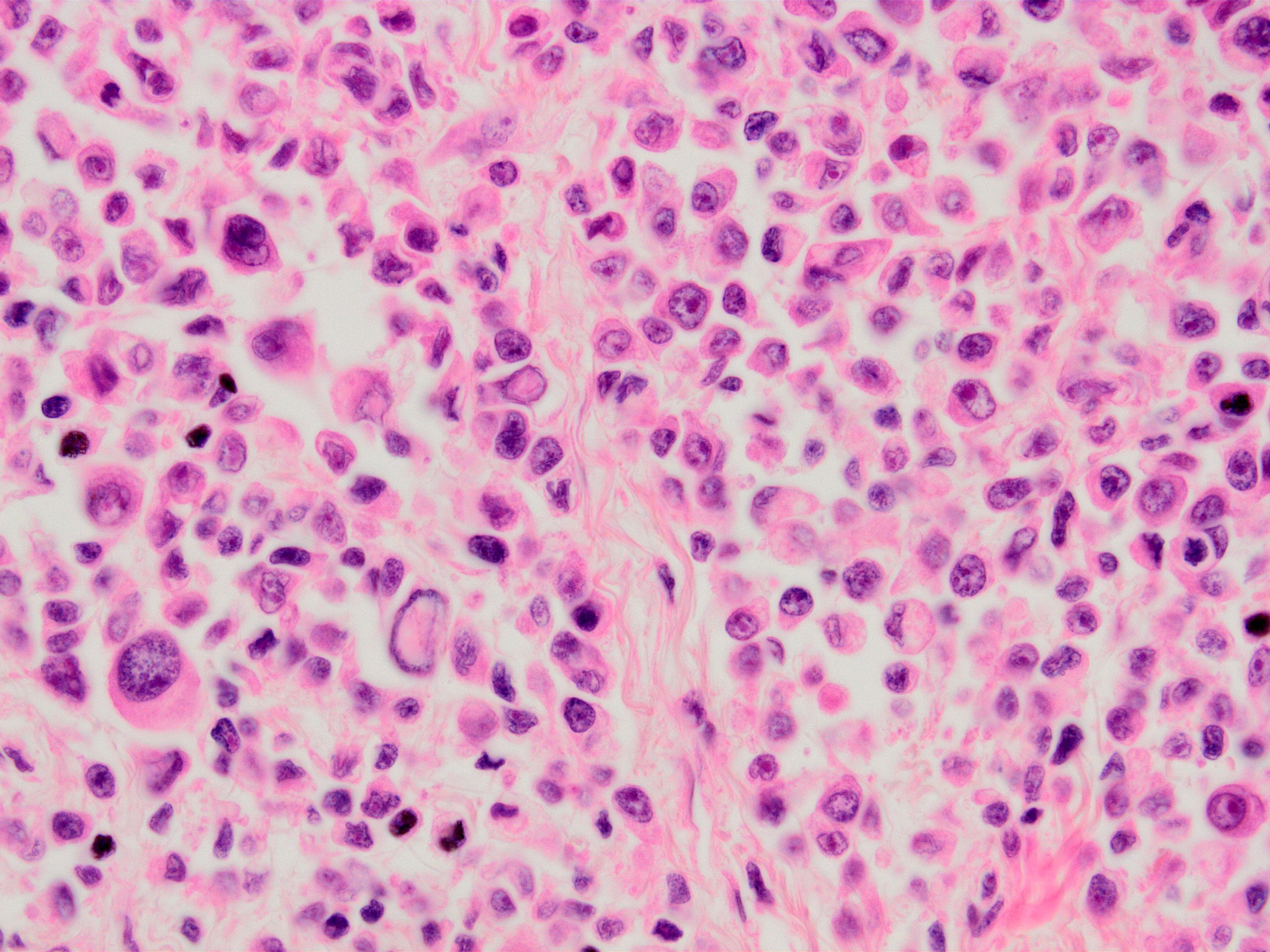
Rare pleomorphic cells
Pseudoalveolar pattern
Melanin pigment
Myxoid / microcystic pattern
Lymph node metastasis
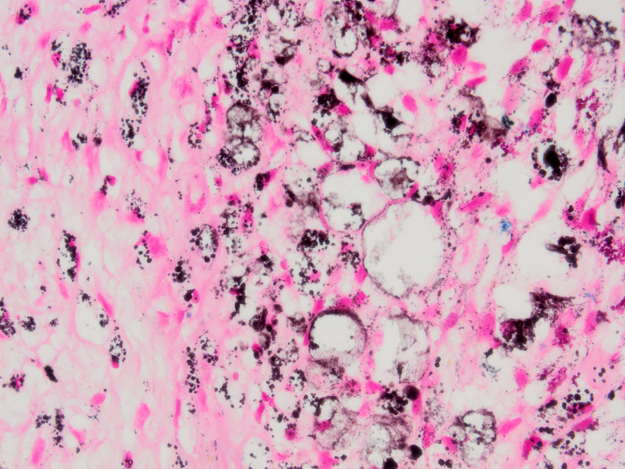
Fontana stain
S100
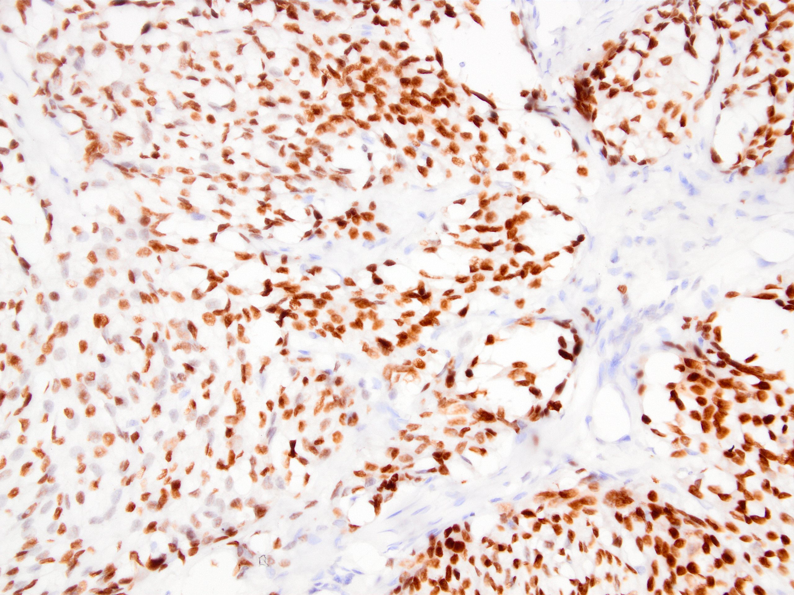
SOX10
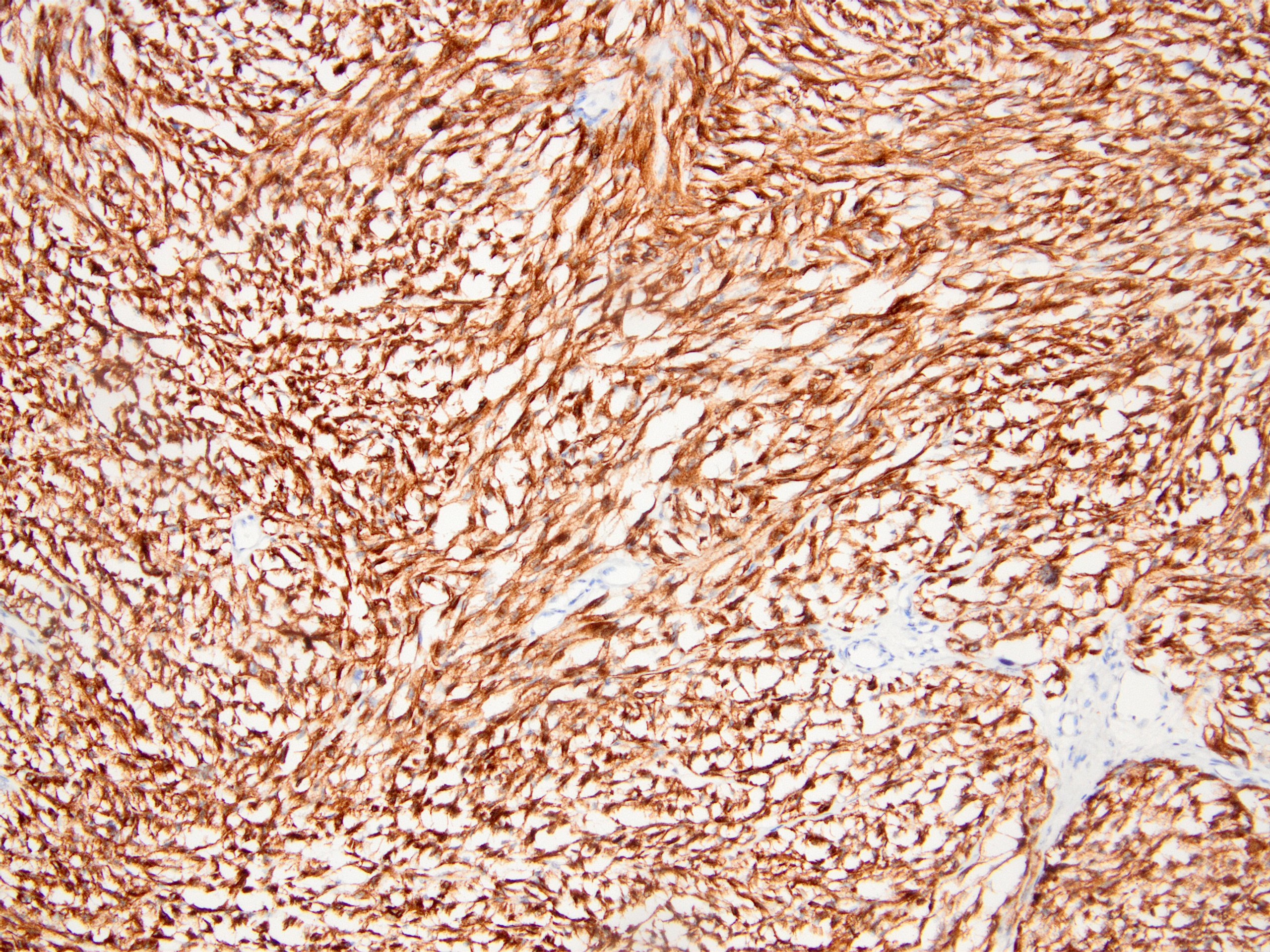
HMB45
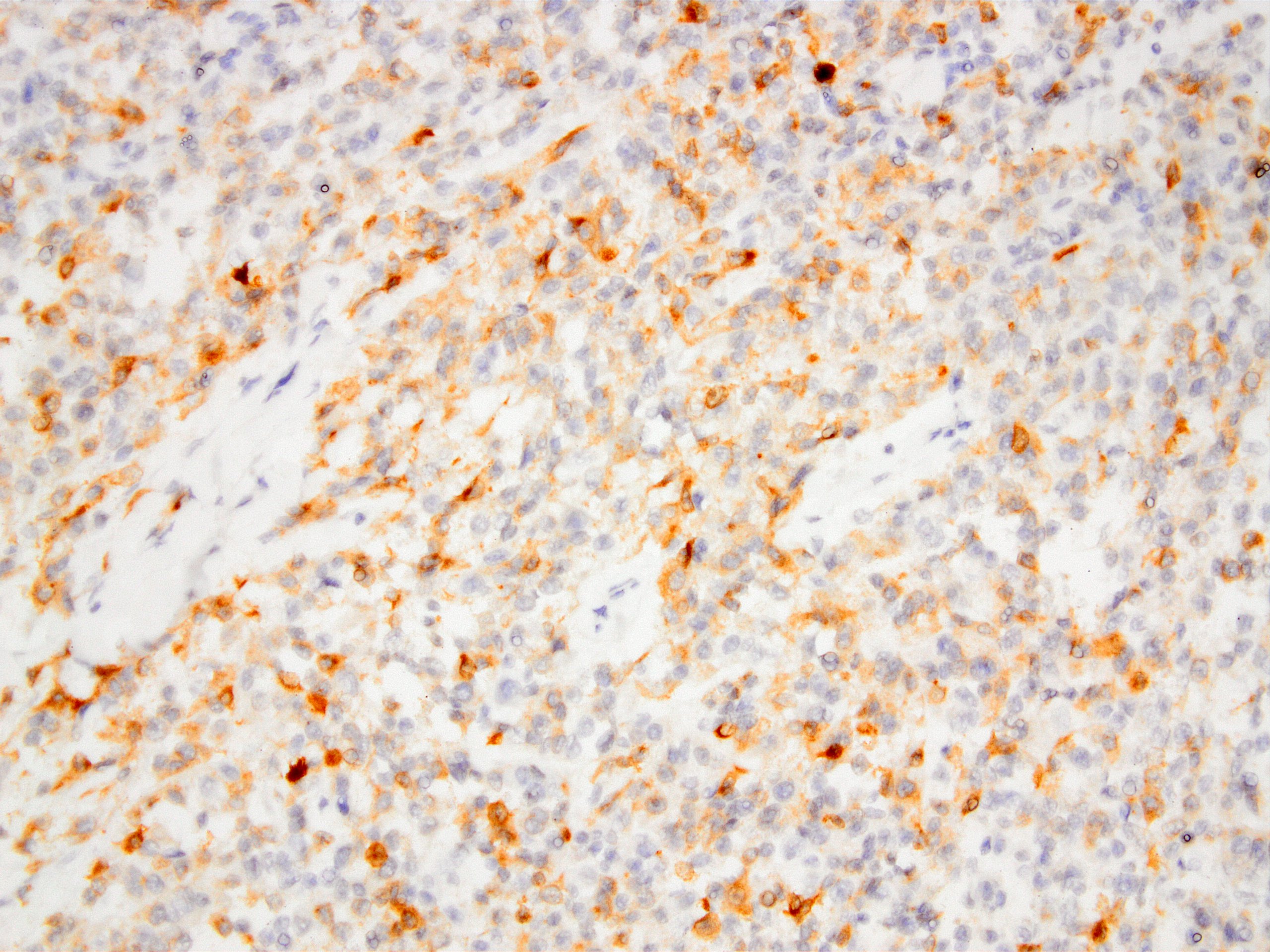
MelanA
Cytology description
- Hypercellular smears composed of monomorphic epithelioid / polygonal cells, including singly scattered or loosely clustered spindle cells, with prominent nucleoli
- Granular to vacuolated, well defined cytoplasm
- Binucleation / multinucleation (50%), abrupt anisonucleosis, racquet shaped cells (20%)
- Tigroid background (10%)
- Focal intracytoplasmic pigment (10%)
- If available, immunohistochemical stains and molecular testing are crucial in reinforcing a diagnosis (Cytopathology 2020;31:280)
Cytology images
Contributed by Bharat Rekhi, M.D.
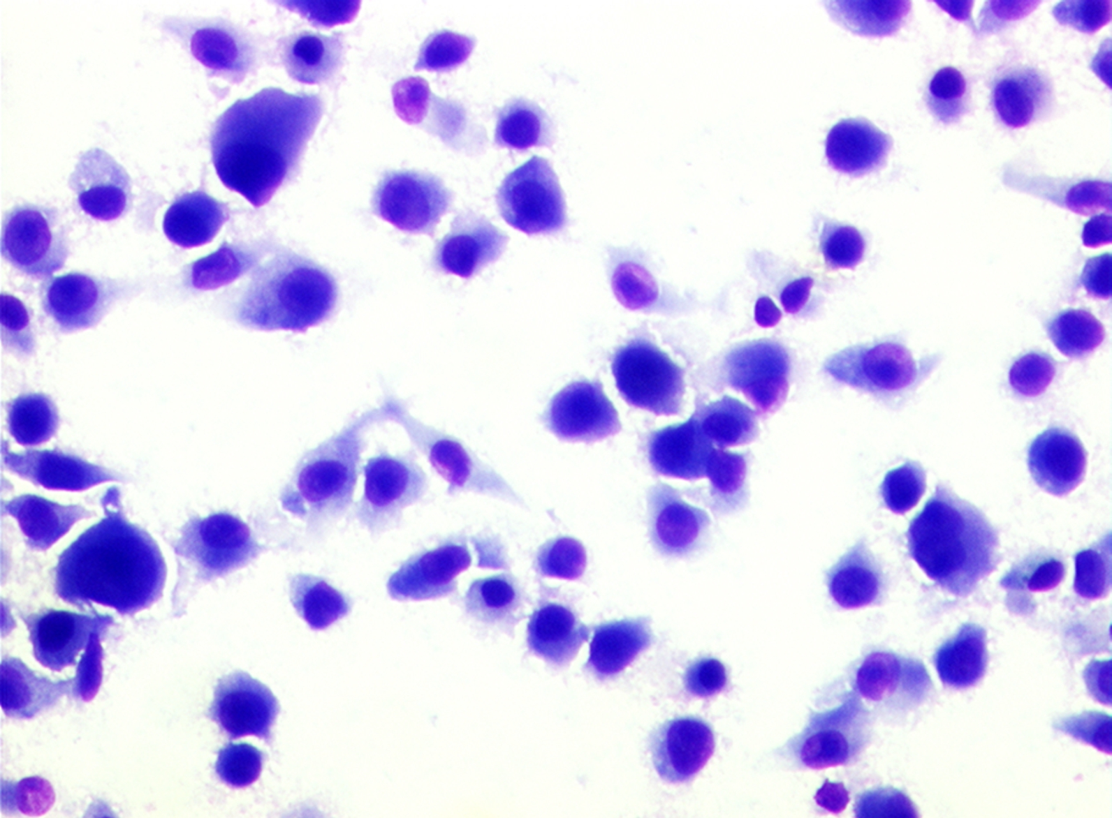
Giemsa
Pap
Positive stains
- S100 (100%), SOX10 (95%) (Am J Surg Pathol 2015;39:826)
- HMB45 (97%, patchy or focal), MITF (81%), MelanA (71%)
- BCL2 (93%)
- CD57 (75%) (Am J Surg Pathol 2008;32:452)
Negative stains
- SMA, desmin, CAM 5.2
- PanCK (3%), CD34 (10%) (Am J Surg Pathol 2008;32:452)
- EMA (30%)
- Synaptophysin (43%), CD56 (21%) (Am J Surg Pathol 2008;32:452)
Electron microscopy description
- Resembles synovial sarcoma given the presence of basement membrane material, a biphasic pattern of cells and the presence of pseudoglandular structures with filopodia
- Presence of melanosomes and interdigitating cellular processes wrapping around cells and extracellular structures are similar to melanoma and nerve sheath tumors
- May contain aggregates of glycogen and inclusions composed of multilayered membrane structures forming myelin-like figures
- References: Am J Pathol 1984;114:264, Virchows Arch A Pathol Anat Histopathol 1983;401:109
Molecular / cytogenetics description
- t(12;22)(q13;q12) for EWSR1-ATF1 [4 splice variants including the most common EWSR1(ex8)-ATF1(ex4)] in 90 - 95%
- t(2;22)(q32.3;q12) for EWSR1(ex7)-CREB1(ex7) in 5 - 10% (Mod Pathol 2009;22:1201)
- Aberrations in chr 2, 3, 7, 8, 12, 13, 14, 15, 17, 21 and 22
- Rare BRAF V599E and NRAS codon 61 mutations (Dermatol Res Pract 2012;2012:984096)
Molecular / cytogenetics images
Contributed by Kemal Kösemehmetoğlu, M.D.
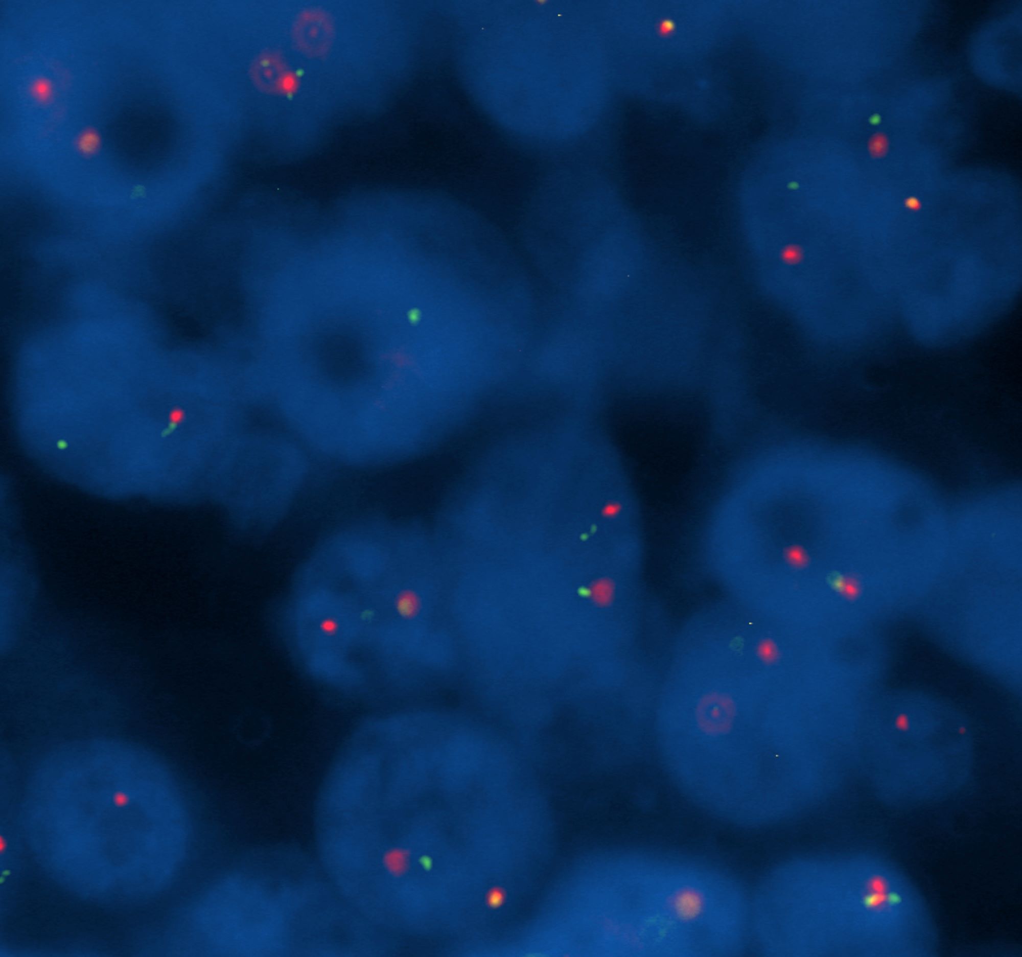
EWSR1 break apart FISH
Images hosted on other servers:
PCR showing EWSR1-ATF1 fusion product (81 bp)
Sequence of PCR product showing EWSR1-ATF1
Videos
Woman in 20s with gluteal mass
Sample pathology report
- Right toe, incisional biopsy:
- Clear cell sarcoma of soft tissue (see comment)
- Comment: Tumor was composed of nests of clear cells with prominent nucleoli and melanocytic differentiation. FISH break apart study revealed the presence of an EWSR1 rearrangement, which excluded the diagnosis of melanoma. These tumors are definitionally regarded as high grade - grade 3 sarcomas. Wide excision is recommended.
Differential diagnosis
- Melanoma:
- More common in the elderly
- With epidermal involvement / component
- No giant cells
- EWSR1-ATF1 / CREB1 negative
- Malignant gastrointestinal neuroectodermal tumor (GNET):
- Epithelioid malignant peripheral nerve sheath tumor:
- Malignant melanotic nerve sheath tumor:
- Carney syndrome
- Paraspinal or nerve plexus related location
- Psammomatous calcifications and abundant melanin pigment
- Nonnested, syncytial growth pattern
- EWSR1-ATF1 / CREB1 negative
- Perivascular epithelioid cell neoplasm (PEComa):
- Cellular blue nevus:
- No cytological atypia
- Dumbbell shaped growth pattern
- EWSR1-ATF1 / CREB1 negative
- Granular cell tumor:
- Alveolar soft part sarcoma:
- Paraganglioma:
- Synovial sarcoma:
- No nested growth
- PanCK+
- Epithelioid sarcoma:
- Carcinoma:
Additional references
Board review style question #1
An 18 year old woman presented with a 1 cm mass in the right great toe. Morphological features are shown above. Which one of the following immunohistochemical panels is the most helpful for reaching the correct diagnosis?
- EMA, SMA, INI1
- HMB45, MelanA, desmin
- PanCK, INI1, S100
- PanCK, synaptophysin, CD31
- SMA, desmin, SOX10
Board review style answer #1
C. PanCK, INI1, S100. If you order just HMB45, MelanA, and desmin and all of them turn out to be positive you may end up with the diagnosis of PEComa. PEComa is in the differential, even desmin is negative. So IHC panel should include an S100 or Sox10 and not necessarily HMB45 and MelanA at first. HMB45 and MelanA are better requested after S100/Sox10 positivity. panCK and INI1 are to exclude epithelioid sarcoma. INI1 could be replaced by HMB45 if requested.
Comment Here
Reference: Clear cell sarcoma
Comment Here
Reference: Clear cell sarcoma
Board review style question #2
An 18 year old woman presented with a 1 cm toe mass. There was diffuse S100 expression plus SOX10, HMB45 and MelanA positivity. What is the next step?
- Diagnose as melanoma
- Order PAX8 and TFE3
- Order whole genome sequencing
- Perform an EWSR1 break apart FISH
- Request PCR for EWSR1-CREB1
Board review style answer #2
D. Perform an EWSR1 break apart FISH. The differential diagnosis is between melanoma and CCS and EWSR1 FISH is less time consuming and cheaper than NGS to reach the correct diagnosis.
Comment Here
Reference: Clear cell sarcoma
Comment Here
Reference: Clear cell sarcoma
