Kidney tumor
Renal cell carcinoma - rare
Mucinous tubular and spindle cell carcinoma
Resident / Fellow Advisory Board: Alcino Pires Gama, M.D.
Deputy Editor-in-Chief: Maria Tretiakova, M.D., Ph.D.


Last author update: 3 April 2024
Last staff update: 3 April 2024
Copyright: 2003-2025, PathologyOutlines.com, Inc.
PubMed Search: Mucinous tubular and spindle cell carcinoma
Table of Contents
Definition / general | Essential features | Terminology | Epidemiology | Sites | Pathophysiology | Etiology | Clinical features | Diagnosis | Radiology description | Radiology images | Prognostic factors | Case reports | Treatment | Gross description | Gross images | Frozen section description | Microscopic (histologic) description | Microscopic (histologic) images | Virtual slides | Positive stains | Negative stains | Electron microscopy description | Molecular / cytogenetics description | Molecular / cytogenetics images | Sample pathology report | Differential diagnosis | Practice question #1 | Practice answer #1 | Practice question #2 | Practice answer #2Cite this page: Valencia A, Gordetsky JB, Craig JC. Mucinous tubular and spindle cell carcinoma. PathologyOutlines.com website. https://www.pathologyoutlines.com/topic/kidneytumormalignantmucinoustubular.html. Accessed August 27th, 2025.
Definition / general
- Distinct polymorphic, malignant renal cell neoplasm composed of bland anastomosing tubules, spindle cell areas and myxoid stroma / extracellular mucin
Essential features
- Compact tubules lined by bland cuboidal or flattened cells merging with low grade spindle cell areas
- Myxoid stroma with basophilic extracellular mucin
- Absence of distinct, well formed papillae
- References: Eur Urol 2022;82:458, Mod Pathol 2021;34:1392, Diagn Pathol 2015;10:168, Arch Pathol Lab Med 2020;144:115
Terminology
- Historic terminology not recommended by WHO 2022
- Low grade tubular mucinous renal neoplasm
- Low grade collecting duct carcinoma
- Low grade myxoid renal epithelial neoplasm with distal nephron differentiation
Epidemiology
- Accounts for < 1% of all renal neoplasms
- Median age: 50 - 60 years
- F > M
- References: Am J Surg Pathol 2018;42:767, Clin Genitourin Cancer 2019;17:268, BJU Int 2015;116:85
Sites
- Kidney
Pathophysiology
- These tumors have a phenotypic expression pattern similar to the loop of Henle region of normal nephrons (Am J Surg Pathol 2018;42:1571)
- Hippo pathway dysregulation with increased nuclear YAP1 protein expression (Am J Surg Pathol 2018;42:767)
- CDKN2A or CDKN2B deletion may be present in high grade tumors (Mod Pathol 2021;34:445)
Etiology
- Unknown
Clinical features
- Most cases are asymptomatic
- Incidental finding on imaging
Diagnosis
- Essential morphologic features include anastomosing tubules lined by low grade cells
- Tubules should merge with areas of bland spindle cells
- Areas of myxoid stroma or extracellular mucin should be present
- References: Eur Urol 2022;82:458, Mod Pathol 2021;34:1392, Diagn Pathol 2015;10:168, Arch Pathol Lab Med 2020;144:115
Radiology description
- Majority are homogeneous
- Small subset may show cystic changes, which is more commonly seen in papillary renal cell carcinoma (RCC)
- Typically isodense or hypodense on unweighted CT
- Hyperintense on T2 due to extracellular mucin
- Appears unencapsulated
- References: Br J Radiol 2021;94:20210548, Abdom Radiol 2021;46:5250
Radiology images
Images hosted on other servers:

Isodense mass in the renal medulla

Hyperintensity on T2 weighted image
Prognostic factors
- Indolent behavior (Clin Genitourin Cancer 2019;17:268)
- Rarely metastatic (e.g., with epithelial anaplasia or sarcomatoid differentiation)
Case reports
- 39 year old man with right lower back pain and 8 cm kidney mass (Urol Case Rep 2021:40:101889)
- 40 year old woman with incidental kidney mass (J Kidney Cancer VHL 2022;9:10)
- 45 year old man with incidental kidney mass (Medicine (Baltimore) 2018;97:e12933)
- 69 year old man with kidney mass and osseous lesions (IJU Case Rep 2021;4:333)
Treatment
- Surgical excision is the mainstay of treatment
- Selected cases received adjuvant therapy with tyrosine kinase inhibitors and PD-1 inhibitors (Clin Genitourin Cancer 2019;17:268)
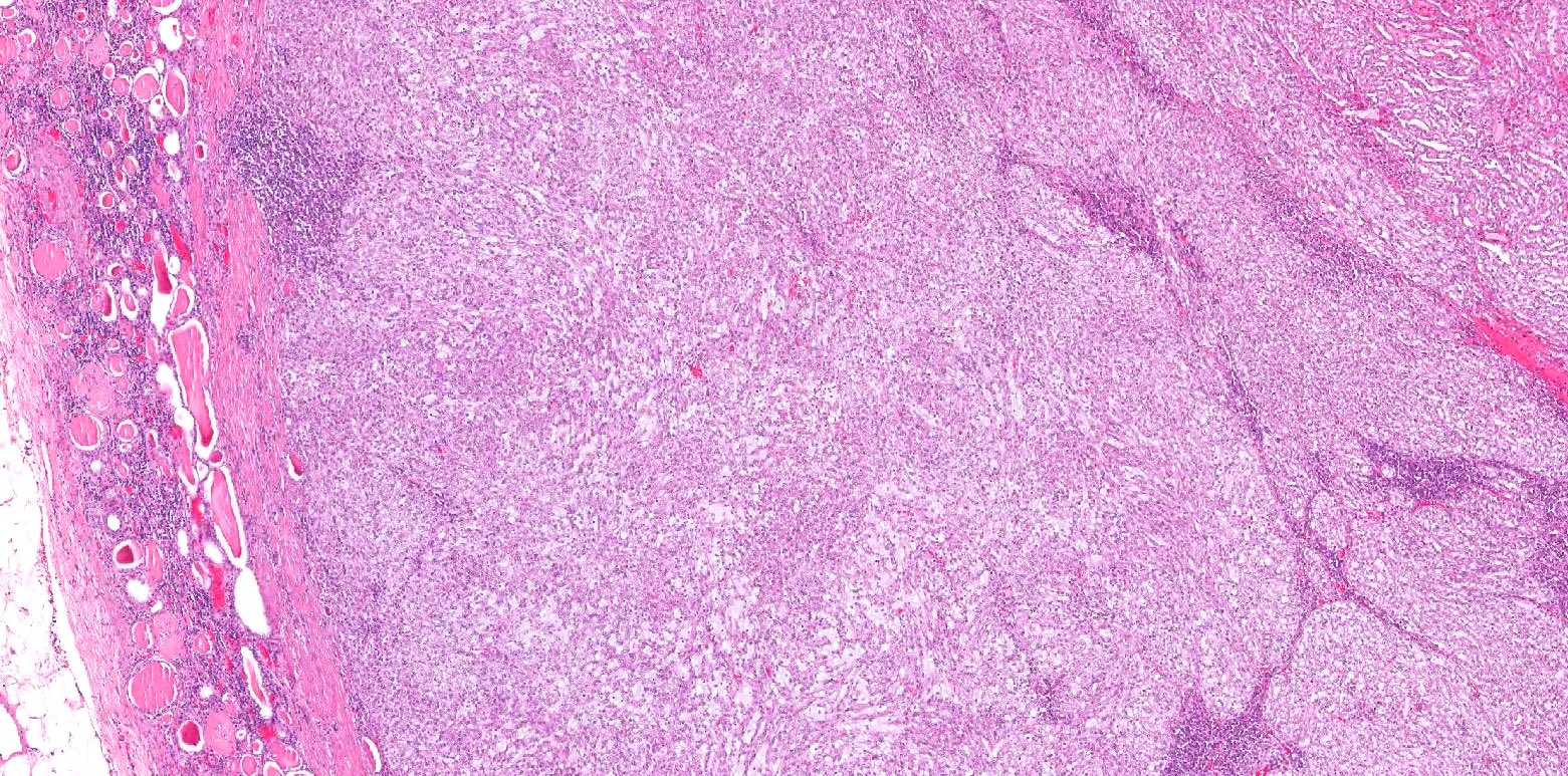
Gross description
- Well circumscribed, solid, homogenous (Arch Pathol Lab Med 2020;144:115)
- Capsule may be present (Arch Pathol Lab Med 2020;144:115)
- Tan, gray-pink or pale yellow cut surface (Arch Pathol Lab Med 2020;144:115)
- Typically intraparenchymal or partially exophytic mass (BMC Cancer 2023;23:815)
- Most masses are small (Arch Pathol Lab Med 2020;144:115)
- Areas of hemorrhage and necrosis are rare (Arch Pathol Lab Med 2020;144:115)
Gross images
Images hosted on other servers:

Cut surface with focal hemorrhage
Frozen section description
- Elongated tubules with low grade epithelial lining merging with areas of low grade spindled cells in a myxoid background
- Extracellular mucin may be present
Microscopic (histologic) description
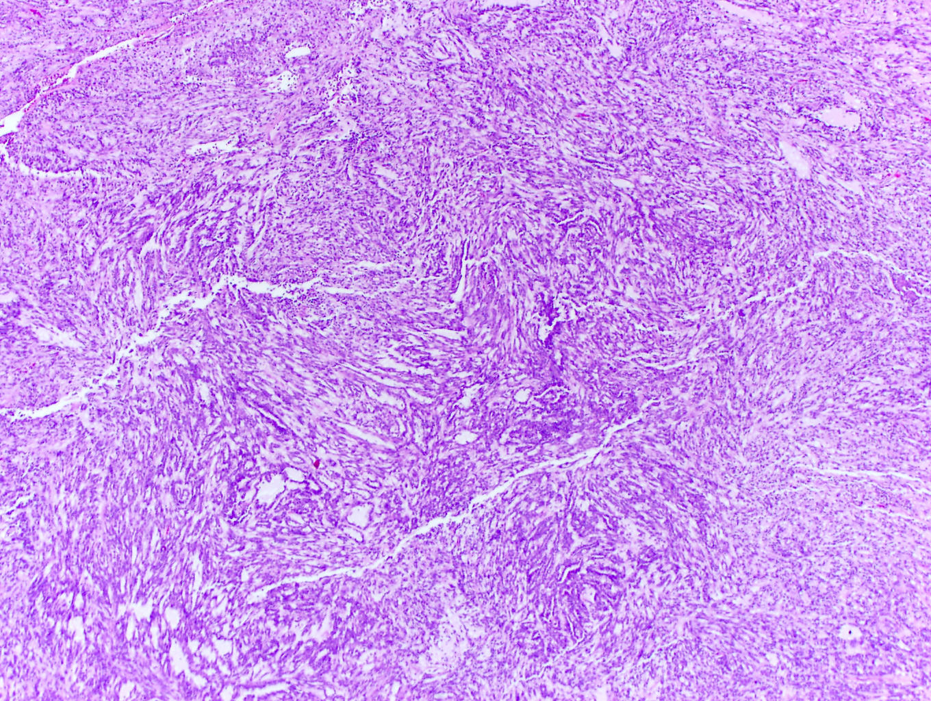
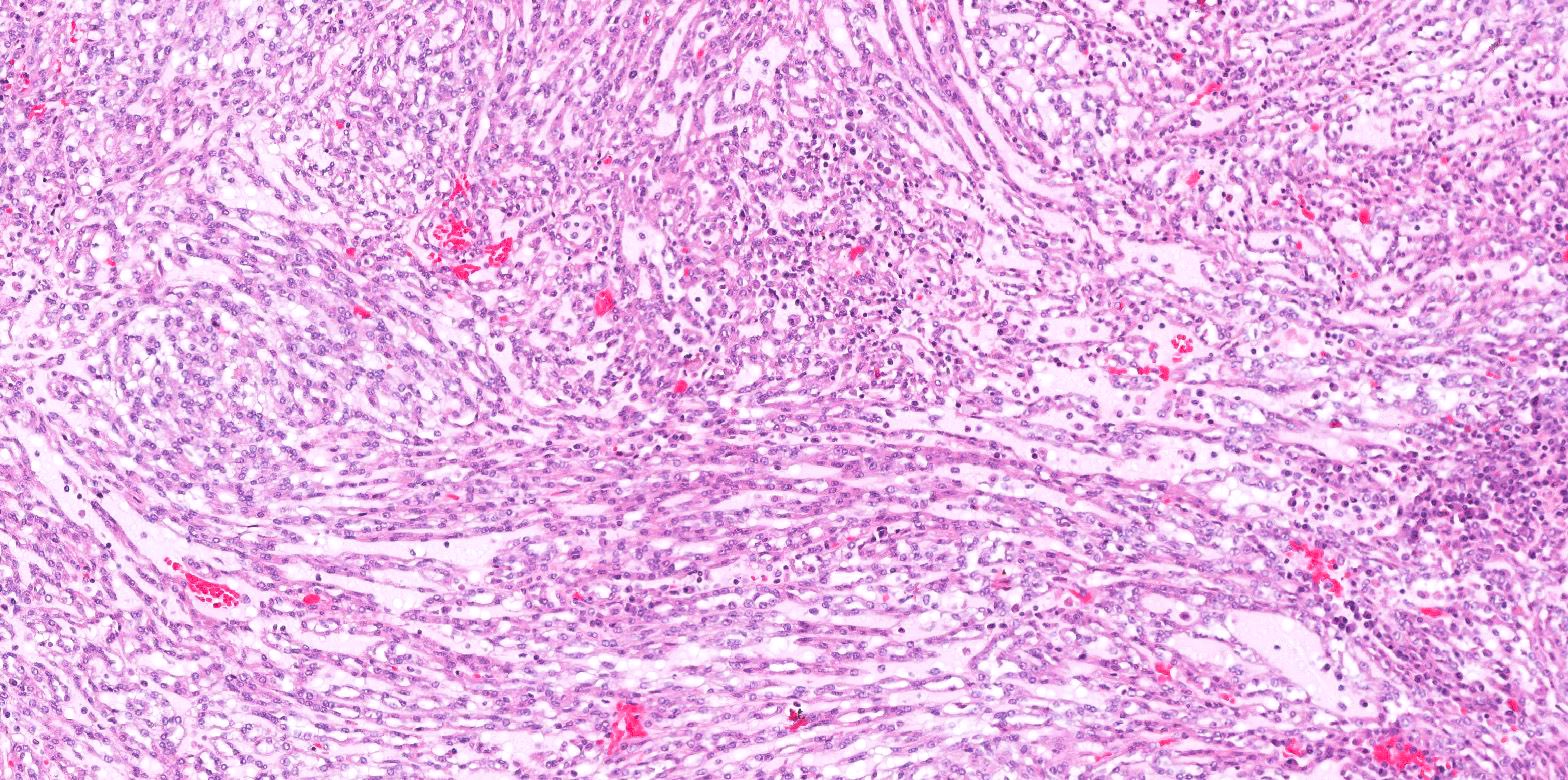
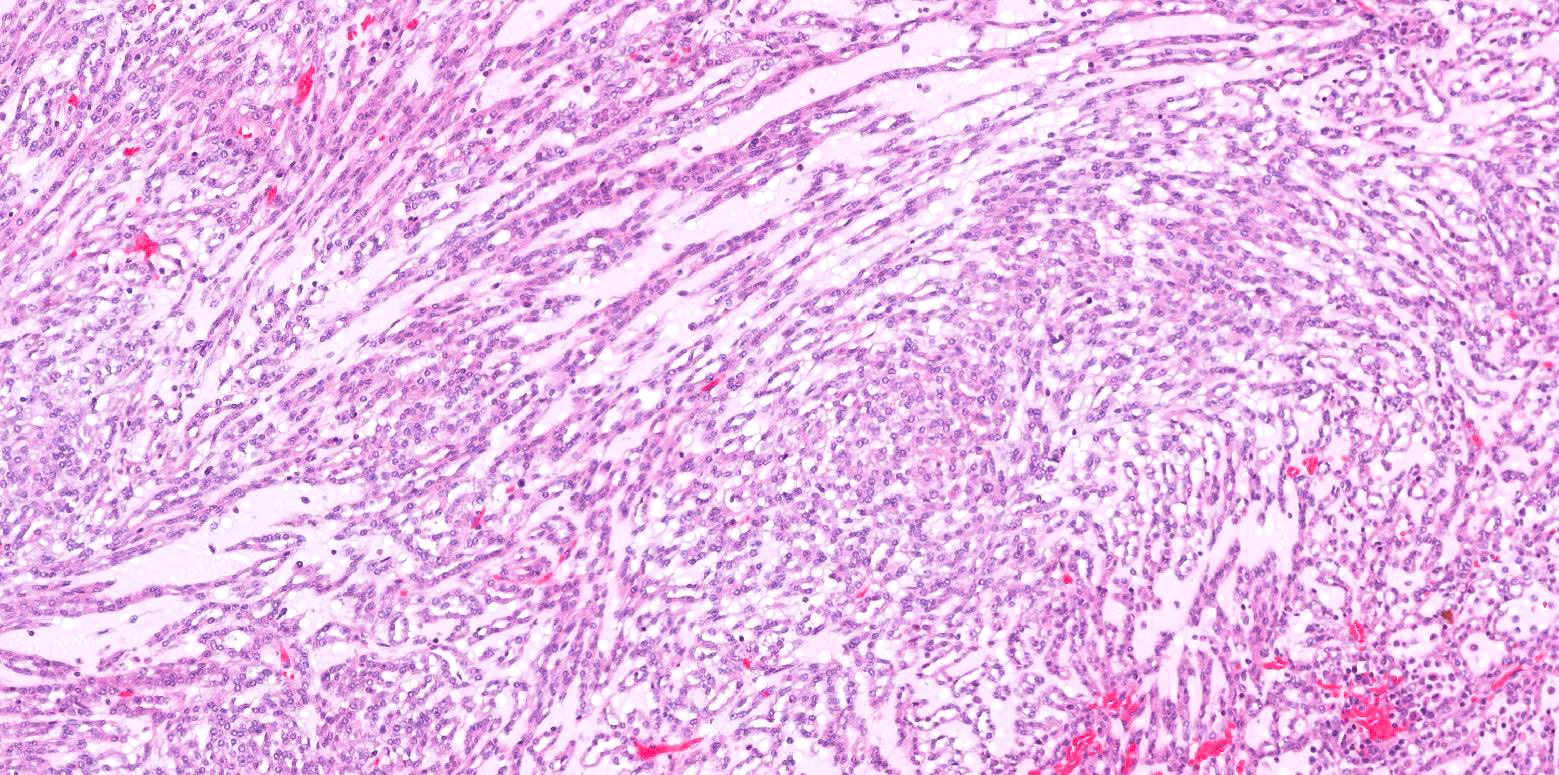
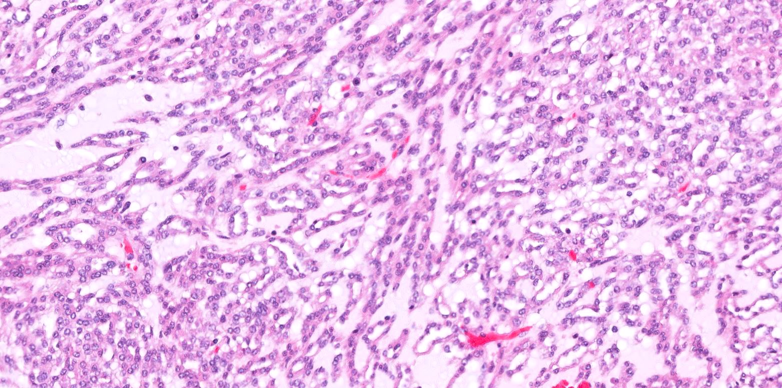
- Bland tubules merging with bland spindle cells in a myxoid stroma
- Absence of distinct, well formed papillae
- Tightly packed tubules and spindle cell areas with smooth lumina
- Compact arrangements of whorled tubules
- Areas of myxoid stroma
- Presence of a capsule or foamy macrophages may be present
- High nucleolar grade or extensive necrosis have been described as occurring in rare cases (Histopathology 2017;71:719)
- References: Eur Urol 2022;82:458, Mod Pathol 2021;34:1392, Diagn Pathol 2015;10:168, Arch Pathol Lab Med 2020;144:115
Microscopic (histologic) images
Contributed by J. Cody Craig, M.D., Aida Valencia, M.D., Jennifer B. Gordetsky, M.D. and @katcollmd on Twitter
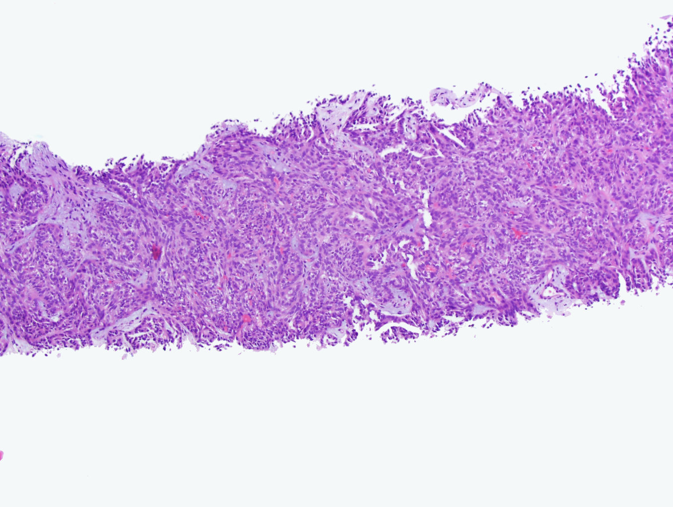
Bland tubules and spindle cells
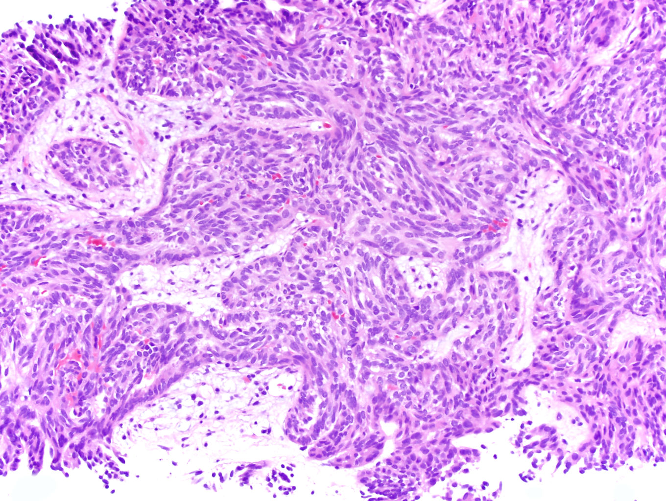
Whorled tubules
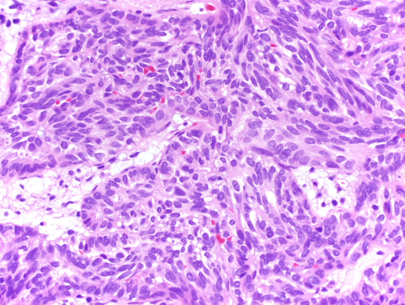
Bland cuboidal and spindle cells
Compact tubules
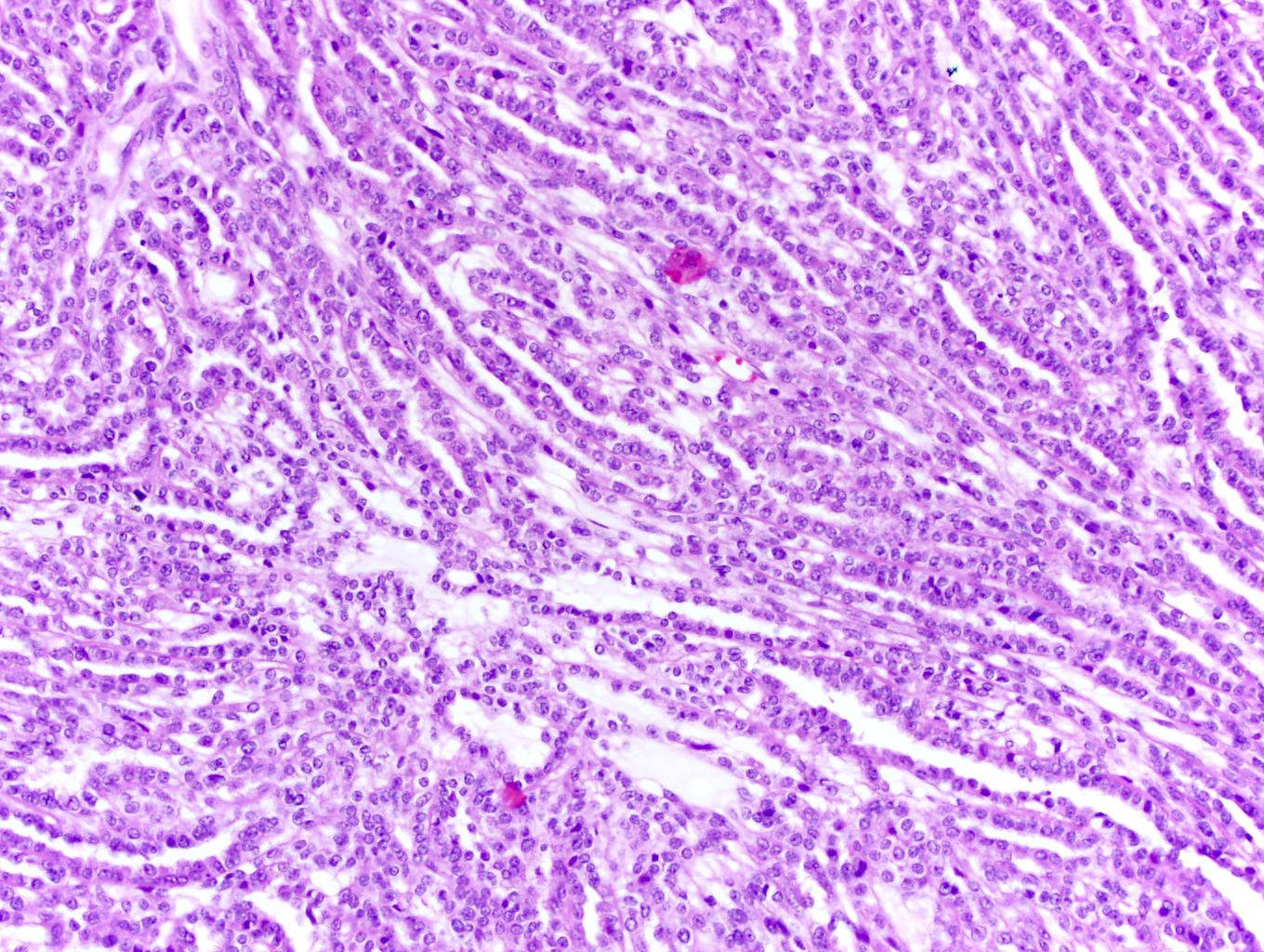
Elongated tubules
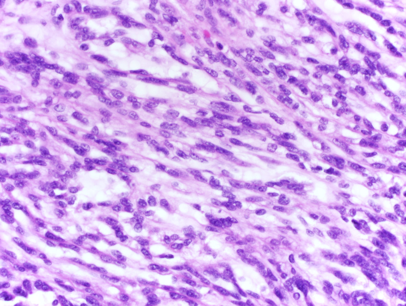
Bland spindle cells
Mucinous tubular and spindle cell carcinoma
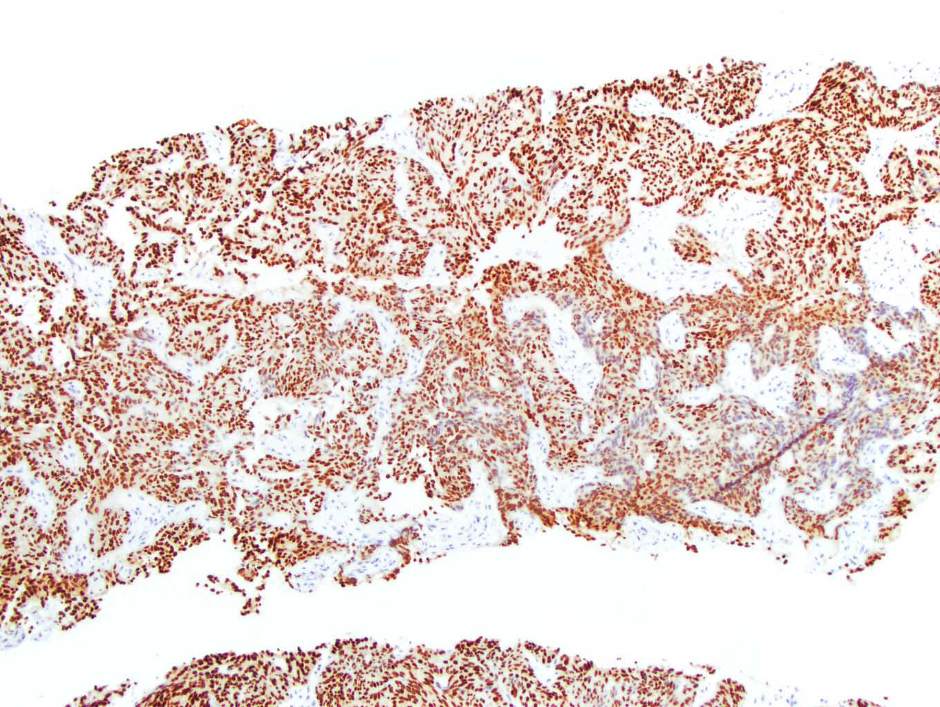
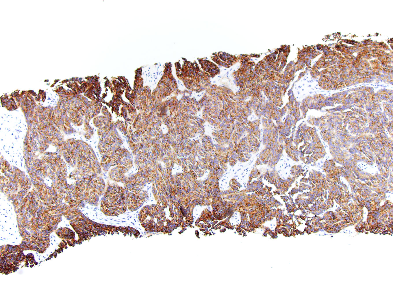
PAX8
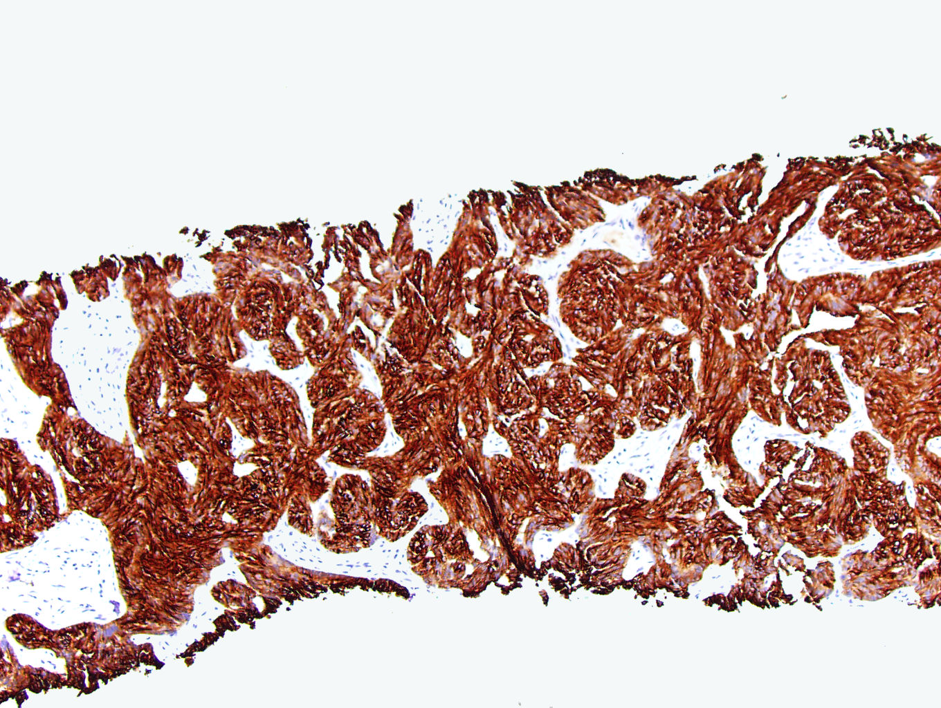
CK7
p504s / AMACR
Virtual slides
Images hosted on other servers:

Mucinous tubular and spindle cell carcinoma
Positive stains
Negative stains
Electron microscopy description
- Tightly packed, often elongated tubules composed of slender attenuated cells, similar to those found in the normal loop of Henle (Int Braz J Urol 2002;28:477)
- Discontinuation of the tubular basement membranes (Int Braz J Urol 2002;28:477)
- Low columnar, cuboidal and spindle cells with a low nuclear to cytoplasmic ratio (Pathol Res Pract 2014;210:454)
- Small nuclei with small nucleoli, dispersed chromatin and smooth nuclear membranes (Pathol Res Pract 2014;210:454)
- Scant cytoplasmic organelles and intermediate filaments (Pathol Res Pract 2014;210:454)
- Short apical microvilli and well developed desmosomes at the site of attachment between the apposing cell membranes (Pathol Res Pract 2014;210:454)
- Foamy macrophages may be present (Pathol Res Pract 2014;210:454)
Molecular / cytogenetics description
- Monosomy of chromosomes 1, 6, 9, 14, 15 and 22 (Am J Surg Pathol 2018;42:767)
- Multiple loss of chromosomes 1, 4, 6, 8, 9, 13, 14, 15 and 22 (Am J Surg Pathol 2018;42:767)
- FISH shows no VHL deletions (Am J Surg Pathol 2018;42:767, Int Braz J Urol 2002;28:477)
- Recurrent chromosomal losses and somatic mutations of genes in the Hippo pathway (Am J Surg Pathol 2018;42:1571)
- Moderate to high expression of VSTM2A via RNA in situ hybridization (Am J Surg Pathol 2018;42:1571)
- Biallelic loss of Hippo pathway tumor suppressor genes (PTPN14, NF2, SAV1) (Cancer Discov 2016;6:1258)
Molecular / cytogenetics images
Images hosted on other servers:

Most common genetic alterations

SNP array analysis
Sample pathology report
- Kidney, right, radical nephrectomy:
- Mucinous tubular and spindle cell carcinoma, 3 cm (see comment)
- Organ confined
- Margins negative for tumor
- No tumor necrosis identified
- No sarcomatoid or rhabdoid features identified
- No metatasis to lymph nodes
- Pathologic stage: pT1a pN0 pMx
- Comment: The specimen shows a kidney composed of low grade tubules merging with areas of spindled cells in a myxoid stroma. No well formed papillary areas are present. Immunohistochemical stains show positivity for PAX8, CK7, racemase and vimentin.
Differential diagnosis
- Papillary renal cell carcinoma (Am J Surg Pathol 2008;32:1353):
- Papillary architecture
- May have necrosis
- Papillary cores show psammoma bodies or foamy macrophages
- Lack of spindled stroma and mucin
- There is no difference in the nuclear expression of YAP / TAZ between mucinous tubular and spindle cell carcinoma and papillary renal cell carcinoma (Am J Surg Pathol 2018;42:767)
- Frequently positive for CD10
- Renal cell carcinoma with sarcomatoid differentiation (Mod Pathol 2021;34:1392):
- High grade cytologic atypia in spindle cells
- Abrupt transition from epithelial to spindle cell component
- Presence of distinct alternative histologic subtype (clear cell, papillary, etc.)
- ALK rearrangement associated renal cell carcinoma (Diagn Pathol 2022;17:52):
Practice question #1
A patient undergoes a partial nephrectomy for a 4 cm renal mass. The above tumor is seen on H&E examination. This tumor should show which immunophenotype?
- PAX8+, BAP1 loss, calretinin+, WT1+
- PAX8+, CA9+, CD10+, CK7-
- PAX8+, CK7+, vimentin+, racemase+
- PAX8+, TTF1+, CK7+
Practice answer #1
C. PAX8+, CK7+, vimentin+, racemase+. The images show a mucinous tubular and spindle cell carcinoma, which should show positive staining for PAX8, CK7, racemase and vimentin. Answer D is incorrect because this immunophenotype is more consistent with metastatic thyroid cancer. Answer B is incorrect because this immunophenotype is more consistent with a clear cell renal cell carcinoma. Answer A is incorrect because this immunophenotype is more consistent with mesothelioma.
Comment Here
Reference: Mucinous tubular and spindle cell carcinoma
Comment Here
Reference: Mucinous tubular and spindle cell carcinoma
Practice question #2
Which of the following features would be most helpful in distinguishing a mucinous tubular and spindle cell carcinoma from a renal cell carcinoma with sarcomatoid features?
- PAX8+, CK7+, vimentin+, racemase+
- Presence of foamy macrophages
- Retained FH and SDHB expression on IHC
- VHL mutation on molecular studies
Practice answer #2
D. VHL mutation on molecular studies. VHL mutations are not found in mucinous tubular and spindle cell carcinomas. This would be more consistent with a clear cell renal cell carcinoma. Answer A is incorrect because this immunophenotype can be found in papillary renal cell carcinoma. Answer C is incorrect because this immunophenotype excludes an SDHB deficient or an FH deficient RCC but many other renal cell neoplasms can have this finding. Answer B is incorrect because mesothelial tumors will express both CK5/6 and D2-40.
Comment Here
Reference: Mucinous tubular and spindle cell carcinoma
Comment Here
Reference: Mucinous tubular and spindle cell carcinoma
