Prostate gland & seminal vesicles
Nonneoplastic
Nephrogenic metaplasia / adenoma
Resident / Fellow Advisory Board: Alcino Pires Gama, M.D.
Deputy Editor-in-Chief: Maria Tretiakova, M.D., Ph.D.

Last author update: 9 August 2023
Last staff update: 9 August 2023
Copyright: 2003-2025, PathologyOutlines.com, Inc.
PubMed search: Prostatic nephrogenic adenoma
Table of Contents
Definition / general | Essential features | ICD coding | Epidemiology | Sites | Pathophysiology | Etiology | Clinical features | Diagnosis | Prognostic factors | Case reports | Treatment | Gross description | Microscopic (histologic) description | Microscopic (histologic) images | Cytology description | Positive stains | Negative stains | Sample pathology report | Differential diagnosis | Additional references | Practice question #1 | Practice answer #1 | Practice question #2 | Practice answer #2Cite this page: Sanguedolce F. Nephrogenic metaplasia / adenoma. PathologyOutlines.com website. https://www.pathologyoutlines.com/topic/prostatenephrogenichyper.html. Accessed September 3rd, 2025.
Definition / general
- Benign polypoid, papillary, fungating or velvety lesion found in the bladder or prostatic urethra after urothelial injury
- Has multiple histologic patterns
Essential features
- Evidence that it is derived from shed renal tubular cells (N Engl J Med 2002;347:653)
- Alternatively, it is postulated that a subset of cases might be a metaplastic reaction (Int Sch Res Notices 2015;2015:704982)
- Tubules lined by flattened, cuboidal or hobnail cells
- Many morphologic variants: papillary, tubular, flat, cysts and microcysts, signet ring cell-like, fibromyxoid
- Hyaline rim surrounds the tubules
- No mitotic activity
- Only occasional solid areas or rare clear cells present
- May focally involve the prostatic parenchyma and mimic prostate adenocarcinoma
ICD coding
- ICD-10: N32.9 - bladder disorder, unspecified
Epidemiology
- Any age (mostly old adults) (Int Sch Res Notices 2015;2015:704982)
- Usually following a urothelial injury (instrumentation, urethral catheterization, recurrent infection, stones, bacillus Calmette-Guérin [BCG] therapy or renal transplant) (Urology 2016;95:29)
Sites
- Prostatic urethra
- Described throughout the entire urothelium (see nephrogenic metaplasia in the bladder chapter)
Pathophysiology
- Evidence that it is derived from shed renal tubular cells (N Engl J Med 2002;347:653)
- Alternatively, it is postulated that a subset of cases might be a metaplastic reaction (Int Sch Res Notices 2015;2015:704982)
Etiology
- Prior injury to the urothelium
Clinical features
- May present as a mass or with irritative lower urinary tract symptoms, hematuria, dysuria, obstruction
- Usually small (< 1 cm) but may be up to 7 cm
Diagnosis
- Biopsy, transurethral resection
Prognostic factors
- May recur; risk factors not known
Case reports
- 39 year old woman with nephrogenic adenoma arising in urethral diverticulum (Cureus 2023;15:e36578)
- 62 year old man with giant prostatic hypertrophy and recurrent nephrogenic adenoma of the prostate (BMC Urol 2013;13:18)
- 67 year old man with asymptomatic macroscopic hematuria and a history of laser photovaporization of the prostate for benign prostate hyperplasia (Urol Case Rep 2020;33:101382)
- 72 year old man with nephrogenic adenoma presenting with irritative voiding symptoms as benign prostatic hyperplasia and mimicking prostatic and bladder carcinomas (Cureus 2023;15:e35998)
Treatment
- Transurethral resection with regular follow ups
Gross description
- Friable soft tissue fragments
Microscopic (histologic) description
- Tubules lined by simple cuboidal, flattened or hobnail cells, forming an exophytic papillary or endophytic lesion (Urology 2016;95:29)
- Associated acute and chronic inflammation and edema in the stroma; lack of desmoplastic reaction
- Many morphologic variants (multiple patterns may occur in the same cases): papillary, tubular, tubulocystic, polypoid, flat, fibromyxoid, signet ring cell-like (Mod Pathol 2013;26:792)
- Hyaline rim surrounds the tubules (PAS positive thickened basement membrane) (Adv Anat Pathol 2019;26:171)
- No mitotic activity
- Scant cytoplasm, finely granular uniform chromatin; usually inconspicuous nucleoli (Ann Diagn Pathol 2019;38:11)
- Only occasionally solid areas, rare clear cells, blue mucin within the tubules present
- Sometimes atrophic tubules filled with eosinophilic colloid-like material mimicking thyroid follicles and mesonephric hyperplasia
- May be associated with radiotherapy
- Rare fibromyxoid variant features, including compressed spindle shaped cells in a prominent fibromyxoid background; it can present as pure fibromyxoid or combined with classic (tubular) morphology (Am J Surg Pathol 2023;47:37)
Microscopic (histologic) images
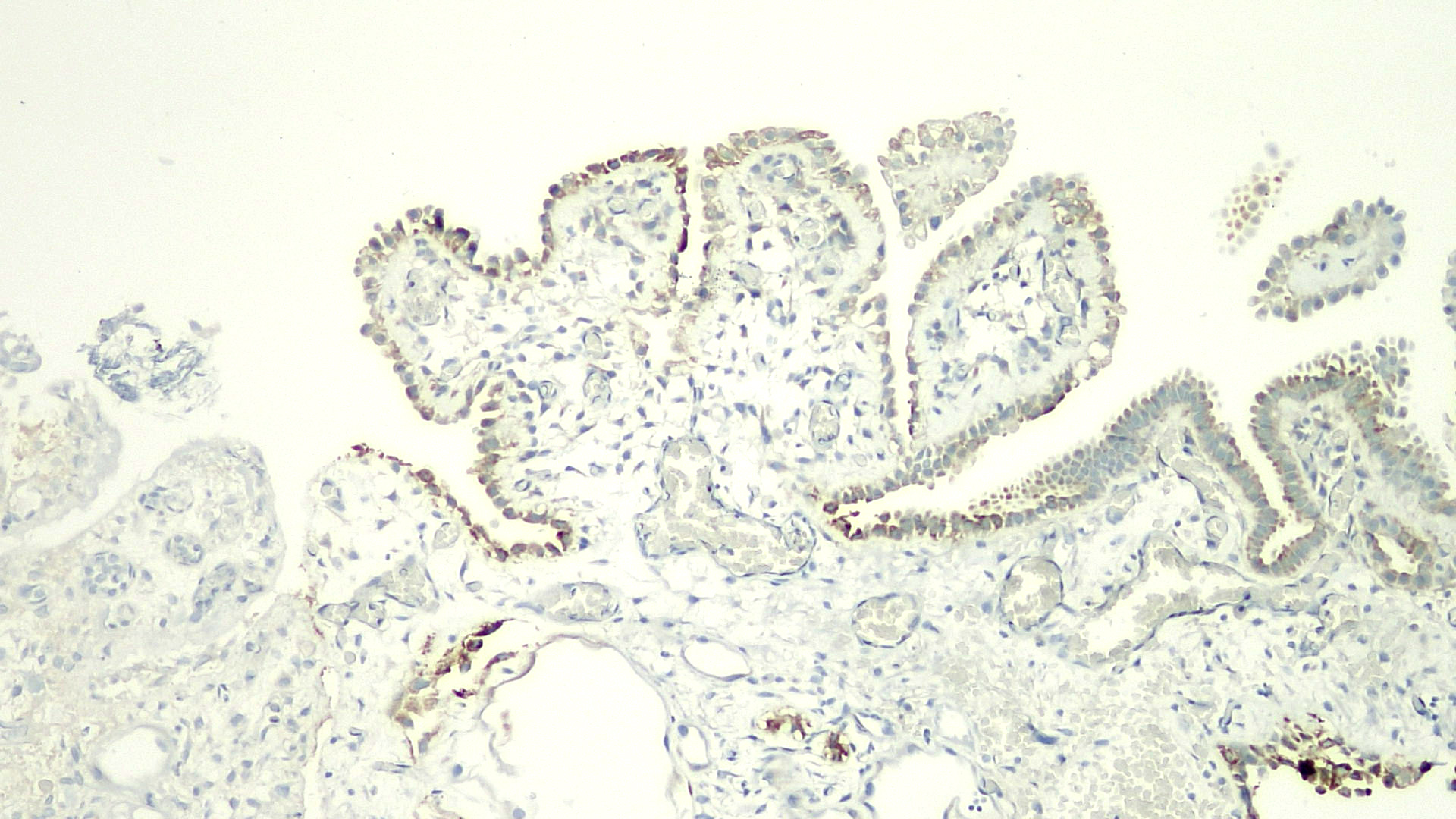
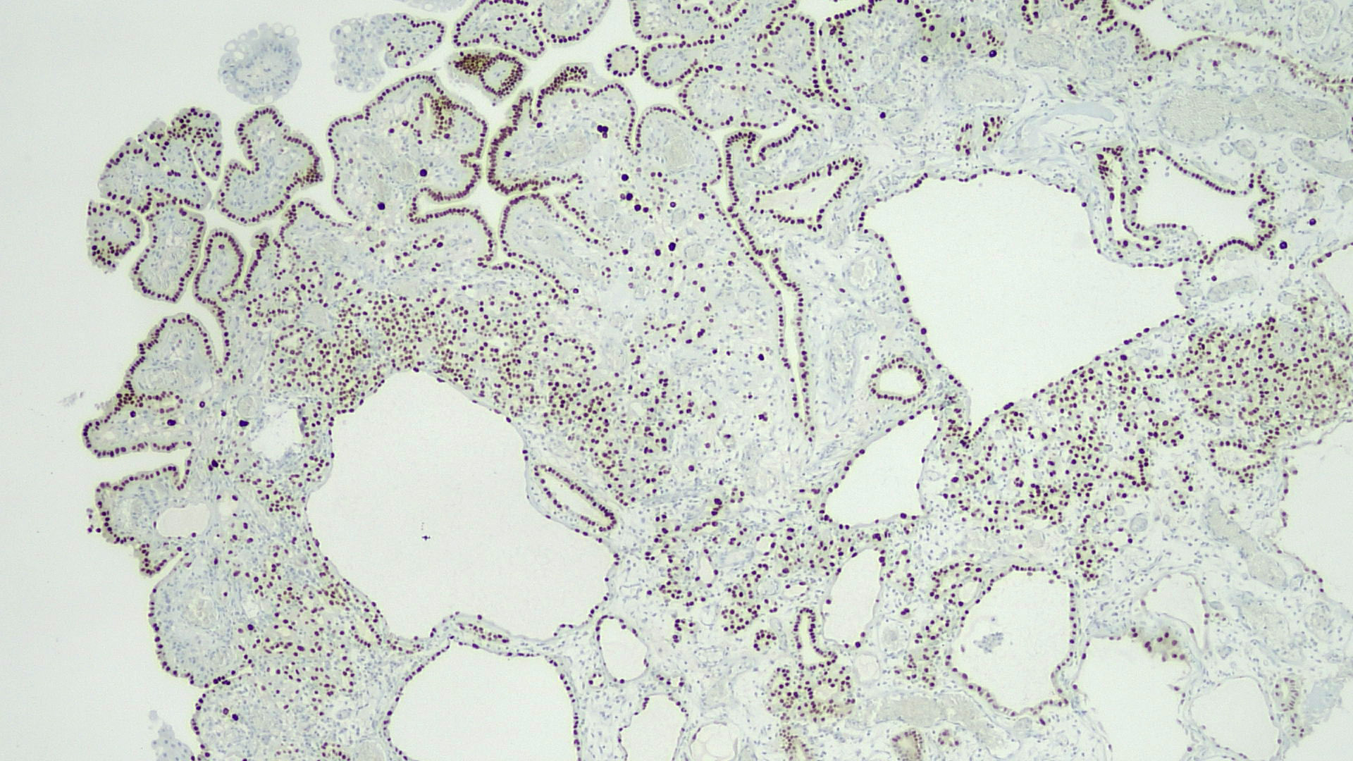
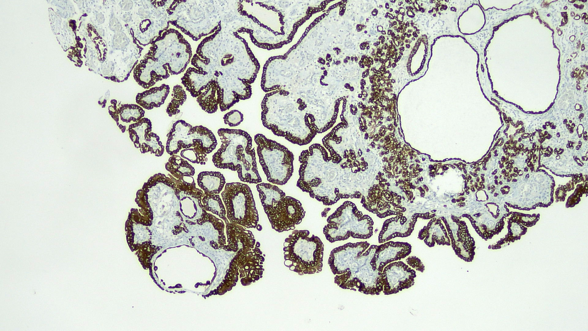
Contributed by Francesca Sanguedolce, M.D., Ph.D.
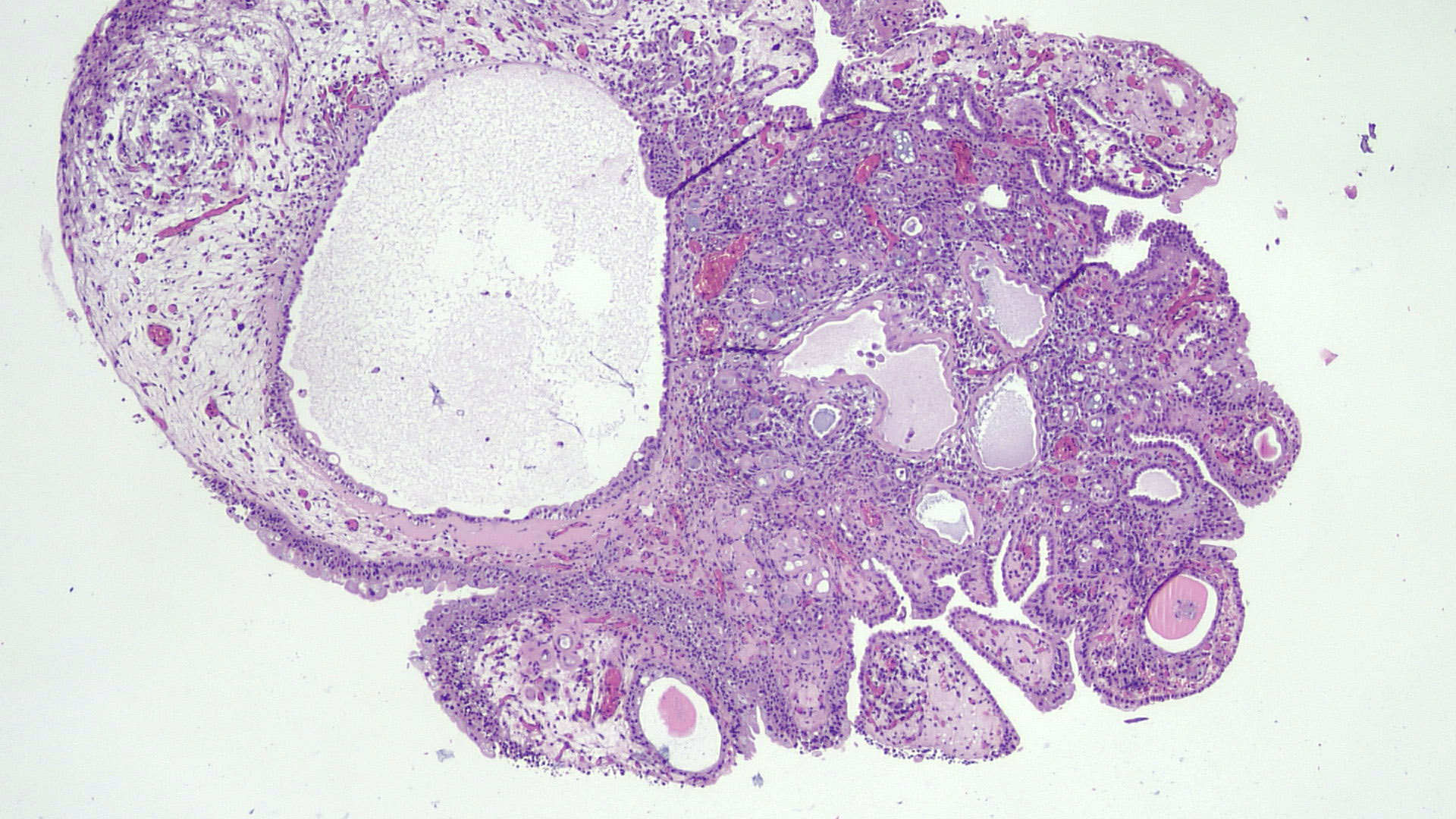
Multiple morphologies
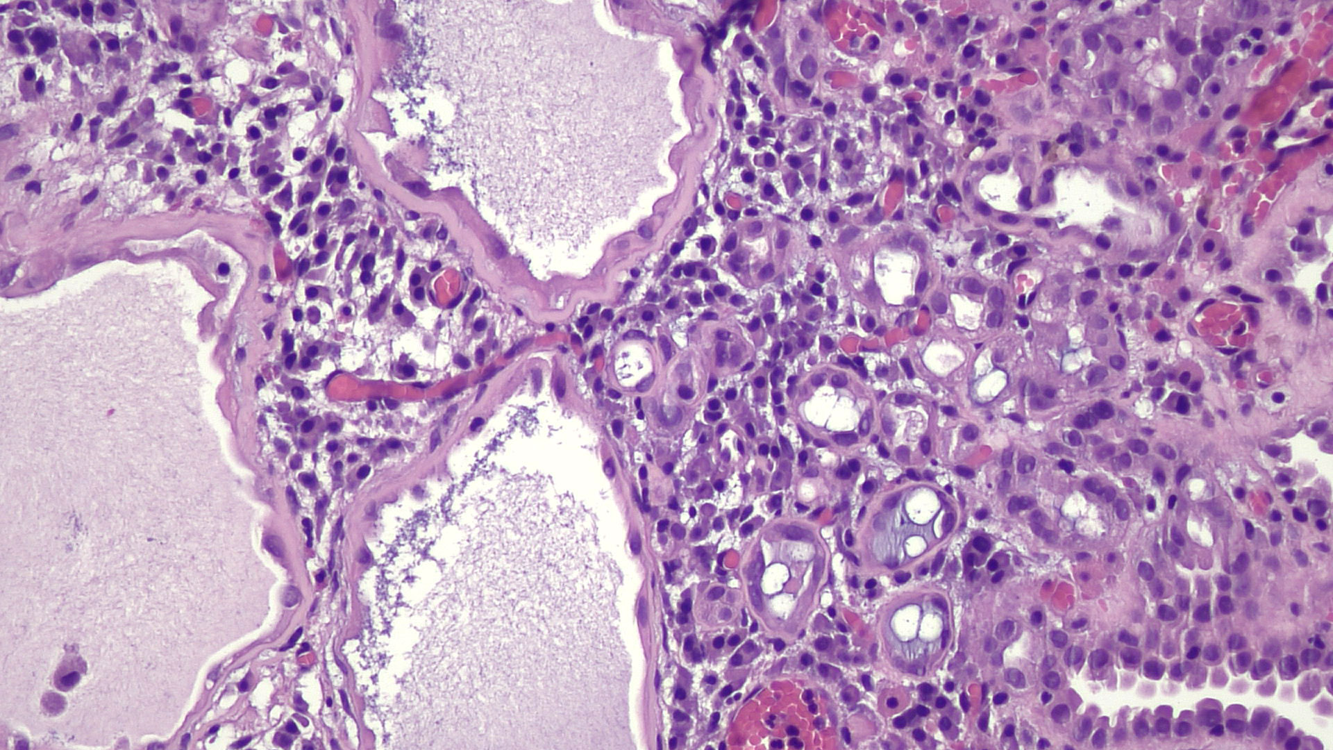
Inflammation, thickened basement membrane
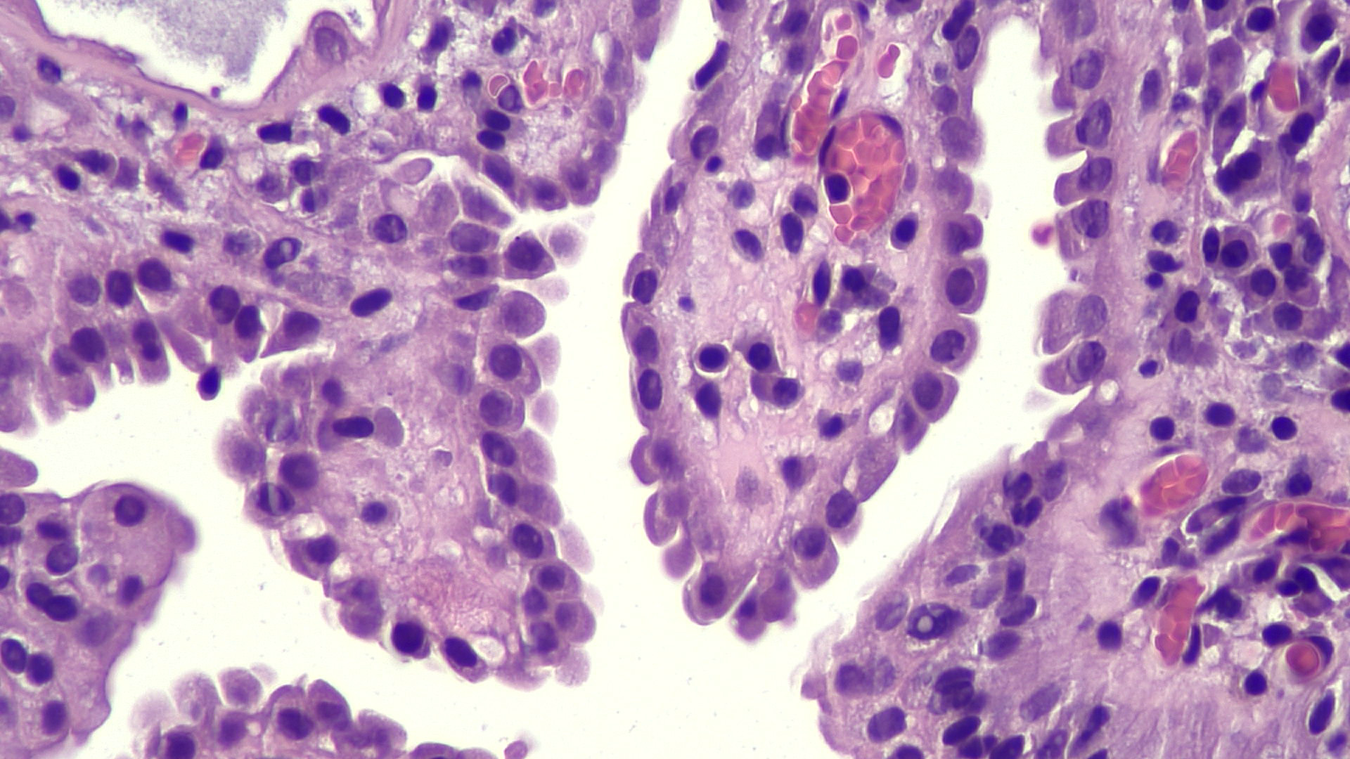
Cytological features
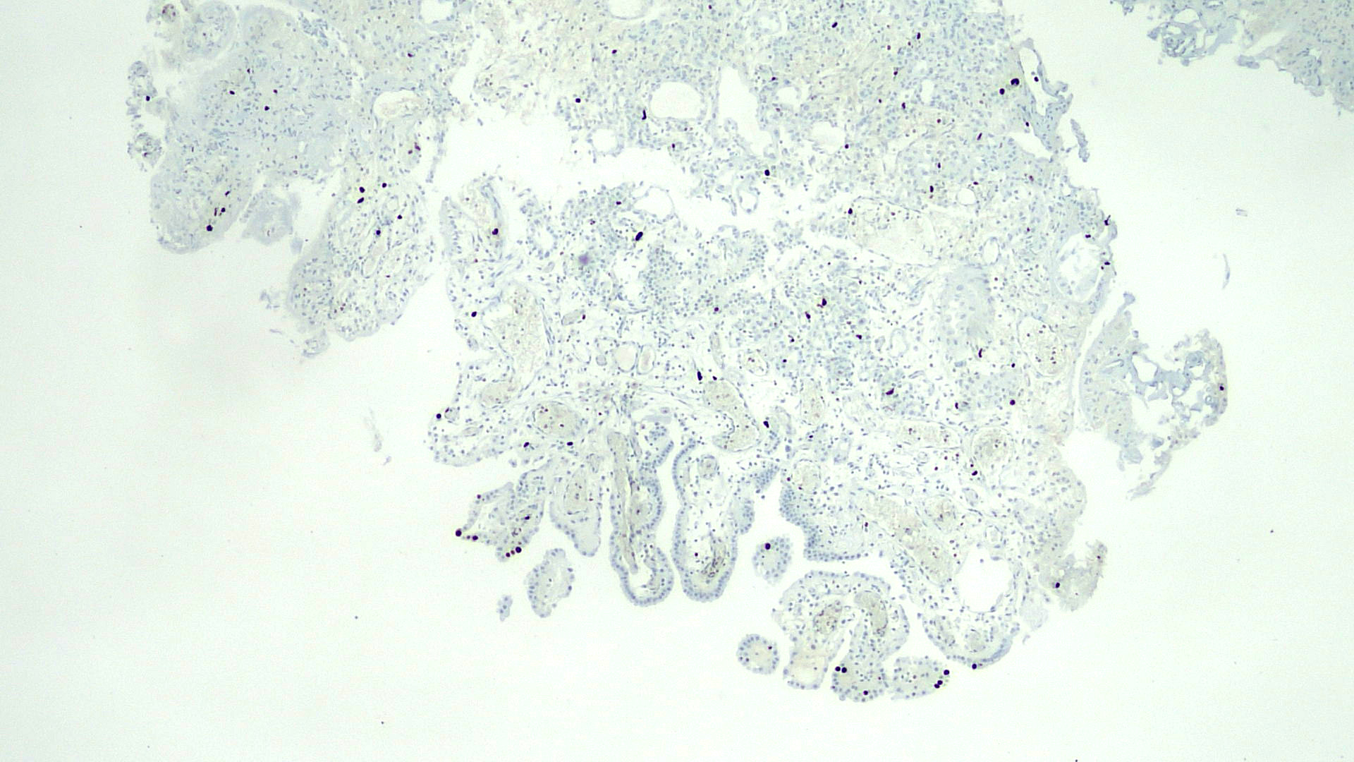
Ki67
AMACR
PAX8
CK7
Cytology description
- Numerous vacuolated, polygonal and columnar shaped cells singly or in small groups
- Papillary fragments resembling low grade papillary urothelial carcinoma (Am J Clin Pathol 2016;145:373)
Positive stains
- PAX2, PAX8, AMACR, CD10, CK7 (Am J Surg Pathol 2014;38:1664)
- CK903, EMA, S100A1 (Ann Diagn Pathol 2013;17:41)
- Napsin A (Hum Pathol 2020;102:23)
- Weakly positive or negative for PSA and PSAP
Negative stains
- p63, CEA (Virchows Arch 2013;463:819)
- GATA3 (positive in 40% of cases) (Am J Surg Pathol 2014;38:1664)
- Very low Ki67 proliferation rate (< 5%) (Ann Diagn Pathol 2019;38:11)
Sample pathology report
- Prostate, biopsy / transurethral resection:
- Nephrogenic adenoma
Differential diagnosis
- Prostatic adenocarcinoma:
- Especially atrophic variant of prostate adenocarcinoma:
- Not associated with genitourinary trauma
- Infiltrative, more atypia
- No adjacent urothelium present, no thyroidization, usually no inflammation (Am J Surg Pathol 2001;25:802)
- Clear cell adenocarcinoma of the bladder:
- Occasional clear cells, more prominent pleomorphism, especially hyperchromatic enlarged nuclei and extensive muscular invasion
- Increased mitotic index and proliferation rate
- More common in women (Hum Pathol 2010;41:594)
- Invasive urothelial carcinoma:
- Especially some subtypes of urothelial carcinoma with bland morphology, such as tubular and microcystic urothelial carcinoma (Int J Surg Pathol 2012;20:123)
- Size and shape variation of the epithelial structures and haphazard infiltrative growth
- Lack of inflamed stroma and presence of desmoplasia
- PAX8 negative and p63 positive
Additional references
Practice question #1
Which of the following statements is true about nephrogenic adenoma?
- Can have multiple morphological patterns
- Carcinoembryonic antigen (CEA) is often positive
- Has a high proliferative index
- Is a metaplastic lesion
Practice answer #1
A. Can have multiple morphological patterns. Nephrogenic adenoma often features multiple different morphologies within the same lesion, the most common being tubular, cystic and papillary. Answer D is incorrect because it has been established that nephrogenic adenoma, which has been thought to be a metaplastic lesion, actually results from shed renal tubular cells. Answer C is incorrect because this entity has a very low proliferative index. Answer B is incorrect because this entity lacks CEA staining.
Comment Here
Reference: Nephrogenic metaplasia / adenoma
Comment Here
Reference: Nephrogenic metaplasia / adenoma
Practice question #2
Which of the following is a variant of nephrogenic adenoma?
- Fibromyxoid
- Glycogen rich
- Nested
- Plasmacytoid
Practice answer #2
A. Fibromyxoid. The rare fibromyxoid variant features compressed, spindle shaped cells in a prominent fibromyxoid background; it can present as pure fibromyxoid or combined with classic (tubular) morphology. Answers B, C and D are incorrect because they all refer to subtypes of urothelial cancer.
Comment Here
Reference: Nephrogenic metaplasia / adenoma
Comment Here
Reference: Nephrogenic metaplasia / adenoma
