

Soft tissue
Adipose tissue tumors
Other benign lesions
Hibernoma


Copyright: 2002-2024, PathologyOutlines.com, Inc.
PubMed Search: Hibernoma
- Rare, benign, adipocytic tumor composed of variable proportions of brown fat cells admixed with white adipose tissue
- Benign tumor comprising multivacuolated eosinophilic granular fat cells with small, central normochromic nuclei (J Pathol Transl Med 2017;51:499)
- Pseudolipoma (not recommended) (J Pathol Transl Med 2017;51:499)
- ICD-O: 8880/0 - hibernoma
- ICD-11: 2E80.0Z & XH1054 - lipoma, unspecified and hibernoma
- Comprises < 2% of benign lipomatous tumors (Case Rep Oncol 2017;10:438, Am J Surg Pathol 2018;42:951)
- Makes up 1% of all adipocytic tumors (Case Rep Oncol 2017;10:438)
- Usually present from 2 - 75 years
- Mean age: 38 years, 60% presenting in third and fourth decades of life (Case Rep Oncol 2017;10:438, Cureus 2017;9:e1124, J Comput Assist Tomogr 2019;43:793, J Pathol Transl Med 2017;51:499)
- Slightly more common in men (Cureus 2020;12:e9111, Am J Surg Pathol 2018;42:951)
- May range from 1 - 24 cm, with an average diameter of 9.3 cm (Iran J Pathol 2017;12:406, Case Rep Oncol 2017;10:438)
- Most common site is the thigh (29%), followed by the shoulder (12%), back (10%), neck (9%) and chest wall (6%) (Case Rep Oncol 2017;10:438, Am J Case Rep 2020;21:e921447, Cureus 2017;9:e1124, Cureus 2020;12:e9111, J Pathol Transl Med 2017;51:499)
- Other sites: intraabdominal, retroperitoneal and thoracic cavity (Virchows Arch 2021;478:527)
- Rare: intraosseous, appears to be more common in women (Skeletal Radiol 2022;51:1325, J Comput Assist Tomogr 2019;43:793, J Pathol Transl Med 2017;51:499)
- Subcutaneous, sometimes intramuscular (20%) (Am J Case Rep 2020;21:e921447)
- Intraosseous hibernoma: osteoblasts and adipocytes have common origin from mesenchymal stem cells
- Brown fat cell differentiation is associated with PRD1-BF1-RIZ1 homologous domain containing 16 (PRDM16) underregulated by bone morphogenetic protein 7 (BMP7), which also stimulates bone formation
- This explains how brown fat cells and sclerotic bony trabeculae are mixed in intraosseous hibernomas (J Pathol Transl Med 2017;51:499)
- Association with MEN type 1 (Proc Natl Acad Sci U S A 2010;107:21122)
- Small, slow growing, painless, mobile mass
- Can cause local pressure effects (Case Rep Oncol 2017;10:438, Am J Case Rep 2020;21:e921447)
- Usually incidental, identified on radiology carried out for other causes (Am J Case Rep 2020;21:e921447, Cureus 2017;9:e1124)
- See Microscopic (histologic) description
- CT scans shows well circumscribed, hyperdense lesion in the subcutaneous tissue, sometimes intramuscular (Am J Case Rep 2020;21:e921447, Cureus 2017;9:e1124, Virchows Arch 2021;478:527)
- MRI: usually contains low signal internal strands, heterogeneous on contrast, commonly enhanced, isointense or hypointense to subcutaneous fat, hyperintense to surrounding muscles on both T1 and T2 (Am J Case Rep 2020;21:e921447, Cureus 2017;9:e1124, Cureus 2020;12:e9111, Virchows Arch 2021;478:527)
- PET also demonstrates a large well defined lesion with heterogeneous low attenuation
- Different subtypes of hibernoma can show different 18F fluorodeoxyglucose (FDG) metabolic activities depending on the proportions of their brown fat contents (Cureus 2017;9:e1124, Cureus 2020;12:e9111)
- Hibernoma has a higher FDG affinity than other fatty lesions (BMC Surg 2021;21:30)
- Intraosseous hibernomas can be sclerotic (63.6%) or lytic (18.2%) (J Comput Assist Tomogr 2019;43:793, J Pathol Transl Med 2017;51:499)
Images hosted on other servers:

Intraosseous hibernoma (MRI, CT and PET)

Posterior cervical triangle hibernoma (MRI)

Well demarcated lesion in right hip joint (MRI)

Mass within the left external oblique muscle (MRI)
- Prognosis is good with no significant potential for recurrence (Case Rep Oncol 2017;10:438, Am J Case Rep 2020;21:e921447)
- Rarely, positive margins can result in recurrence (Am J Case Rep 2020;21:e921447, Am J Surg Pathol 2018;42:951)
- 24 year old woman with lipoma-like hibernoma on the left flank (Case Rep Oncol 2017;10:438)
- 25 year old gravida 2 para 1 woman presented with rare case of vulvar hibernoma treated with resection (Cureus 2020;12:e9111)
- 30 year old man with a rare case of adipocytic tumor in head and neck (BMC Ear Nose Throat Disord 2017;17:13)
- 31 year old man with intramuscular abdominal hibernoma (J Surg Case Rep 2021;2021:rjaa304)
- 34 year man with intramuscular hibernoma of the scapular region, misdiagnosed on cytology as a malignant lesion (Iran J Pathol 2017;12:406)
- 46 year old man with mediastinal hibernoma (Virchows Arch 2021;478:527)
- 52 year old woman with intraosseous hibernoma of the appendicular skeleton (Skeletal Radiol 2022;51:1325)
- 73 year old woman with lung cancer in a rare case of hibernoma identified by FDG PET / CT (Cureus 2017;9:e1124)
- Wide local excision with negative margins (Am J Case Rep 2020;21:e921447, Virchows Arch 2021;478:527)
- For patients not fit for surgery, routine surveillance may be considered (Cureus 2020;12:e9111)
Images hosted on other servers:

Lower limb subcutaneous hibernoma

Hibernoma in proximal thigh
- Well demarcated, thin capsule, lobulated with prominent feeding vessels, tan-yellow to reddish brown and greasy, soft and spongy on cut surface (Am J Case Rep 2020;21:e921447, Virchows Arch 2021;478:527, BMC Surg 2021;21:30)
Contributed by Nasir Ud Din, M.B.B.S. and @Andrew_Fltv on Twitter
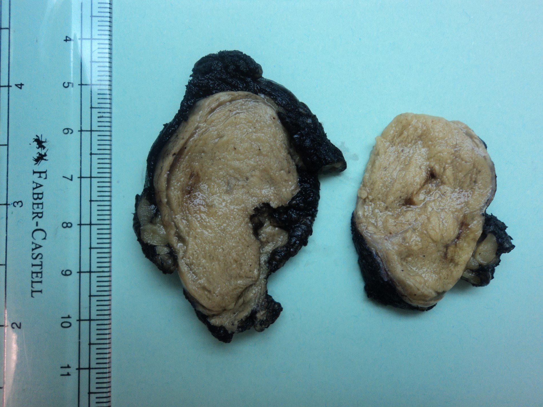
Well defined, encapsulated hibernoma
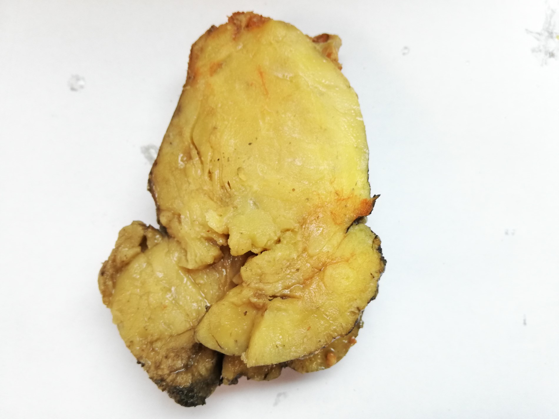
Lipoma-like hibernoma
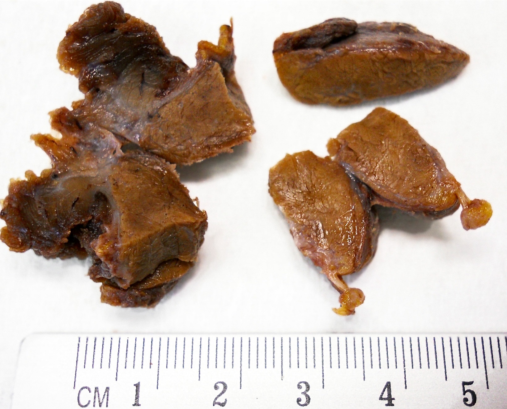
Hibernoma
- Frozen section usually reveals no evidence of malignancy (Hand (N Y) 2015;10:547)
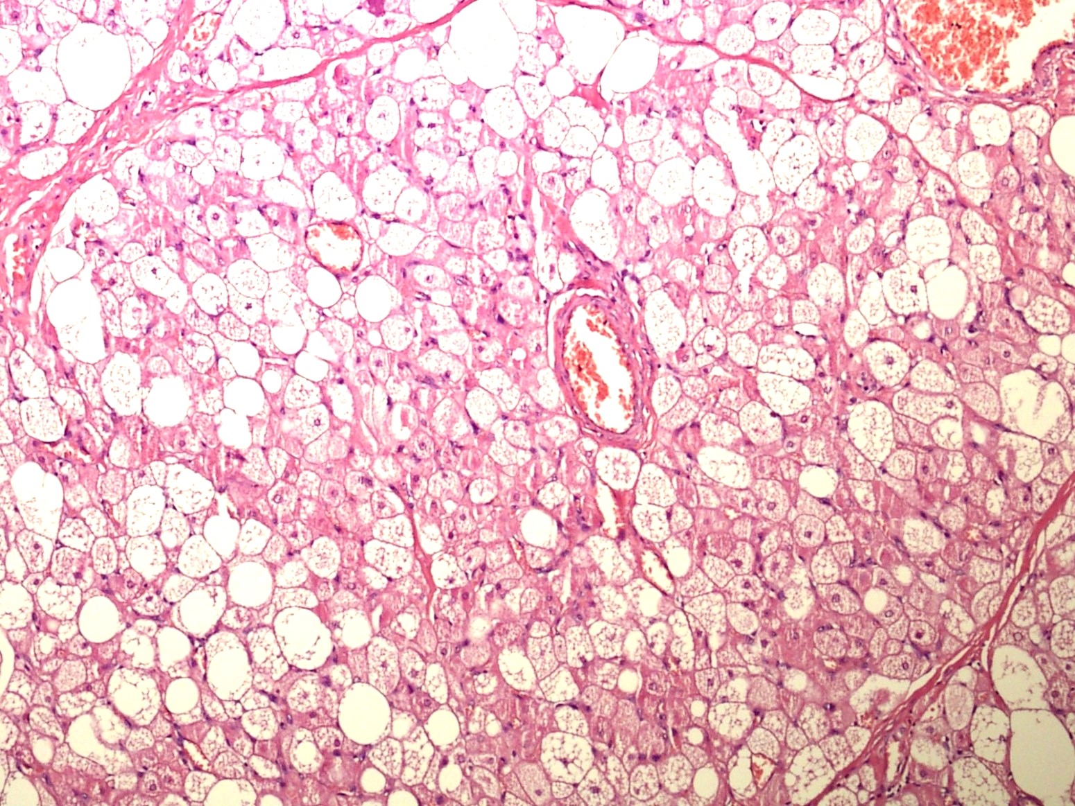
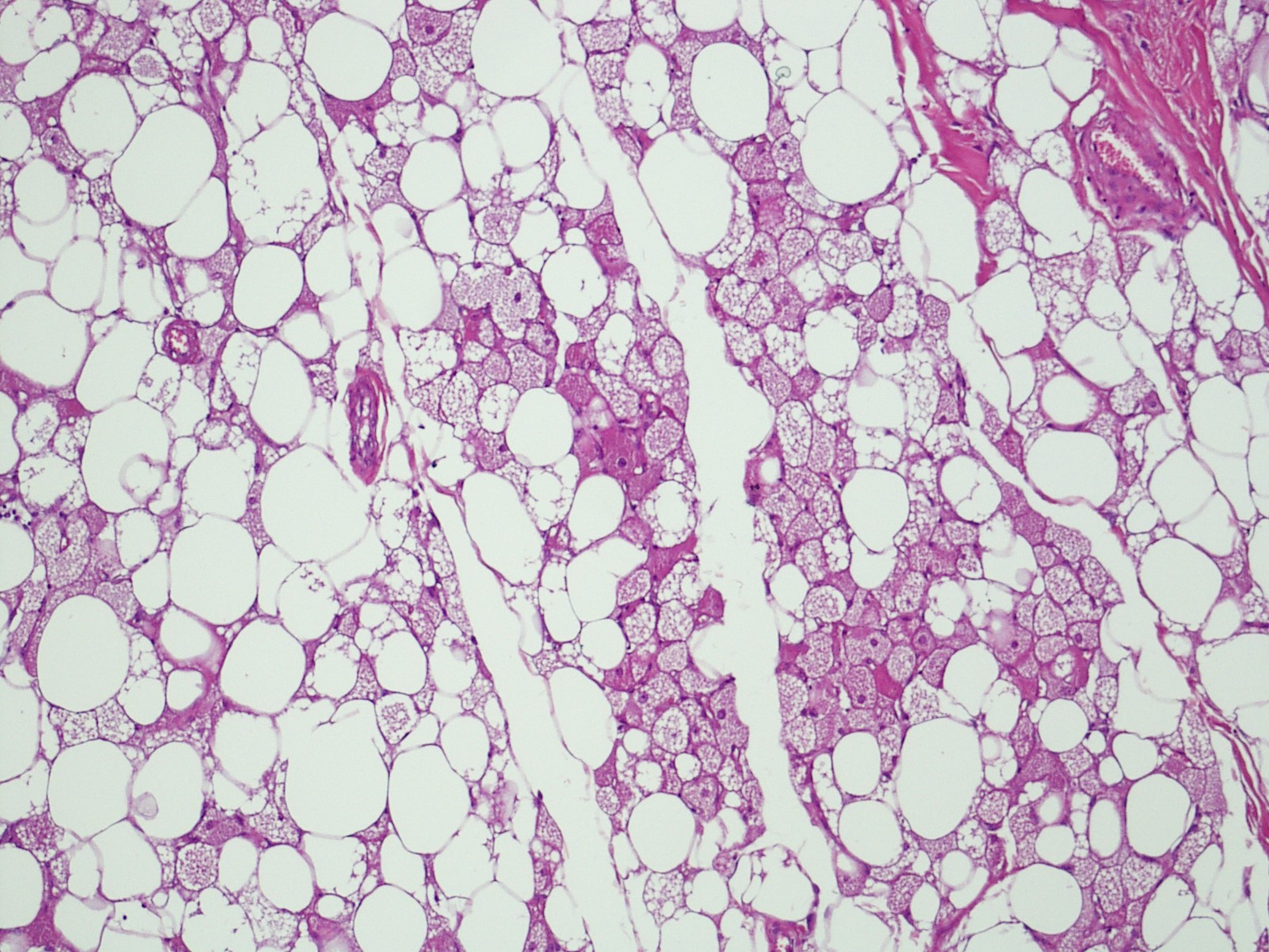
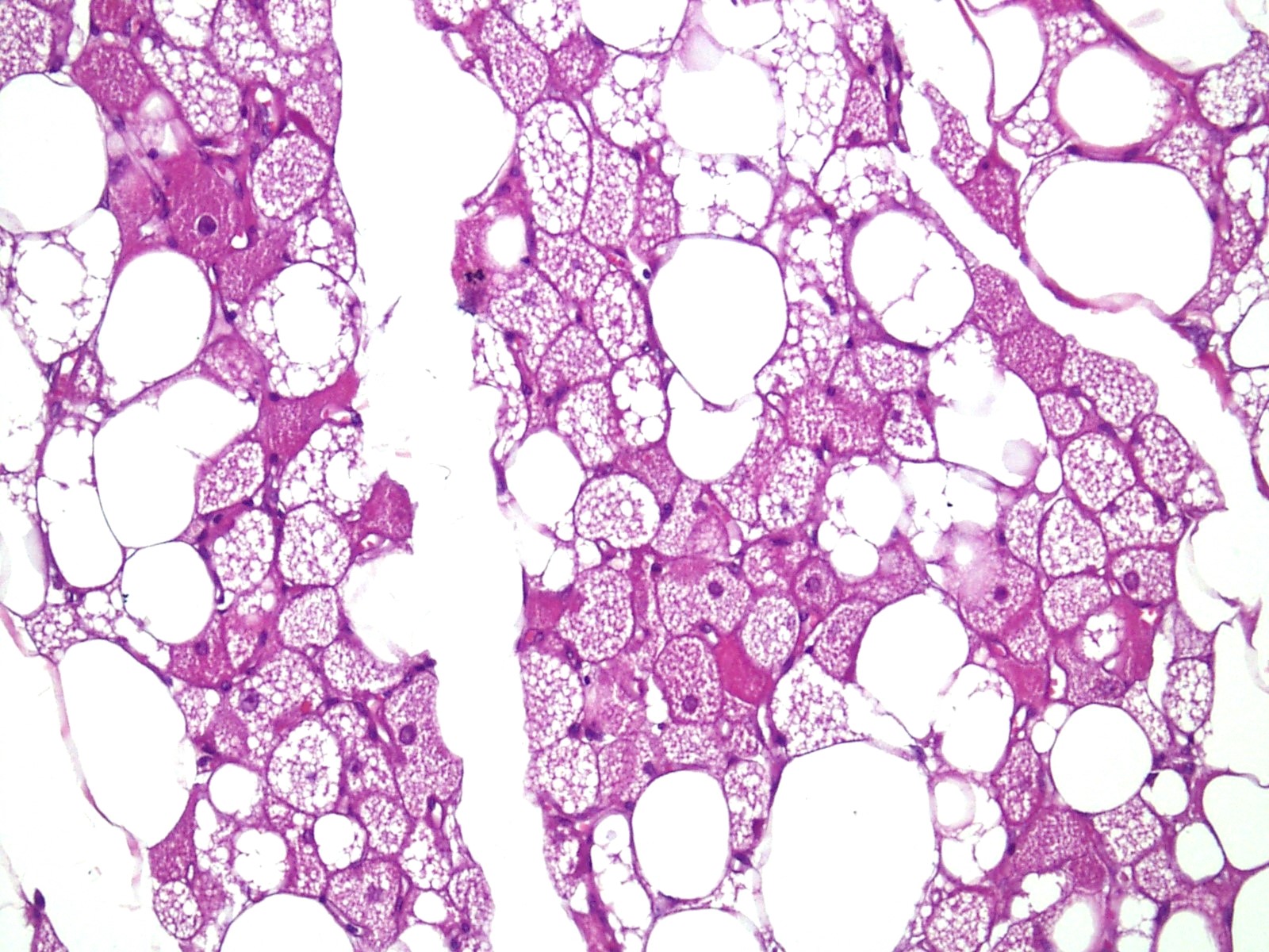
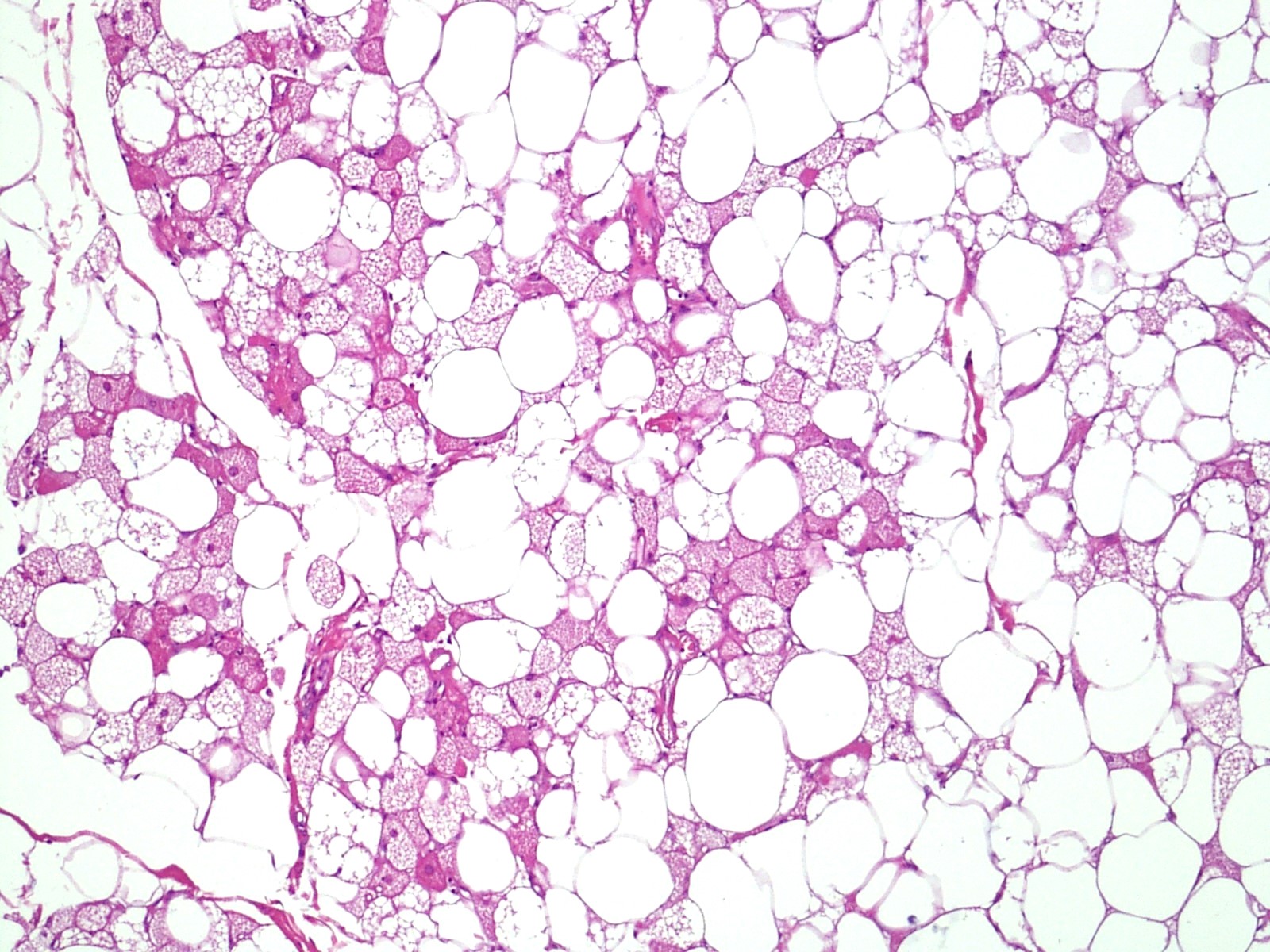
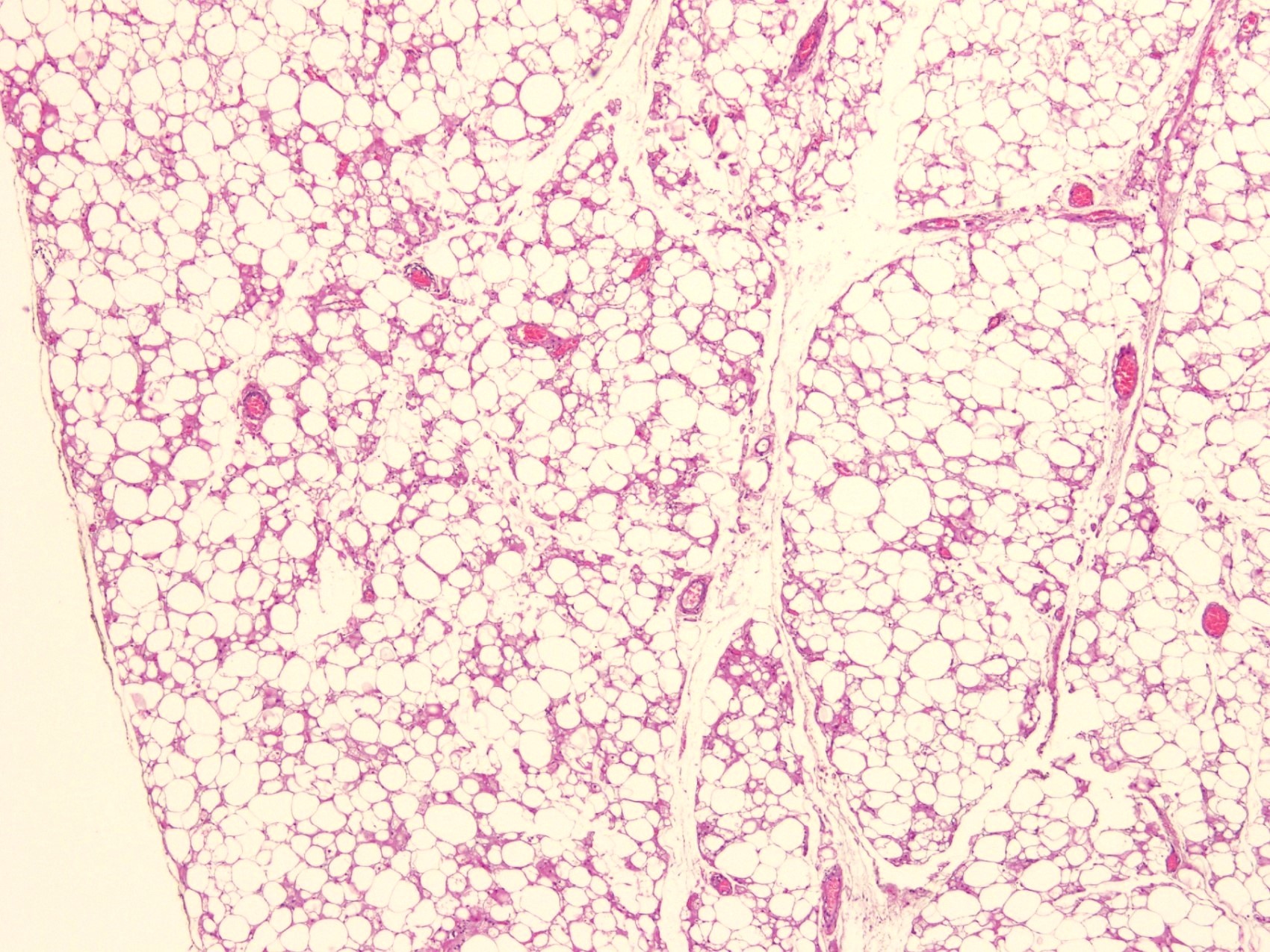
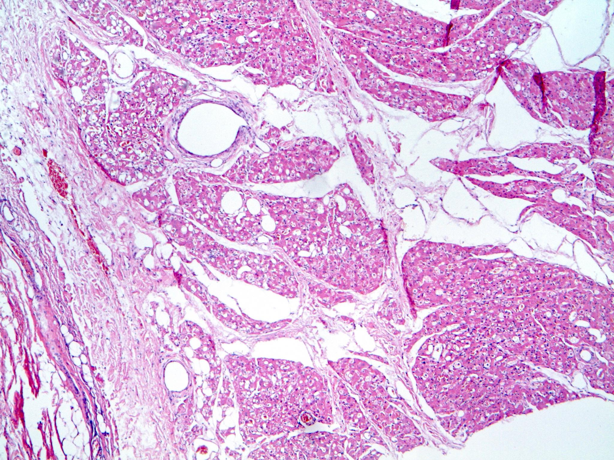
- Neoplastic lesion composed of polygonal brown fat cells with stromal cells in the background (Case Rep Oncol 2017;10:438)
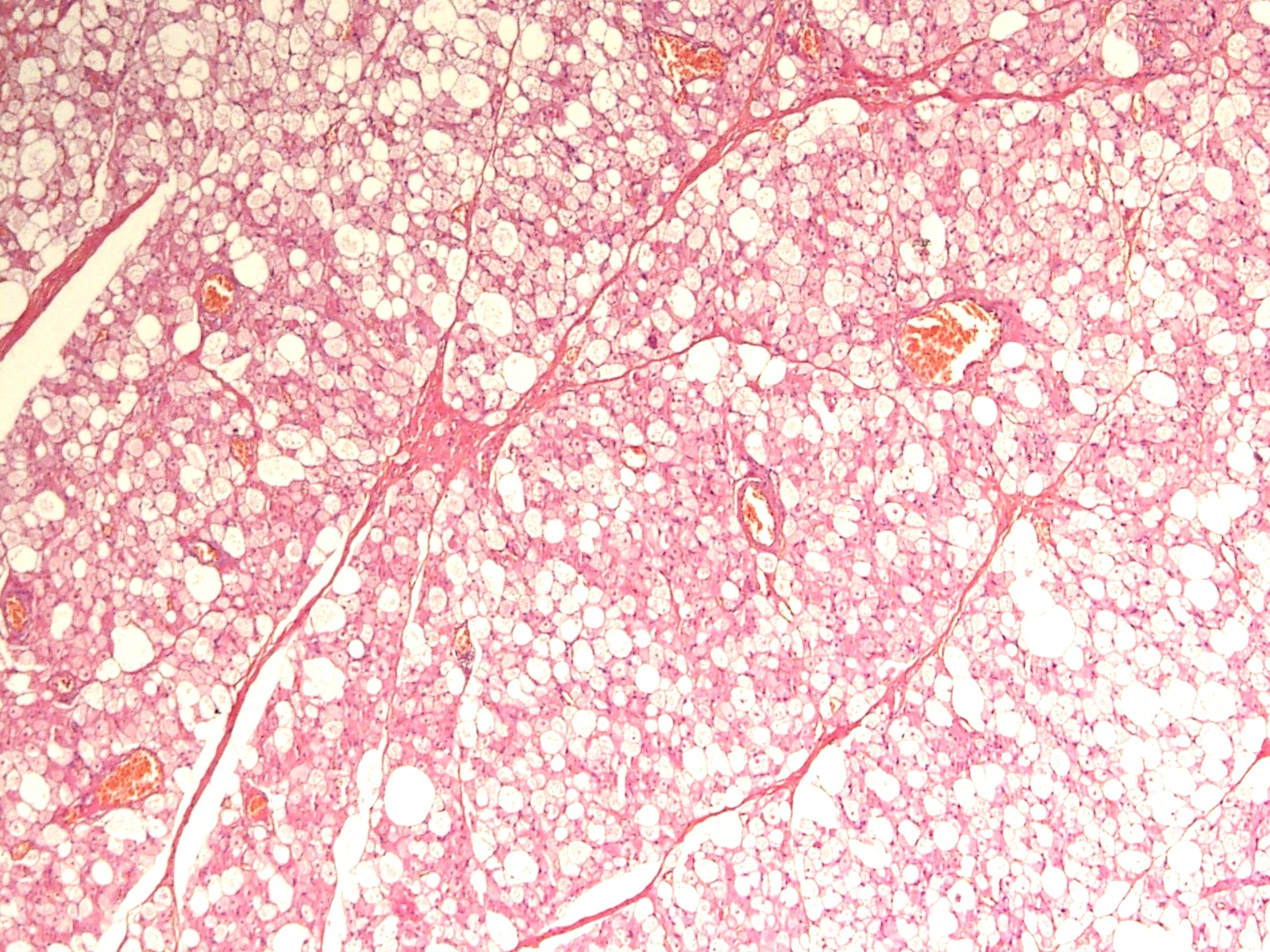
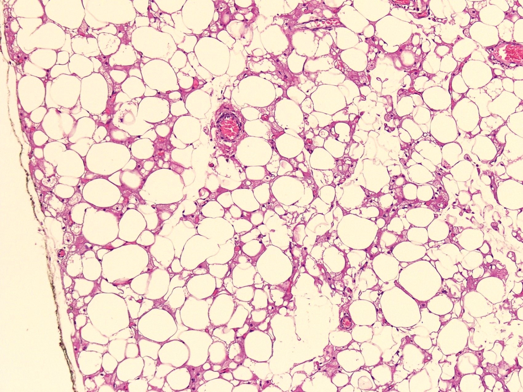
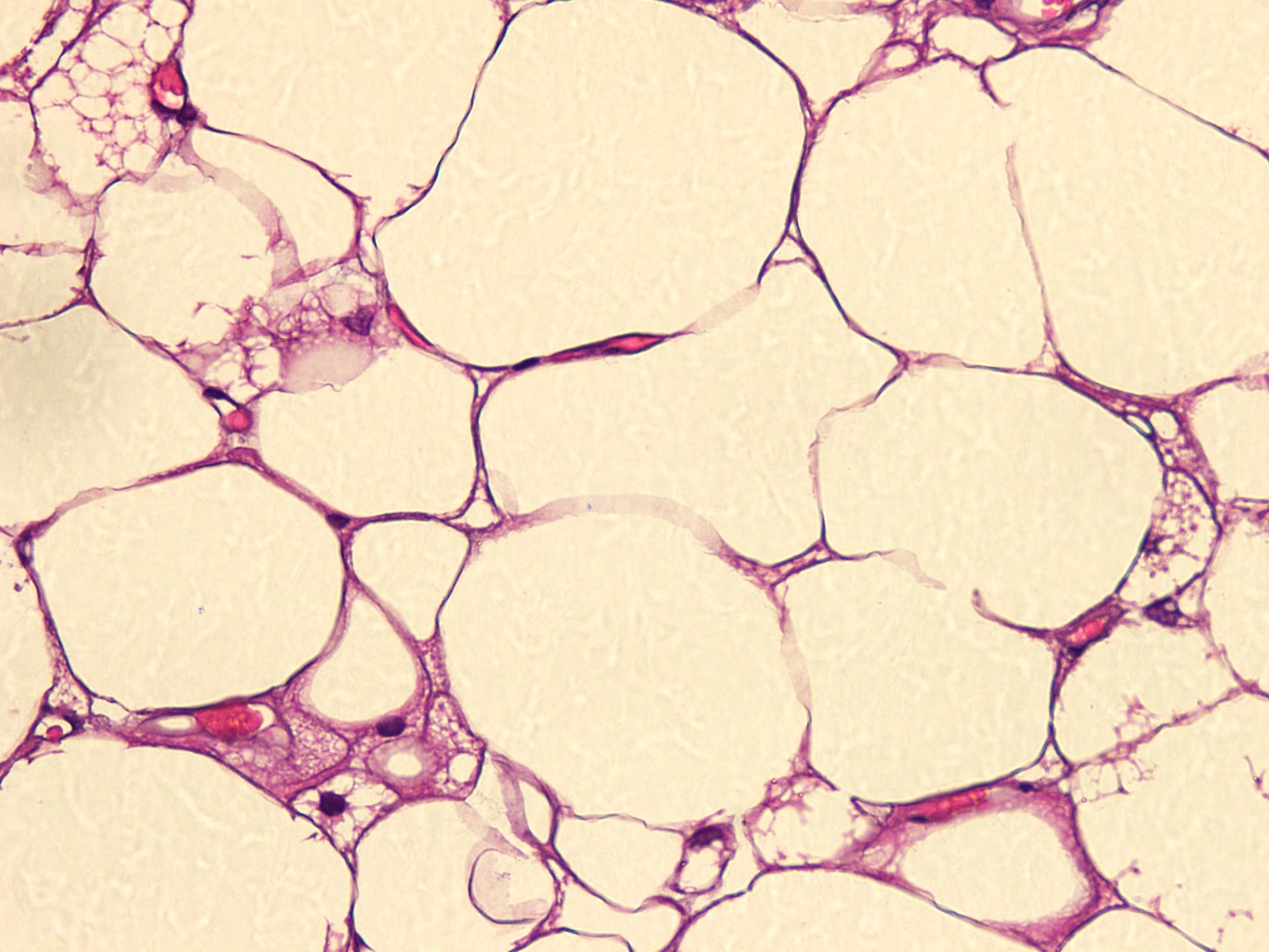
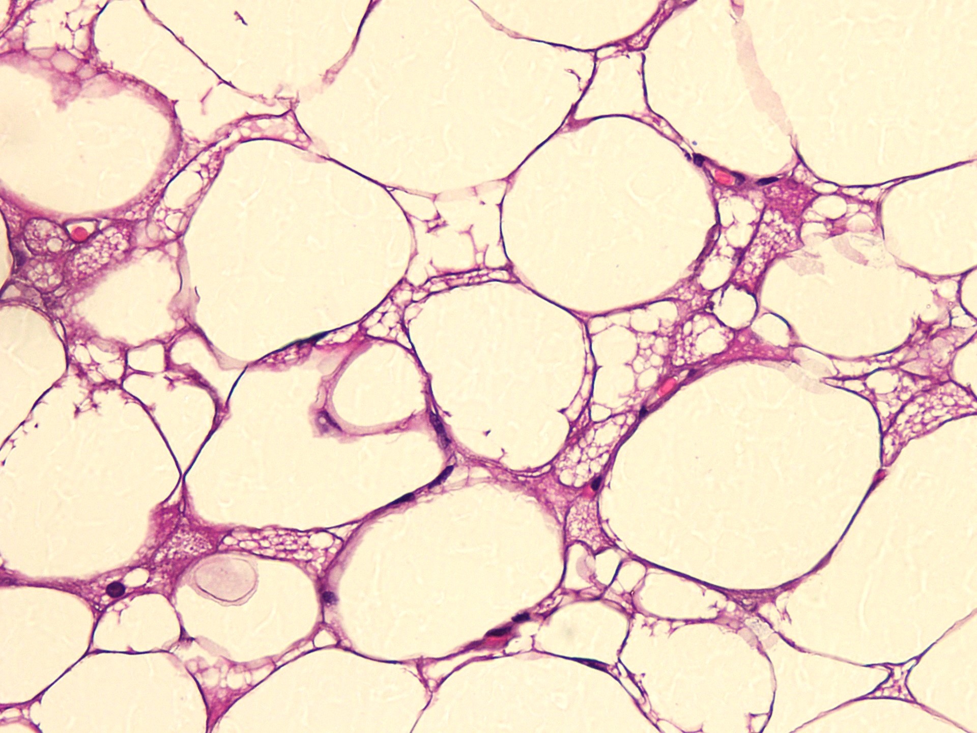
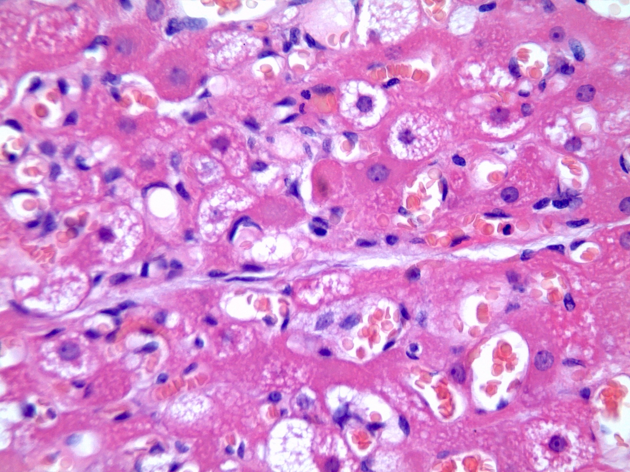
- Large number of pale and eosinophilic brown fat cells with multivacuolated, eosinophilic granular cytoplasm and small central nucleus (about 70%) admixed with variable amount of univacuolated white cells (Case Rep Oncol 2017;10:438, Am J Case Rep 2020;21:e921447, J Comput Assist Tomogr 2019;43:793, J Pathol Transl Med 2017;51:499, Am J Surg Pathol 2018;42:951, Virchows Arch 2021;478:527)
- Multivacuolations resemble lipoblasts
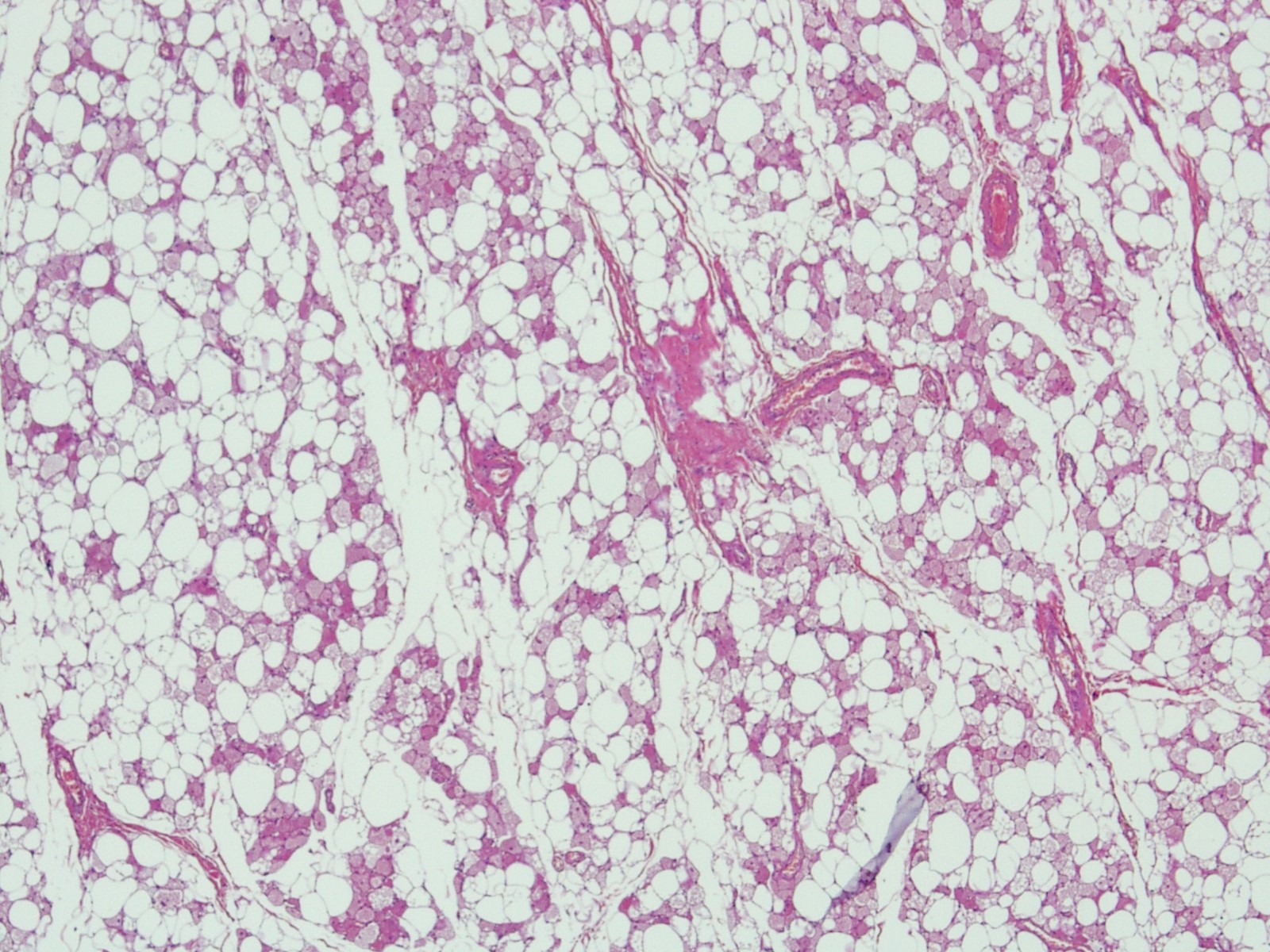
- Morphological variations or subtypes: typical, myxoid (9%), lipoma-like (7%), spindle cell (2%), thick bundles of collagen fibers, presence of mast cells and exclusively containing brown fat cells (Case Rep Oncol 2017;10:438, Am J Case Rep 2020;21:e921447, Virchows Arch 2021;478:527)
- Cytological atypia, necrosis and mitosis is unusual (J Pathol Transl Med 2017;51:499, Am J Surg Pathol 2018;42:951, BMC Surg 2021;21:30)
Contributed by Nasir Ud Din, M.B.B.S. and @Andrew_Fltv on Twitter
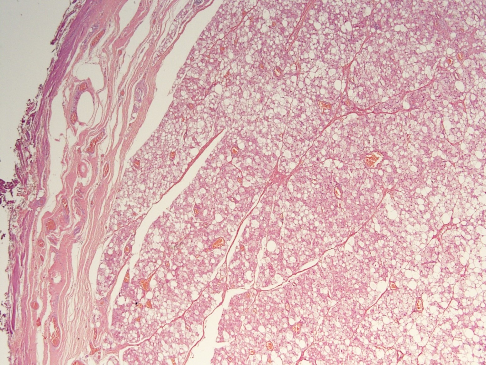
Peripheral well defined borders
Benign adipocytes
Sheets of brown cells and vasculature

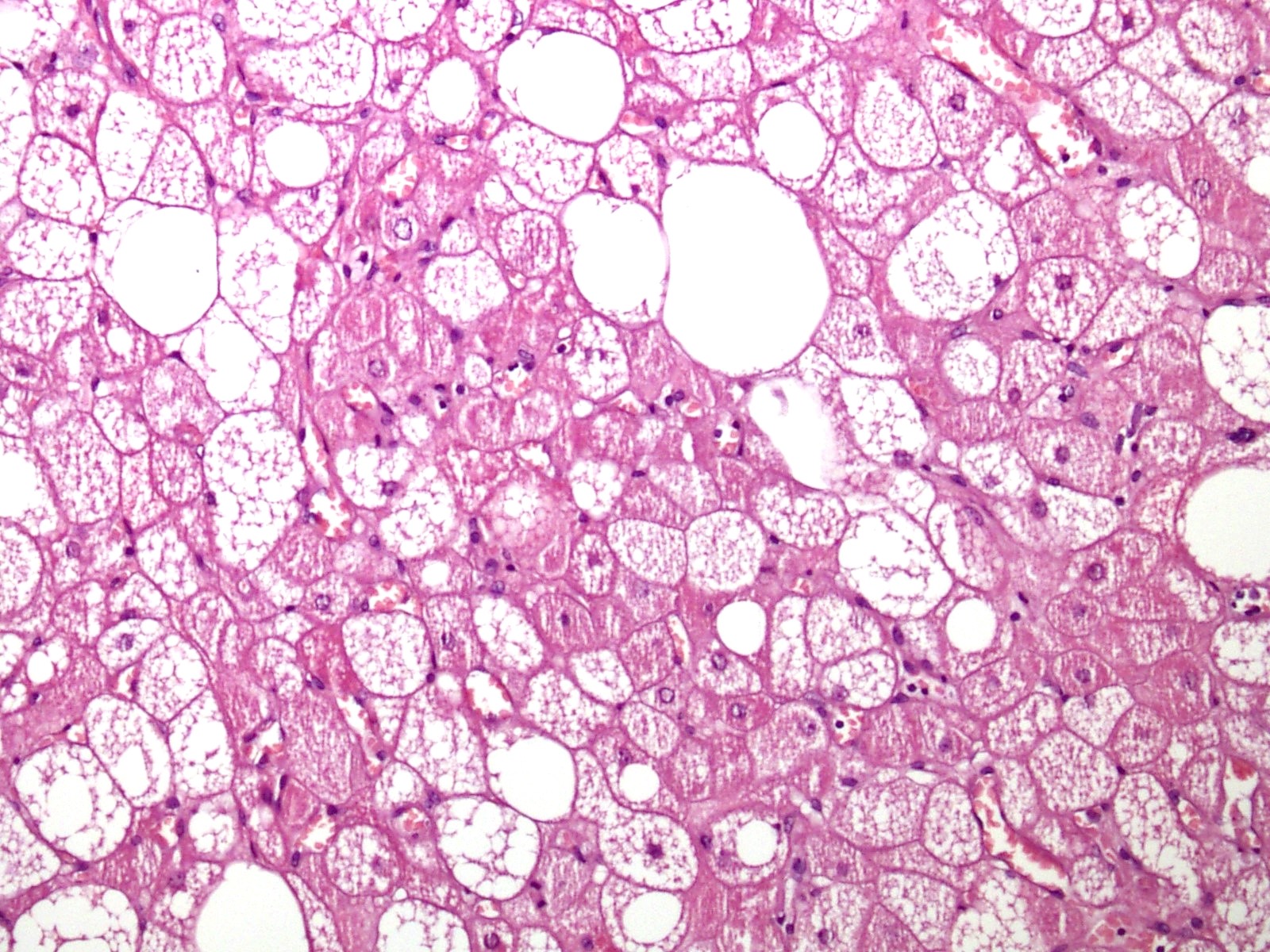
Eosinophilic granulated cells with central nuclei
Hibernoma with all benign features
Rich vascularization
Granular vacuolated cytoplasm
Multivacuolated and univacuolated cells
Brown cells and mature adipocytes
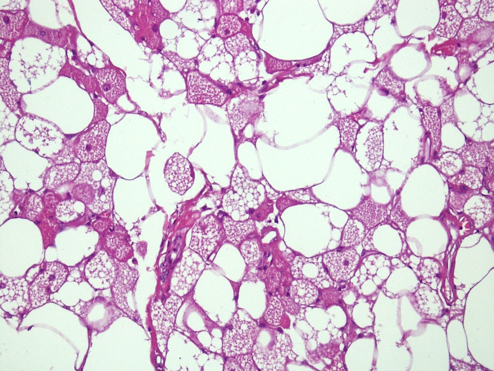
Mixture of brown and white fat cells
Lipoma-like hibernoma
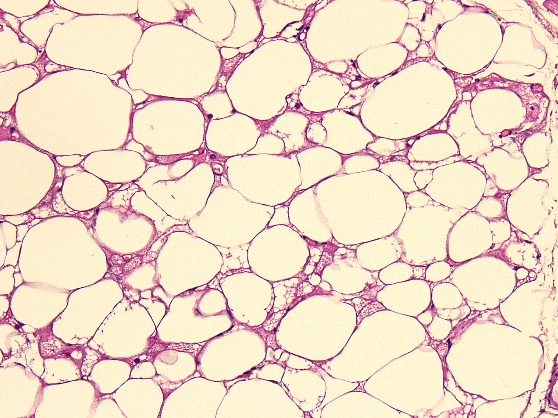
Lipoma-like hibernoma
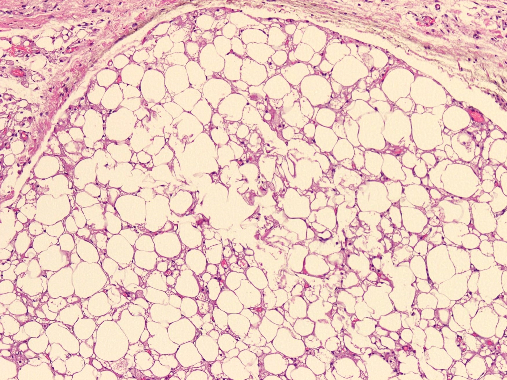
Well defined border
No signs of malignancy
Univacuolated and granular eosinophilic cells
S100

Hibernoma
- Uniform, polygonal multivacuolated cells with finely abundant granular cytoplasm, bland round nucleus and inconspicuous to prominent nucleoli
- Occasional spindle shaped cells can be present
- No nuclear atypia, mitosis or necrosis
- Benign granular cell tumor is a close differential on cytology (J Clin Diagn Res 2017;11:ED01)
Images hosted on other servers:

Regular / eccentric
nuclei, granular
cytoplasm
(Giemsa / Pap)
- S100 (Skeletal Radiol 2022;51:1325, J Pathol Transl Med 2017;51:499)
- Ki67 absent to very low (Case Rep Oncol 2017;10:438)
- CD34 (can be positive in spindle cell component) (BMC Surg 2021;21:30)
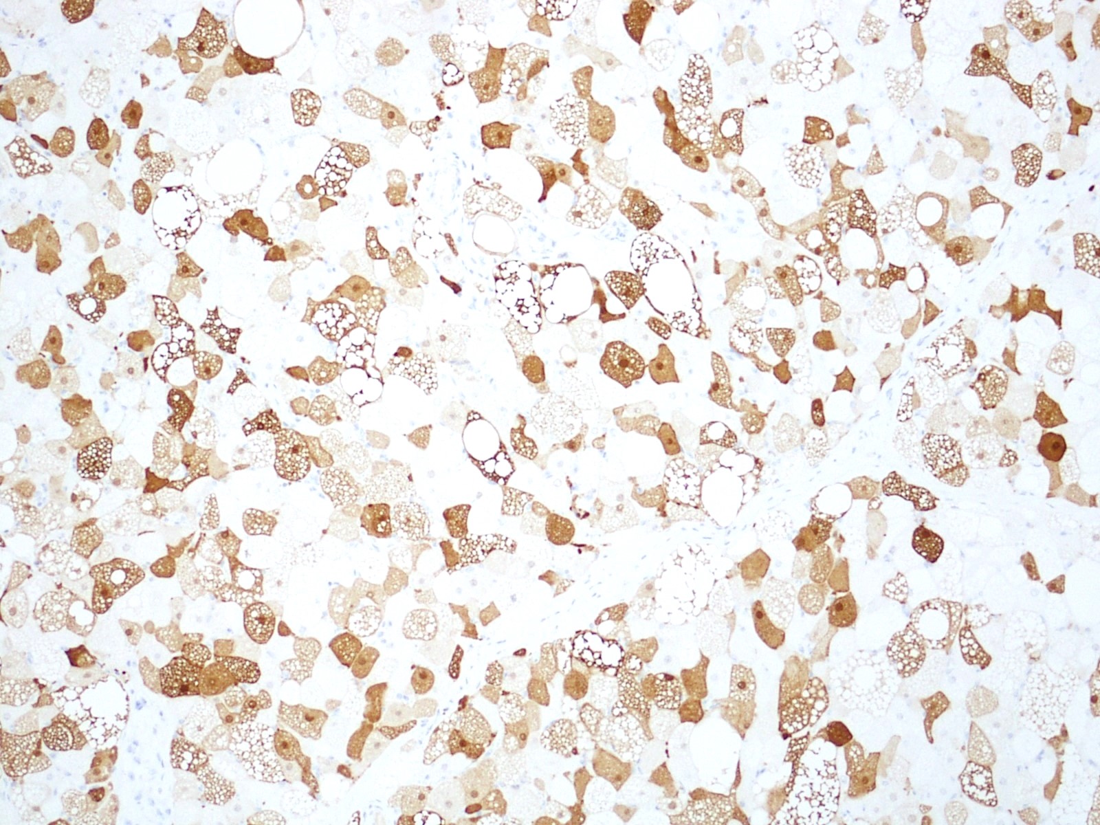
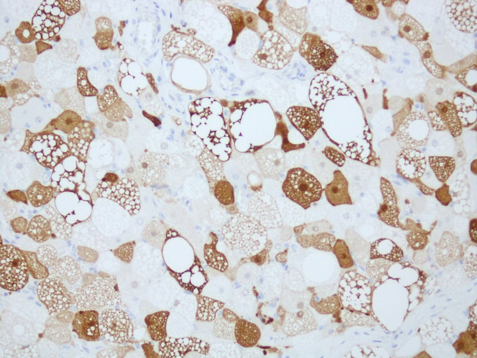
- Brown fat cells in hibernoma stain strongly with the anti-UCP1 antibody (Clin Sarcoma Res 2019;9:8)
- Periodic acid-Schiff with diastase digestion (PASD) reveals PAS positive, diastase resistant cytoplasmic granules (J Clin Diagn Res 2017;11:ED01)
- CD10 (Hum Pathol 2021;110:12)
- Hibernoma cells have several large mitochondria with high electron density matrix and dense cristae, lipid droplets, undulating plasmalemmal invaginations, pinocytic vesicles, free ribosomes, high glycogen content and small amount of smooth endoplasmic reticulum (J Clin Diagn Res 2017;11:ED01)
- Translocations and deletions involving chromosome 11q13-21, most commonly at 11q13.1 (MEN1 and AIP gene locus) (Proc Natl Acad Sci U S A 2010;107:21122)
- UCP1 expression (Clin Sarcoma Res 2019;9:8)
- One case report identified a t(9;11)(q34;q13) translocation (Case Rep Oncol 2017;10:438)
Hibernoma: 5 minute pathology pearls
- Thigh lump, excision:
- Hibernoma (see comment)
- Microscopic description: The sections reveal a well circumscribed neoplastic lesion comprising large polygonal cells arranged in sheets. These cells have multivacuolated, granular cytoplasm with small central nucleus admixed with variable amount of univacuolated cells. There is no significant cytological atypia, necrosis or mitosis.
- Comment: Hibernoma is a benign neoplasm with no significant potential for recurrence.
- Lipoma:
- Occurs in adults aged 40 - 60 years
- Preferably subcutaneous, deep or on bone surface
- Lobules of mature adipocytes (Radiol Case Rep 2022;17:2477)
- Xanthoma:
- Usually occurs in adults
- Smaller cells than hibernoma
- Presence of foamy histiocytes, fibroblasts, inflammatory cells and local Touton giant cells
- No lobulations
- Granular cell tumor:
- Presents in adults in fourth to sixth decade
- Females slightly more than males
- Preferred sites are head and neck, breast, proximal extremities
- Polygonal cells with abundant eosinophilic granular cytoplasm but lack lipid vacuoles
- Adult rhabdomyoma:
- Commonly in head and neck
- Cells with granular cytoplasm
- Phosphotungstic acid hematoxylin and Masson staining show cytoplasmic striae, crystals and rod inclusions in the tumor cells
- Desmin and myogenin positive (J Oral Maxillofac Pathol 2019;23:54)
- Lipoblastoma:
- Found in infants and young children
- Occurs in trunk and extremities
- Contains mature and immature adipocytes separated by connective tissue septa of varying thickness
- PLAG1 rearrangement by FISH
- Atypical lipomatous tumor / well differentiated liposarcoma:
- Occurs in middle aged adults
- Usually at deep soft tissue extremities, retroperitoneum, paratesticular region and mediastinum
- Exhibits nuclear hyperchromasia, atypia, larger cytoplasmic vacuoles and lipoblasts
- Carries 12q13-15 region (MDM2 / CDK4) amplifications (Case Rep Oncol 2017;10:438, Am J Surg Pathol 2018;42:951, Cureus 2020;12:e9111)
- Myxoid liposarcoma:
- Presents in young adults, peak incidence in fourth to fifth decade of life
- Involves deep soft tissue extremities, preferably thigh
- Resembles myxoid variant of hibernoma
- Prominent plexiform capillary pattern
- Molecular translocation t(12;16)(q13;p11.2) (FUS::DDIT3) (Am J Case Rep 2020;21:e921447, BMC Surg 2021;21:30, Am J Surg Pathol 2018;42:951, J Surg Case Rep 2021;2021:rjaa304)
- Intraosseous hibernoma differential diagnosis includes:
A 32 year man presented with a mass in his right thigh; MRI revealed circumscribed lobulated lesion and microscopy is shown in the picture above. What is the diagnosis?
- Atypical lipomatous tumor
- Fat necrosis
- Hibernoma
- Lipoblastoma
- Lipoma
- Anders syndrome
- Gardner syndrome
- Lynch syndrome
- MEN1 syndrome
- MEN2 syndrome
