

Soft tissue
Fibroblastic / myofibroblastic
Calcifying fibrous tumor
Editor-in-Chief: Debra L. Zynger, M.D.


Last author update: 9 April 2020
Last staff update: 9 April 2020
Copyright: 2002-2024, PathologyOutlines.com, Inc.
PubMed Search: Calcifying fibrous tumor[TI]
Table of Contents
Definition / general | Essential features | Terminology | ICD coding | Epidemiology | Sites | Pathophysiology | Etiology | Diagrams / tables | Clinical features | Diagnosis | Radiology images | Prognostic factors | Case reports | Treatment | Clinical images | Gross description | Gross images | Microscopic (histologic) description | Microscopic (histologic) images | Positive stains | Negative stains | Electron microscopy description | Sample pathology report | Differential diagnosis | Board review style question #1 | Board review style answer #1Cite this page: Mruthyunjayappa S, Dhall D. Calcifying fibrous tumor. PathologyOutlines.com website. https://www.pathologyoutlines.com/topic/softtissuecalcifyingfibrous.html. Accessed May 12th, 2024.
Definition / general
- Benign fibrous lesion with abundant hyalinized collagen, psammomatous or dystrophic calcifications and lymphoplasmacytic infiltration
Essential features
- Paucicellular
- Bland spindle cells in a collagenous tissue
- Calcifications and inflammatory infiltrate
Terminology
- First described as childhood fibrous pseudotumor with psammoma bodies in 1988 (Arch Pathol Lab Med 1988;112:798)
- Calcifying fibrous pseudotumor
ICD coding
- ICD-10: D21.9 - benign neoplasm of connective and other soft tissue, unspecified
Epidemiology
- Adolescents / young adults
- M:F = 1:1.27
Sites
- Most common sites: tubular gastrointestinal tract, solid organs, peritoneal and pleural surfaces but can occur anywhere
Pathophysiology
- Calcifying fibrous tumor may represent different stages of IgG4 related disease and fits with the unifying concept of IgG4 related pseudotumor
- Recently, gastrointestinal calcifying fibrous tumor has been thought to be a gastrointestinal lesion of immunoglobulin 4 (IgG4) related disease (Surg Case Rep 2019;5:150)
Etiology
- Previous infection, history of trauma and surgical intervention
- No definitive mechanisms or causes have been confirmed
Diagrams / tables
Images hosted on other servers:

Sites
Clinical features
- Patients can present with various symptoms depending on the location of the tumor
Diagnosis
- Histologic examination of tissue
Radiology images
Images hosted on other servers:

CT scan

Gastrohepatic ligament tumor

Esophageal tumor
Prognostic factors
- Local recurrence rate is ~10%
Case reports
- 20 year old man with incidentally found mass in the ileal mesentery (J Cancer Res Ther 2011;7:500)
- 23 year old woman with recurrent pleural effusions (Case Reports 2018;2018:bcr-2018-226282)
- 32 year old man with incidental adrenal gland lesion (Mol Clin Oncol 2016;5:252)
- 43 year old man presenting with pain in abdomen (APMIS 2015;123:72)
Treatment
- Excision
Clinical images
Images hosted on other servers:

Endoscopy of gastric tumor

Endoscopy of ileum tumor
Gross description
- Single or multiple
- Well circumscribed but unencapsulated
- Spherical to lobulated mass with a solid, white to gray, gritty cut surface
- Variable size, may infiltrate into surrounding tissue (Biomed Res Int 2019;2019:5026860)
Gross images
Contributed by Mary Wong, M.D.
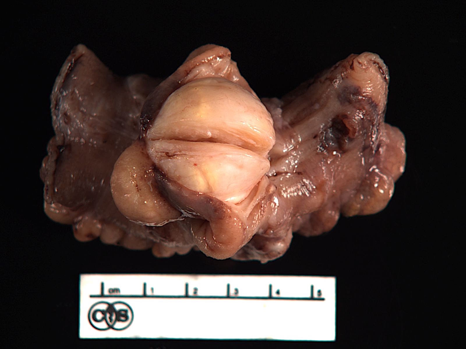
Ileum tumor
Images hosted on other servers:

Gastrohepatic ligament tumor

Ileum tumor
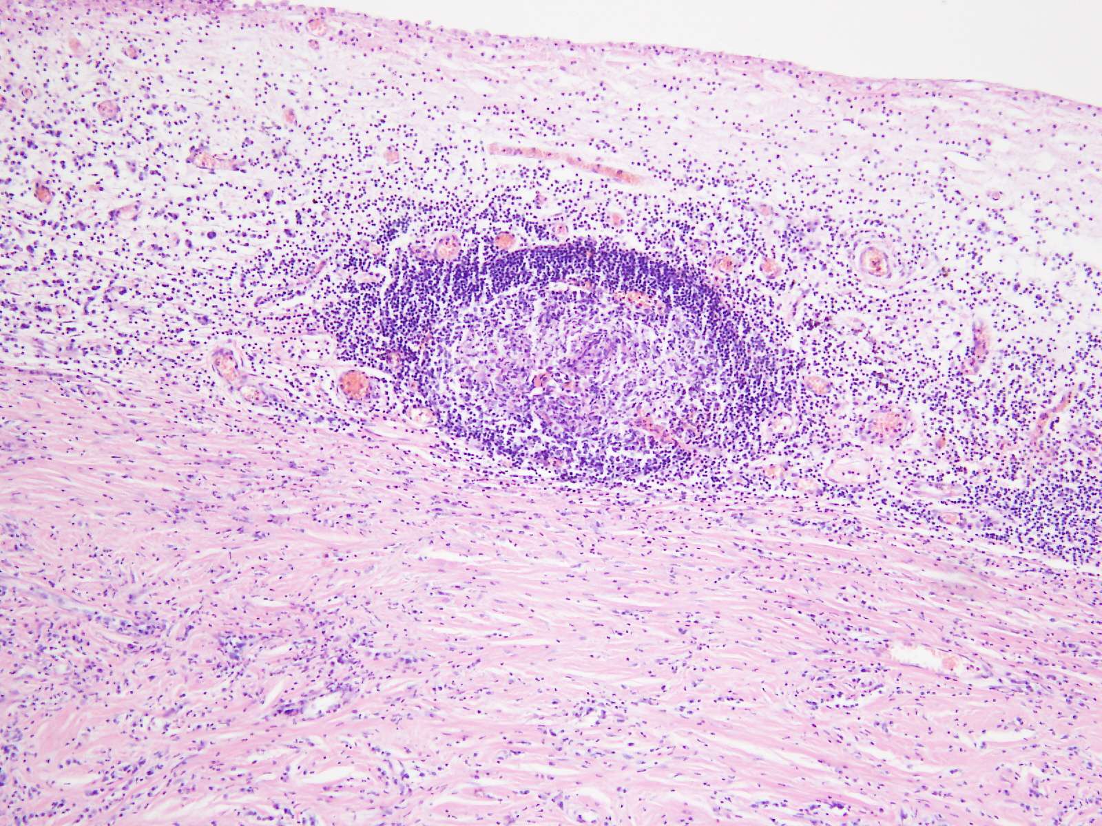
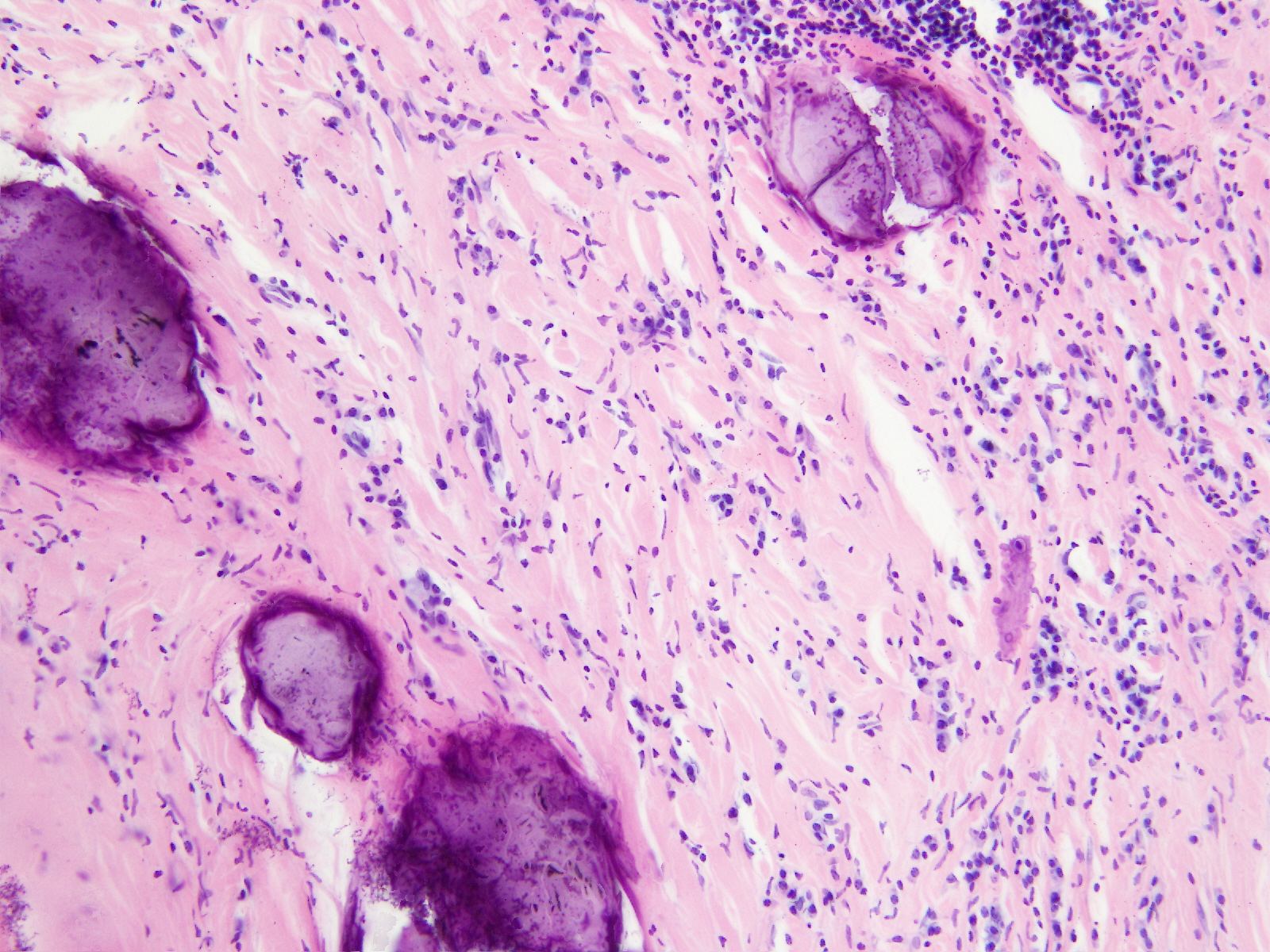
Microscopic (histologic) description
- Paucicellular fibroblastic proliferation with bland spindle cells embedded in dense collagenous tissue
- Varying degrees of lymphocytes (possibly lymphoid follicles), plasma cells
- Scattered dystrophic or psammomatous calcification (Biomed Res Int 2019;2019:5026860)
Microscopic (histologic) images
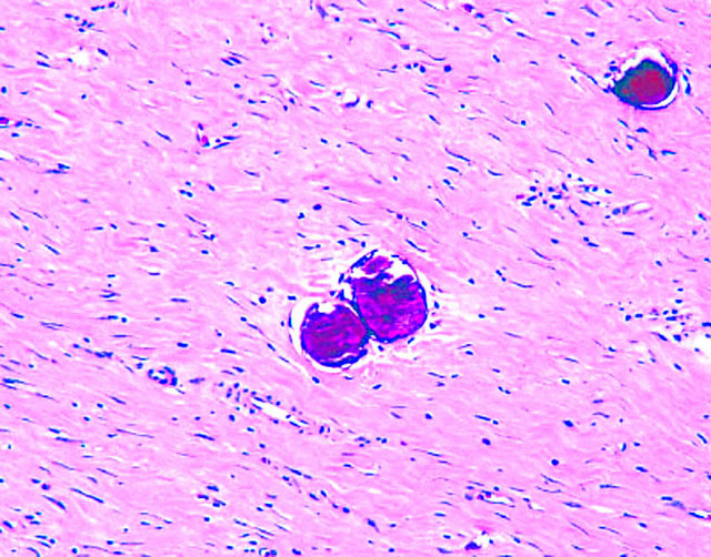
Contributed by Deepti Dhall, M.D.
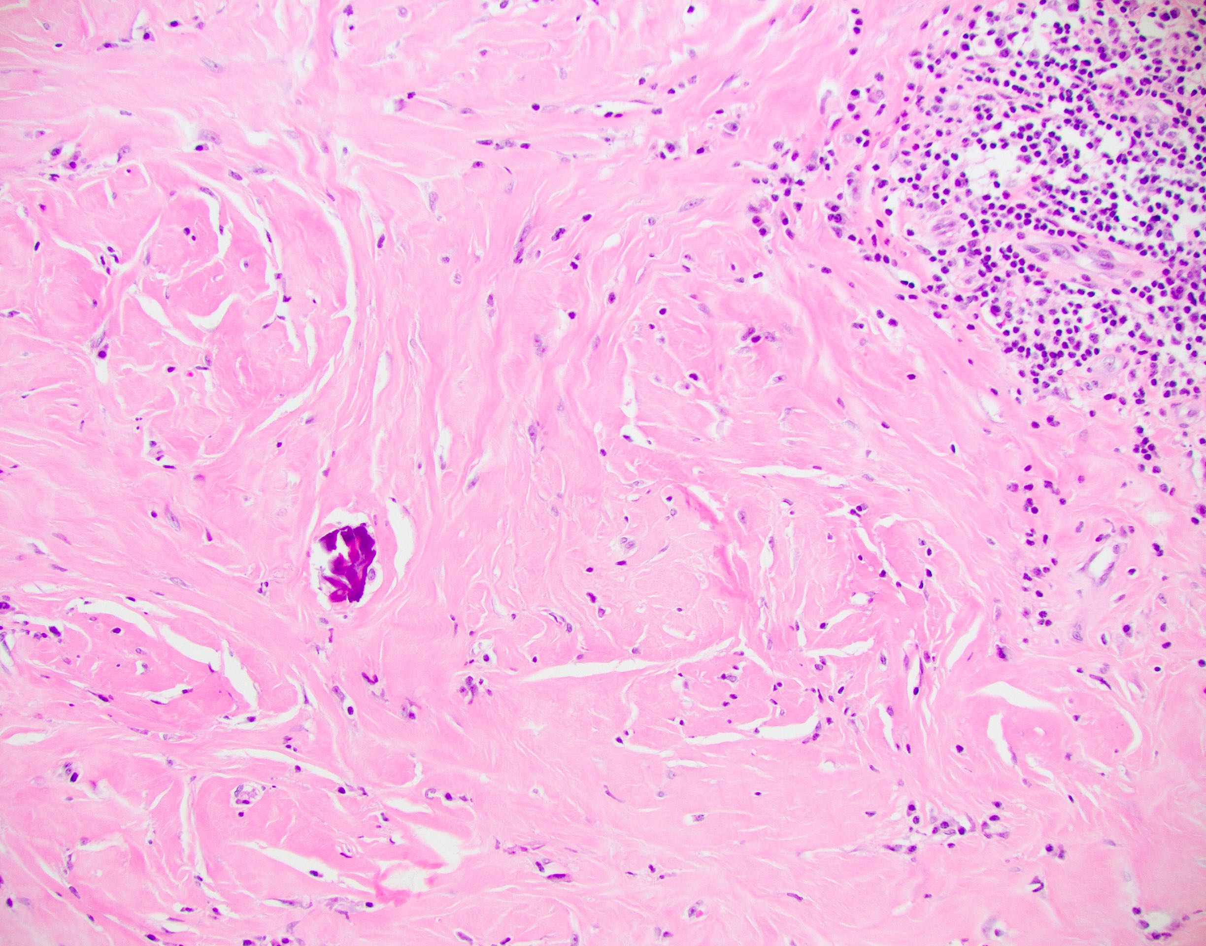
Bland spindle cells and inflammation

Bland spindle cells and calcification
Case #249


Omental tumor
Positive stains
Negative stains
Electron microscopy description
- Immature fibroblastic cells, collagen fibrils, dystrophic and psammomatous calcifications
Sample pathology report
- Ileum, resection:
- Calcifying fibrous tumor, 3.5 cm in greatest dimension
- Surgical margins are negative
Differential diagnosis
- Inflammatory myofibroblastic tumor:
- More cellular, no calcifications
- ALK+, actin+, CD34+ and focally factor XIIIa+ (Mod Pathol 2001;14:784, Int J Surg Pathol 2002;10:189)
- Gastrointestinal stromal tumor (GIST):
- Calcifying aponeurotic fibroma:
- Usually distal location, usually smaller lesion
- More cellular
- Desmoplastic fibroblastoma:
- Older patients
- Low cellularity, larger prominent fibroblasts, no microcalcifications, no prominent inflammatory infiltrate
- Idiopathic retroperitoneal fibrosis and related sclerosing fibroinflammatory lesions:
- More inflammation, especially plasma cells and eosinophils
Board review style question #1
A 30 year old man presents with abdominal pain and a CT reveals a mass within the gastrointestinal system, which is resected. Histopathologic examination reveals paucicellular spindle cells with dystrophic calcifications and lymphoid aggregates. Immunohistochemical stains for CD34 and vimentin are positive; CD117, DOG1, SMA and ALK1 are negative. Which of the following is the diagnosis for this patient?
- Calcifying fibrous tumor
- Desmoplastic fibroblastoma
- Gastrointestinal stromal tumor
- Inflammatory myofibroblastic tumor
Board review style answer #1
