Skin nonmelanocytic tumor
Neural tumors
Palisaded encapsulated neuroma
Authors: Elaine Kunzler, M.D., Bethany R. Rohr, M.D.
Board of reviewers: Caroline I. Mullins, M.D.


Last author update: 3 April 2025
Last staff update: 3 April 2025
Copyright: 2003-2025, PathologyOutlines.com, Inc.
PubMed search: Palisaded encapsulated neuroma
Table of Contents
Definition / general | Essential features | Terminology | ICD coding | Epidemiology | Sites | Etiology | Clinical features | Diagnosis | Prognostic factors | Case reports | Treatment | Clinical images | Microscopic (histologic) description | Microscopic (histologic) images | Virtual slides | Cytology description | Positive stains | Negative stains | Electron microscopy description | Videos | Sample pathology report | Differential diagnosis | Additional references | Practice question #1 | Practice answer #1 | Practice question #2 | Practice answer #2Cite this page: Kunzler E, Rohr BR. Palisaded encapsulated neuroma. PathologyOutlines.com website. https://www.pathologyoutlines.com/topic/skintumornonmelanocyticpalisadedencapsulatedneuroma.html. Accessed September 18th, 2025.
Definition / general
- Palisaded encapsulated neuroma (PEN) is a benign, well circumscribed tumor consisting of Schwann cells and axons
- The term is misleading as nuclear palisading is not always present and the capsule may be partial or inconspicuous
Essential features
- Palisaded encapsulated neuromas are superficial dermal tumors, which often present as solitary lesions on the face or near mucocutaneous junctions
- Histopathologic features include a well circumscribed dermal tumor of bland spindled cells arranged in bundles
- Clefting is often seen between the tumor and surrounding dermis
- Palisaded encapsulated neuromas can be differentiated from schwannomas by their superficial depth and lack of Antoni A and B areas
- They can be differentiated from neurofibromas due to their fascicular arrangement and presence of clefting
Terminology
- PEN is also known as solitary circumscribed neuroma
- This name is preferred by some due to the misleading terminology of PEN
ICD coding
Epidemiology
- Most common in middle aged adults (Arch Dermatol 1972;106:865)
- No gender predilection
- Rare pediatric cases have been reported (Pediatr Dermatol 2014;31:e107)
Sites
- Lower face, near mucocutaneous borders and genitalia
- Occasionally reported on the trunk and acral sites
Etiology
- Usually sporadic with unknown etiology
- Rare cases diagnosed in patients with genodermatoses including Cowden syndrome and neurofibromatosis (Pediatr Dermatol 2017;34:e219, Int J Surg Pathol 2023;31:734)
Clinical features
- Usually a solitary, sessile, skin colored papule on the face (Indian Dermatol Online J 2018;9:262)
- Often asymptomatic and slow growing
- Rarely presents with multiple papules
Diagnosis
- Skin biopsy is required
Prognostic factors
- Recurrence rates are minimal after excision (Indian Dermatol Online J 2018;9:262)
Case reports
- 50 year old man with a solitary papule on the vermillion of the upper lip (JAAD Case Rep 2024;49:1)
- 50 and 56 year old women with left facial papules (Oxf Med Case Reports 2024;2024:omae003)
- 52 year old man with a papule on the glans penis (Dermatol Online J 2020;26:13030)
- 56 year old man with multiple skin colored acral papules (J Cutan Pathol 2022;49:82)
Treatment
- Benign; no treatment required
- Residual PEN can be observed or excised
Clinical images
Images hosted on other servers:

Papule on the lip

Multiple facial papules
Microscopic (histologic) description
- Well circumscribed superficial dermal tumor of spindled cells (Arch Dermatol 1972;106:865)
- Clefting is often seen between the tumor and dermis
- Spindled Schwann cells and axons are arranged in bundles with minimal atypia
- Occasionally, spindled cells are epithelioid or demonstrate nuclear palisading
- Occasionally, a thin, delicate capsule can be observed or dense fibrous tissue surrounds the tumor
- Stroma is variably myxoid
- Close association with adnexal structures in facial lesions
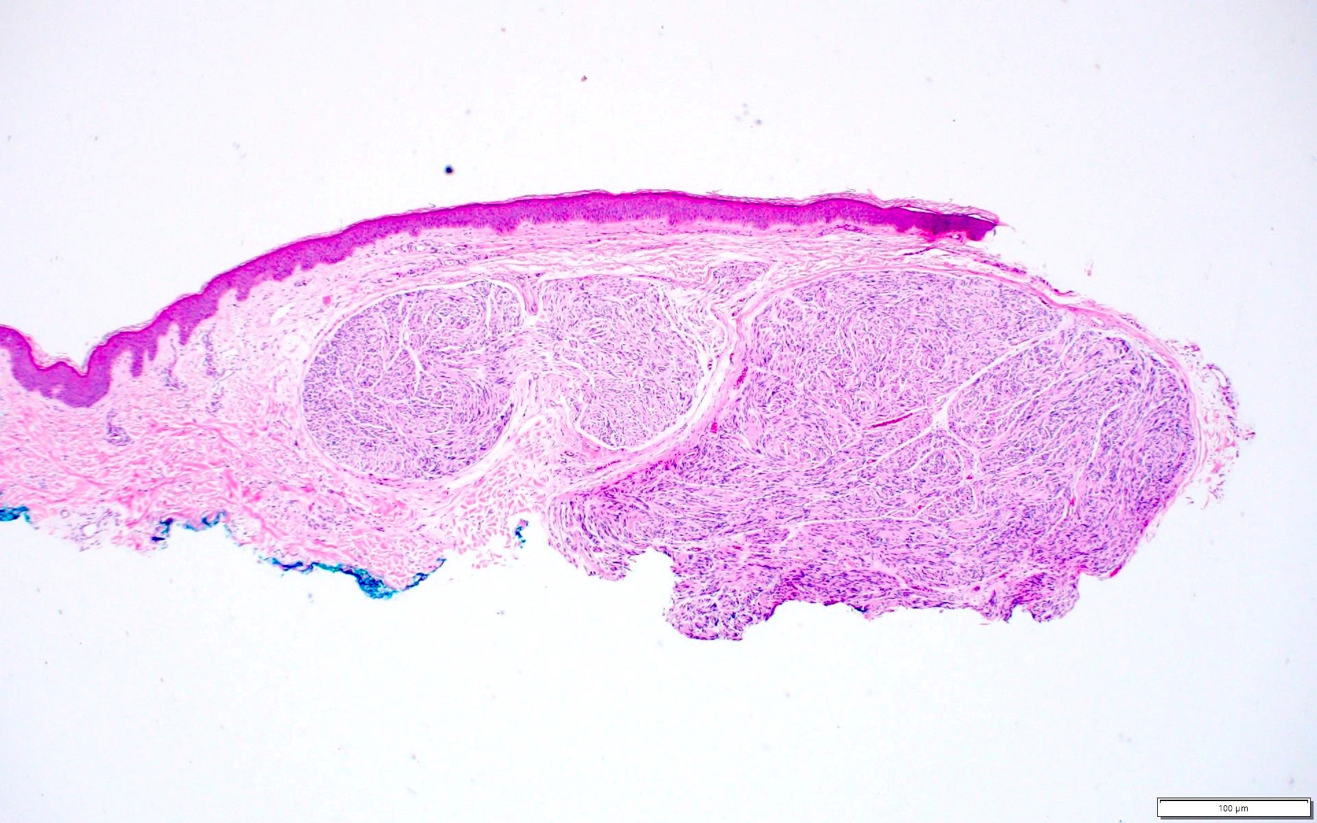
- Plexiform and multilobular patterns have been described (Int J Surg Pathol 2019;27:506)
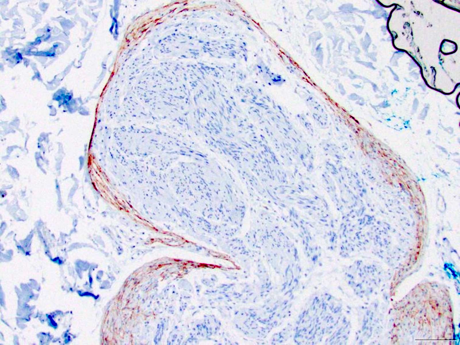
Microscopic (histologic) images
Contributed by Elaine Kunzler, M.D. and Bethany R. Rohr, M.D.
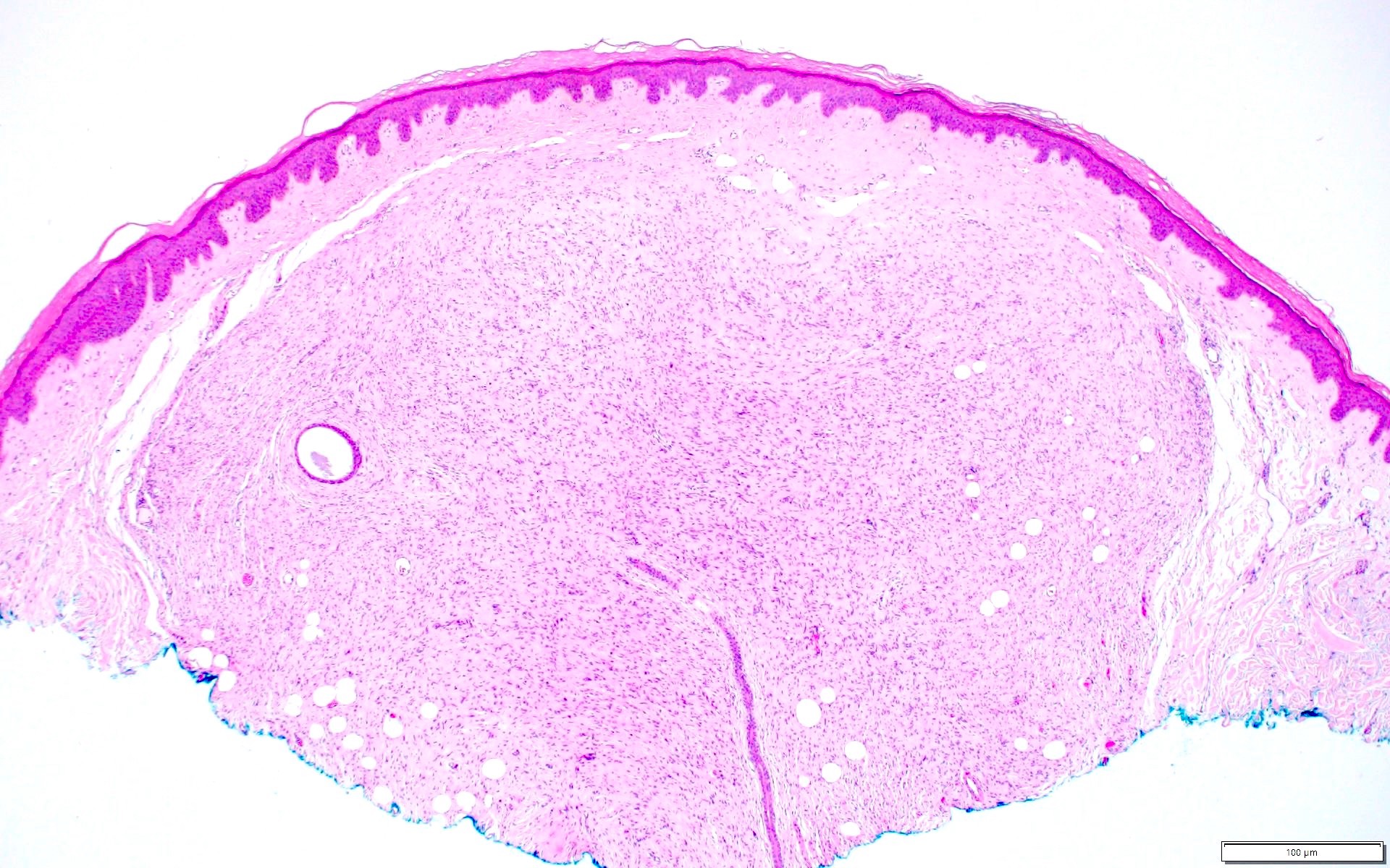
Sharply circumscribed dermal tumor
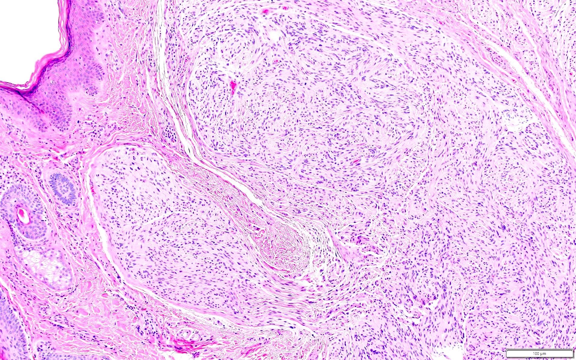
Schwann cells
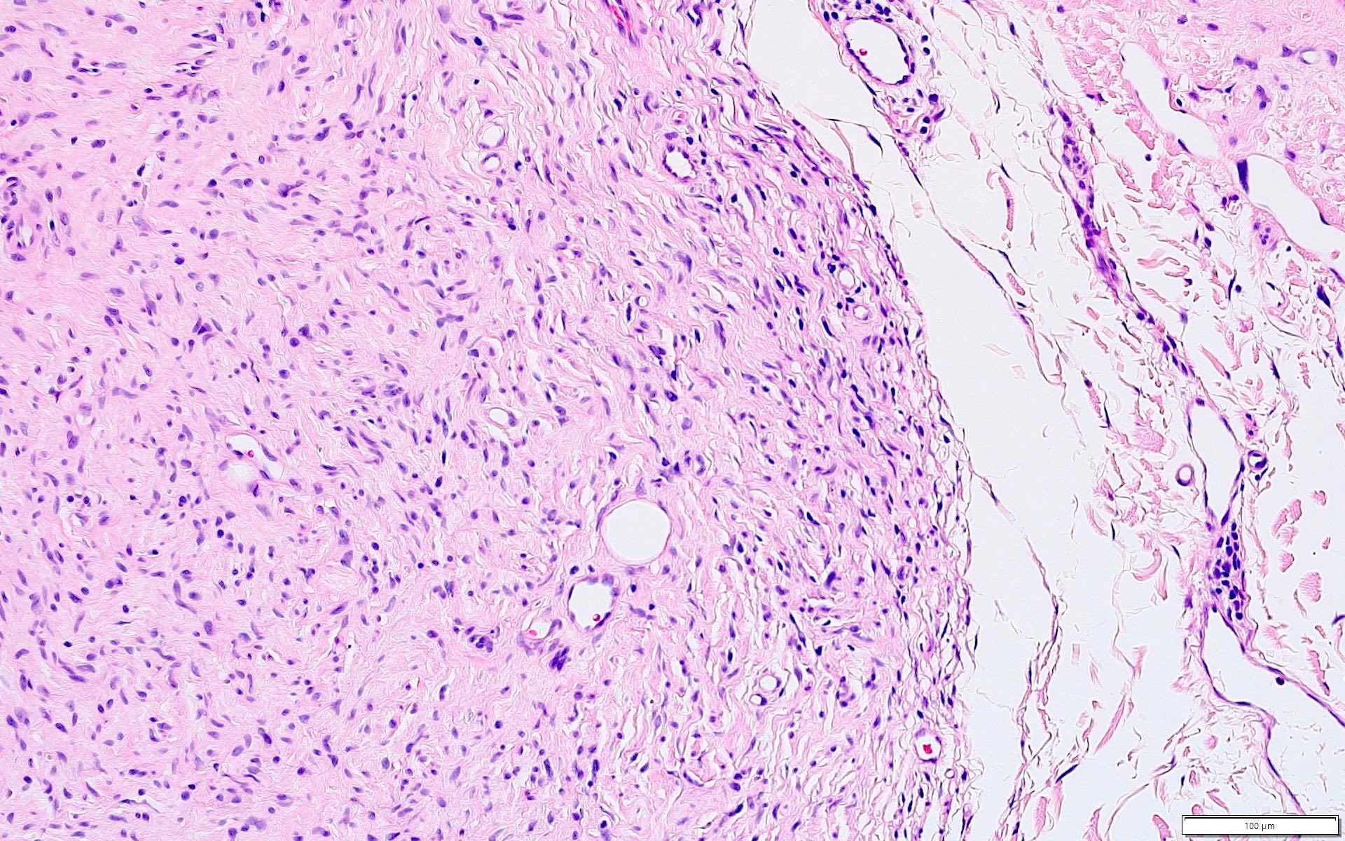
Clefting
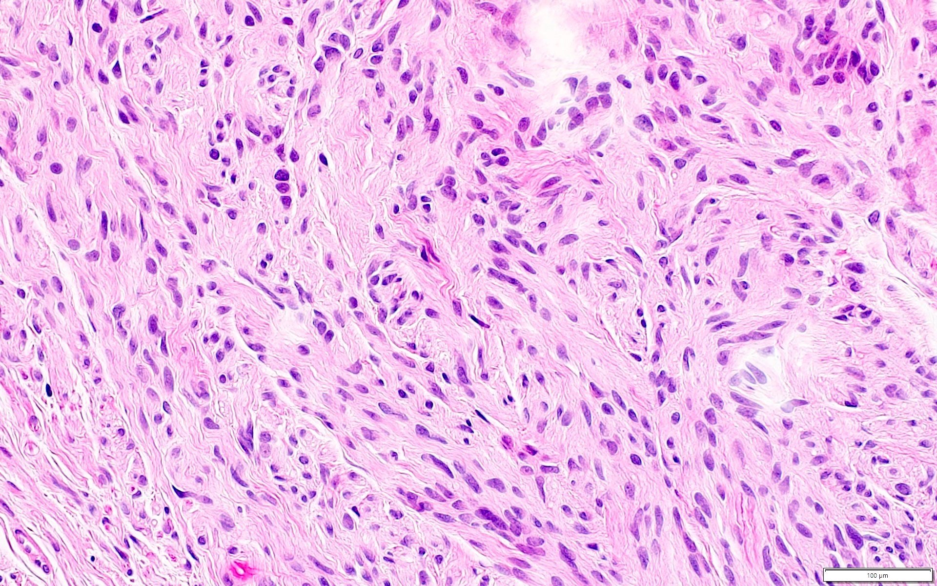
Bland spindled cells
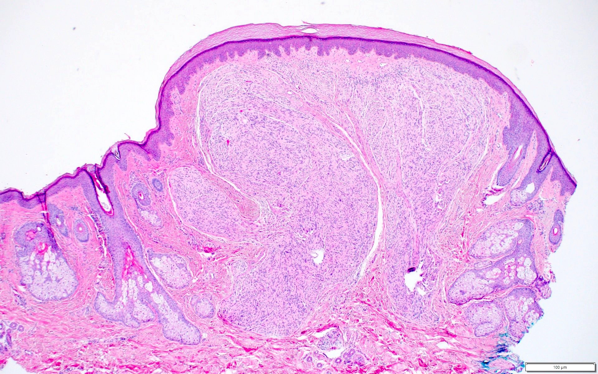
Dome shaped papule
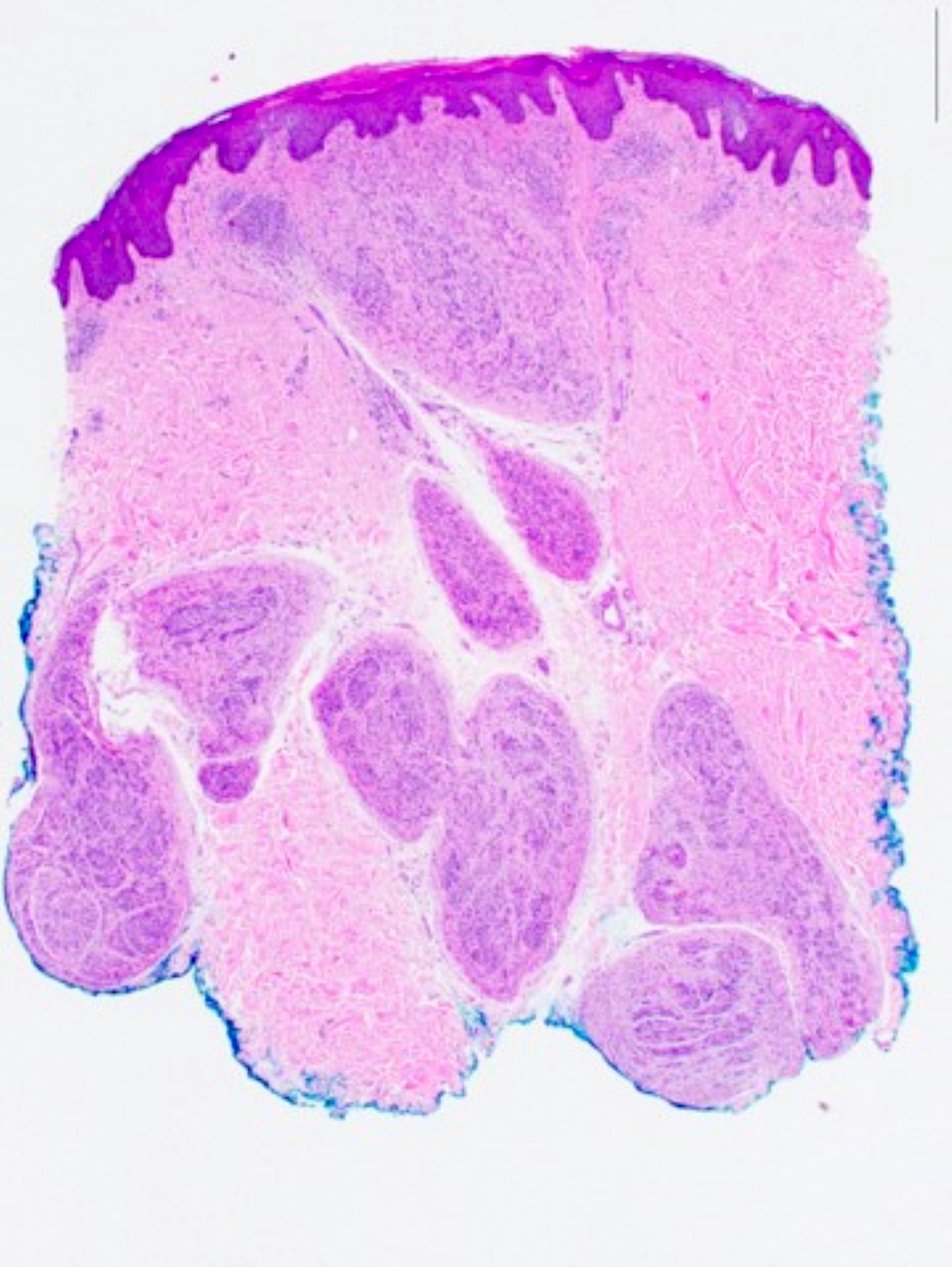
Plexiform PEN
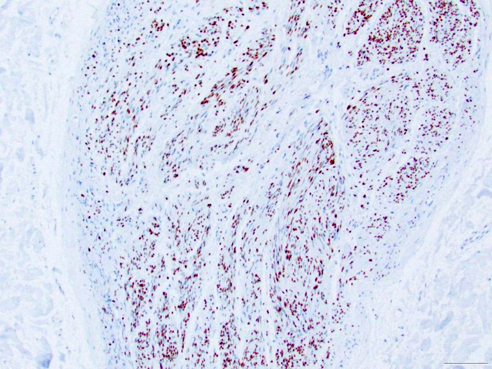
SOX10
EMA
Virtual slides
Images hosted on other servers:

Circumscribed
dermal tumor of
fascicles of Schwann
cells and axons
Cytology description
- Bland spindled cells with oval or elongated nuclei
Positive stains
- S100
- SOX10
- EMA: perineural cells
- Neurofilament: axons
- Reference: J Cutan Pathol 2022;49:82
Negative stains
- Cytokeratin stains
- SMA
- MelanA
- Reference: J Dermatol 2016;43:560
Electron microscopy description
- Axonal profiles and myelin density can be evaluated with electron microscopy (Arch Dermatol 1989;125:386)
Videos
Palisaded encapsulated neuroma
by Dr. Jerad Gardner
Sample pathology report
- Skin, chin, shave biopsy:
- Palisaded encapsulated neuroma / solitary circumscribed neuroma (see comment)
- Comment: There is a dermal tumor of bland spindled cells arranged in bundles. Clefting is observed between the tumor and surrounding dermis.
Differential diagnosis
- Neurofibroma:
- Peripheral nerve sheath tumor
- May be sporadic or associated with neurofibromatosis type 1
- May have ill defined or infiltrative margins
- Plexiform variant is essentially pathognomonic for neurofibromatosis
- Schwannoma:
- Encapsulated and well circumscribed tumor
- Antoni A areas with Verocay bodies and hypocellular Antoni B areas
- Less common in the superficial dermis than PEN
- Mucosal neuroma:
- Can be reactive / traumatic or associated with multiple endocrine neoplasia MEN2B syndrome
- Similar histopathologic findings to PEN, well circumscribed tumor of spindled cells arranged in bundles
- Leiomyoma:
- Traumatic neuroma:
- History of trauma
- May see dermal fibrosis / scar formation
- Histopathology shows haphazard nerve bundles in dermal fibrosis
- Neural hamartoma:
- Composed of tissues of mixed differentiation
Additional references
Practice question #1
A 55 year old man presents with an asymptomatic, 0.3 x 0.3 cm, dome shaped, skin colored papule on the chin. Histopathologic examination reveals a sharply circumscribed dermal nodule of spindled cells arranged in bundles with clefting. The tumor is positive for S100. What is the most likely diagnosis?
- Leiomyoma
- Neurofibroma
- Palisaded encapsulated neuroma
- Schwannoma
Practice answer #1
C. Palisaded encapsulated neuroma (PEN) is a benign spindle cell tumor commonly arising near the chin.
Answer B is incorrect because the findings of a sharply circumscribed tumor with clefting and bundles of spindled cells favor PEN over neurofibroma.
Answer D is incorrect because schwannomas contain Antoni A and B areas, while PEN lacks these features.
Answer A is incorrect because leiomyomas may demonstrate spindled cells arranged in bundles / fascicles but are S100 negative. Angioleiomyomas may be sharply circumscribed and encapsulated.
Comment Here
Reference: Palisaded encapsulated neuroma
Comment Here
Reference: Palisaded encapsulated neuroma
Practice question #2
Which of the following histologic features is most characteristic of palisaded encapsulated neuroma (PEN)?
- Clefting between tumor and dermis
- Collagen trapping
- Infiltrative growth pattern
- Presence of Verocay bodies
Practice answer #2
A. Clefting between tumor and dermis is a common histopathologic feature of PEN.
Answer C is incorrect because infiltrative growth pattern is not a feature of PEN.
Answer D is incorrect because Verocay bodies are observed within Antoni A areas of schwannomas.
Answer B is incorrect because collagen trapping is a feature of dermatofibroma.
Comment Here
Reference: Palisaded encapsulated neuroma
Comment Here
Reference: Palisaded encapsulated neuroma
