

Skin nonmelanocytic tumor
Neural tumors
Granular cell tumor
Authors: Jarish Cohen, M.D., Ph.D., Philip LeBoit, M.D.
Editorial Board Member: Hillary Rose Elwood, M.D.
Deputy Editor-in-Chief: Jonathan D. Ho, M.B.B.S., D.Sc.
Editor-in-Chief: Debra L. Zynger, M.D.


Last author update: 25 January 2023
Last staff update: 7 February 2023
Copyright: 2002-2024, PathologyOutlines.com, Inc.
PubMed Search: Cutaneous granular cell tumor
Table of Contents
Definition / general | Essential features | Terminology | ICD coding | Epidemiology | Sites | Pathophysiology | Etiology | Clinical features | Diagnosis | Case reports | Treatment | Clinical images | Gross description | Gross images | Microscopic (histologic) description | Microscopic (histologic) images | Positive stains | Negative stains | Electron microscopy description | Molecular / cytogenetics description | Videos | Sample pathology report | Differential diagnosis | Board review style question #1 | Board review style answer #1 | Board review style question #2 | Board review style answer #2Cite this page: Cohen J, LeBoit P. Granular cell tumor. PathologyOutlines.com website. https://www.pathologyoutlines.com/topic/skintumornonmelanocyticgct.html. Accessed April 28th, 2024.
Definition / general
- Dermal or subcutaneous tumor with abundant granular cytoplasm and Schwannian differentiation
- Nonneural variant (primitive polypoid granular cell tumor) shows similar granular cytomorphology but does not have immunohistochemical evidence of Schwannian differentiation
Essential features
- A predominantly dermal based tumor composed of cells with abundant eosinophilic to basophilic granular cytoplasm
- Often have an infiltrative configuration
- Tumors demonstrate Schwannian differentiation (S100+, SOX10+)
- Epidermis can exhibit pseudocarcinomatous hyperplasia in a subset of cases
Terminology
- Also known as Abrikossoff tumor and granular cell schwannoma (Arch Otolaryngol 1968;88:174)
- Originally described as granular cell myoblastoma, given the resemblance to skeletal muscle myocytes (Am J Pathol 1947;23:721)
ICD coding
- ICD-10: D23.9 - other benign neoplasm of skin, unspecified
Epidemiology
- Slightly higher incidence in women (Cancer 1987;60:220)
- Mainly adults but has wide age range of presentation (J Surg Oncol 1980;13:301)
Sites
- Common sites are trunk, upper limbs and head / neck
- May also arise on feet, hands, anogenital region and breast
Pathophysiology
- Dysregulation of endosomal pH due to inactivation of accessory proteins of the vacuolar H+ ATPase can give rise to conventional granular cell tumors (Nat Commun 2018;9:3533)
- A subset of nonneural granular cell tumors (S100-) harbor ALK gene fusions (Am J Surg Pathol 2018;42:1133)
Etiology
- Harboring multiple tumors has been associated with Noonan syndrome and LEOPARD syndrome (Eur J Pediatr Surg 2013;23:257, Clin Genet 2009;75:185)
Clinical features
- Slowly growing papule or nodule; rarely ulcerates (Adv Anat Pathol 1999;6:186)
- Rare cases can exhibit multiple lesions (Cutis 2002;69:343)
Diagnosis
- Fanberg-Smith et al. proposed diagnostic criteria for atypical and malignant tumors (Am J Surg Pathol 1998;22:779):
- Increased N:C ratio
- Nuclear pleomorphism
- Necrosis, tumor type
- Spindling of tumor cells
- Vesicular nuclei with prominent nucleoli
- > 2 mitoses per 10 high power fields
- Atypical: 1 - 2 features
- Malignant: > 2 features
- Nasser et al. proposed diagnostic criteria for granular cell tumor of uncertain malignant potential and malignancy (Pathol Res Pract 2011;207:164):
- Necrosis, single cell or en masse
- > 2 mitoses per 10 high power fields
- Uncertain malignant potential: 1 - 2 features
- Malignant: evidence of metastatic disease
Case reports
- 48 year old woman with upper back nodule (Indian J Dermatol 2015;60:322)
- 51 year old woman with left posterior chest wall mass and lymph node metastases (Am J Dermatopathol 2015;37:334)
- 53 year old woman with an enlarging left lower abdominal nodule (Am J Dermatopathol 2010;32:370)
- 54 year old patient with toe lesion (Dermatol Res Pract 2010;2010:184125)
- 69 year old woman with a rapidly enlarging firm neck mass (Am J Dermatopathol 2017;39:e50)
Treatment
- Benign and malignant tumors are typically treated with wide local excision (J Surg Oncol 1980;13:301)
- Mohs micrographic surgery is an option for anatomic sites in which conservative re-excision is desired (Case Rep Oncol Med 2012;2012:453569)
Clinical images
Images hosted on other servers:

Pedunculated buttock lesion

Subcutaneous waist lesion

Soft tissue swelling on the back

Hyperchromic nodule on right cubital fossa
Gross description
- Grayish white to pale yellow
- Oftentimes, not well circumscribed
Gross images
Images hosted on other servers:

Rough surface, ulceration and necrosis

Lobulated, solid leg mass

Infiltrative paraspinal tumor
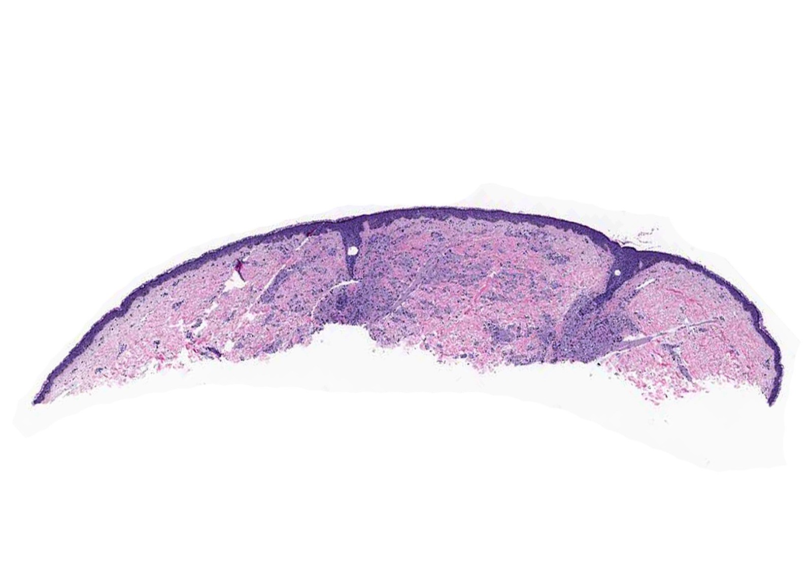
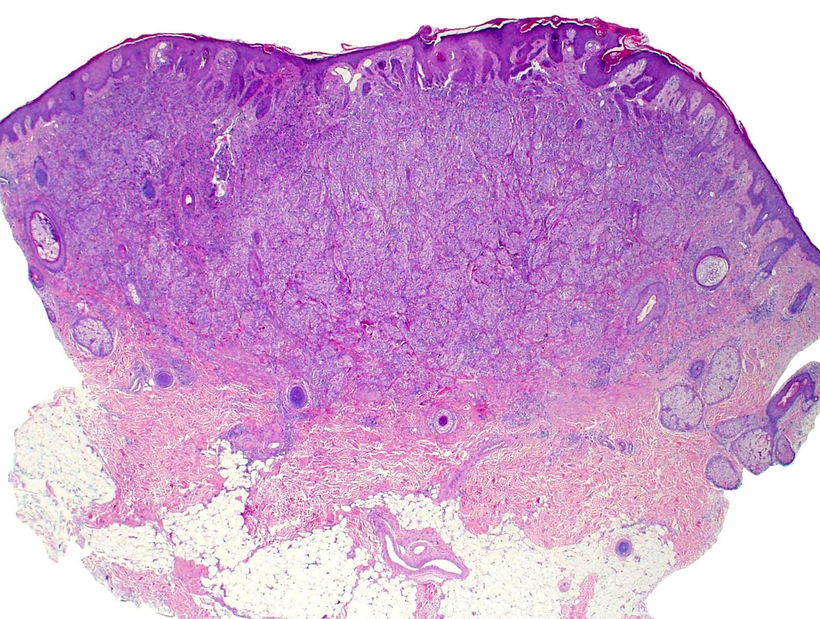
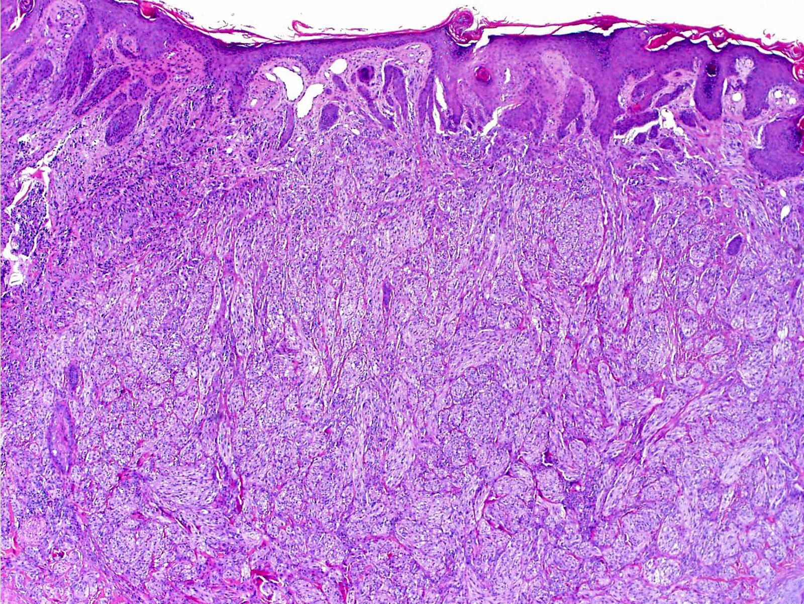
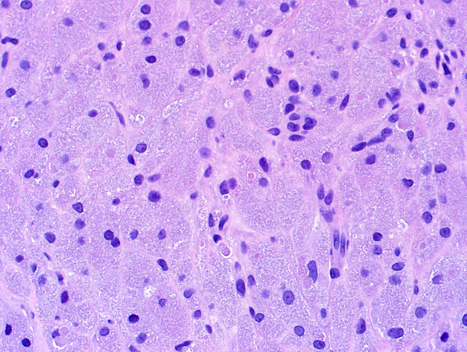
Microscopic (histologic) description
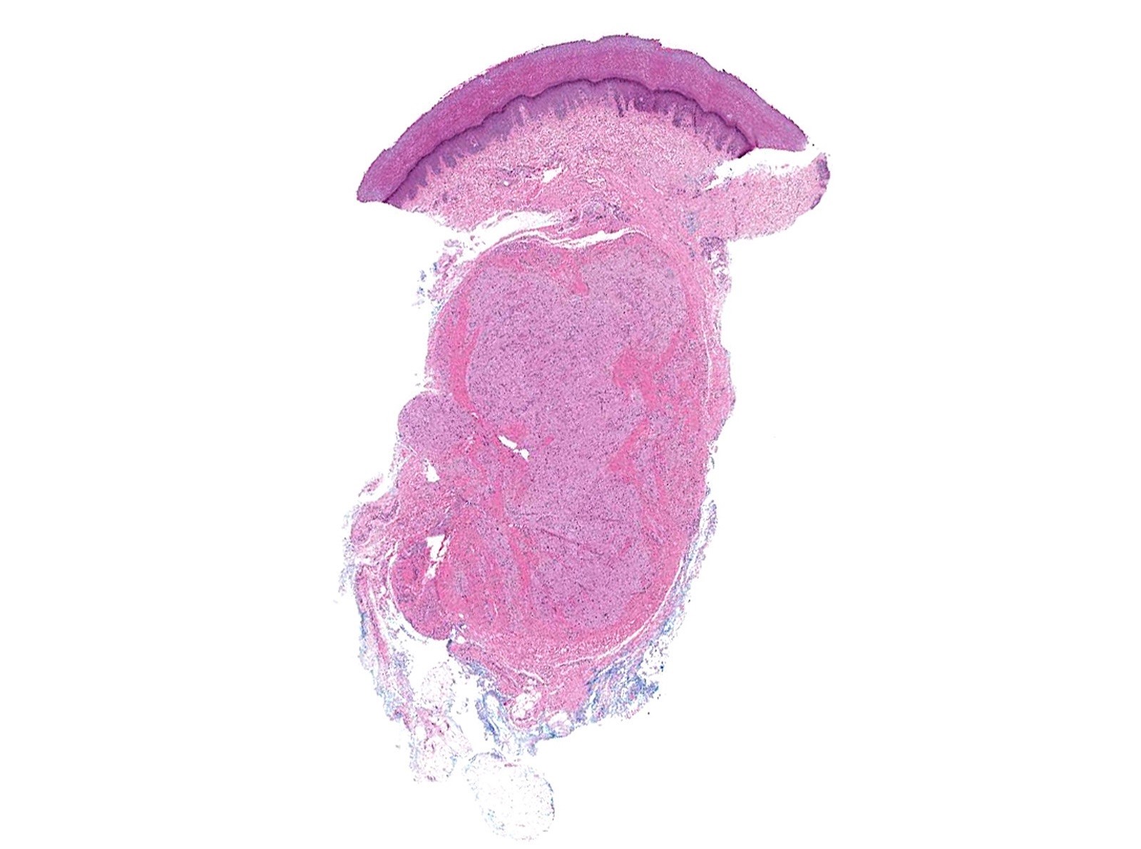
- Infiltrative or circumscribed architecture
- Can involve the subcutis
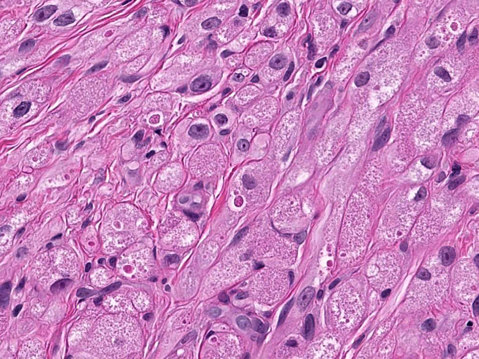
- Large polygonal cells with abundant eosinophilic granular cytoplasm and small, central nuclei
- Epidermis can show pseudocarcinomatous hyperplasia
- Lysosomal macroinclusions (pustulo-ovoid bodies of Milian) are usually present (J Cutan Pathol 2007;34:405)
- Can exhibit accentuation around arrector pili muscles or peripheral nerves (J Clin Pathol 2014;67:19)
- Nonneural granular cell tumors (S100-) can exhibit nucleomegaly, pleomorphism and variable mitotic activity (Am J Surg Pathol 1991;15:48, Histopathology 2005;47:179)
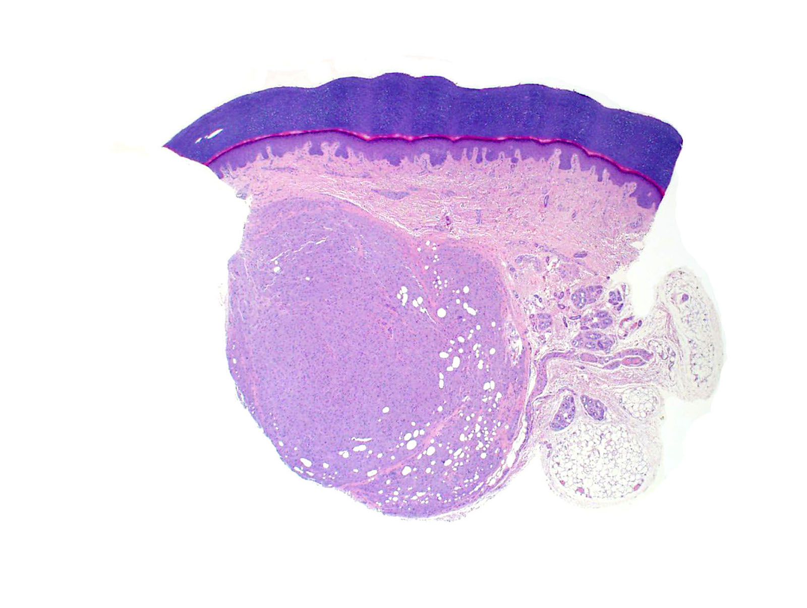
Microscopic (histologic) images
Contributed by Jarish Cohen, M.D., Ph.D.
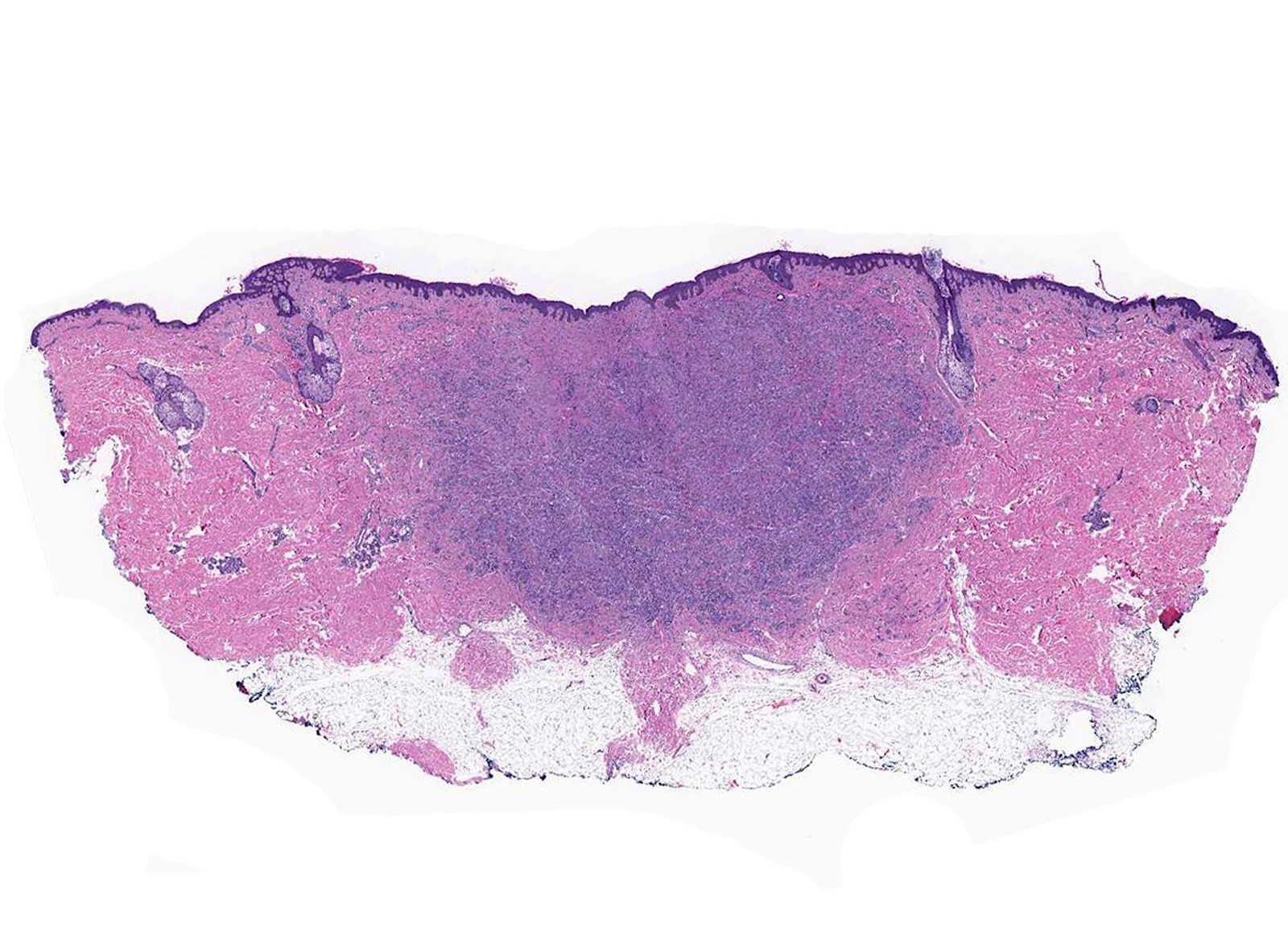
Dermal wedge shaped tumor
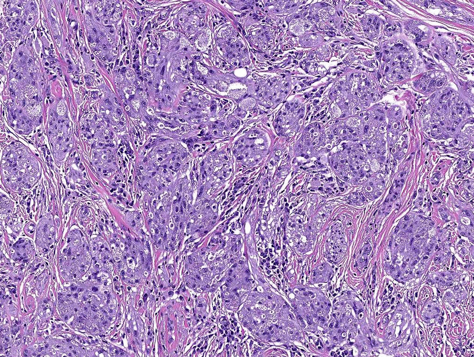
Nests and cords
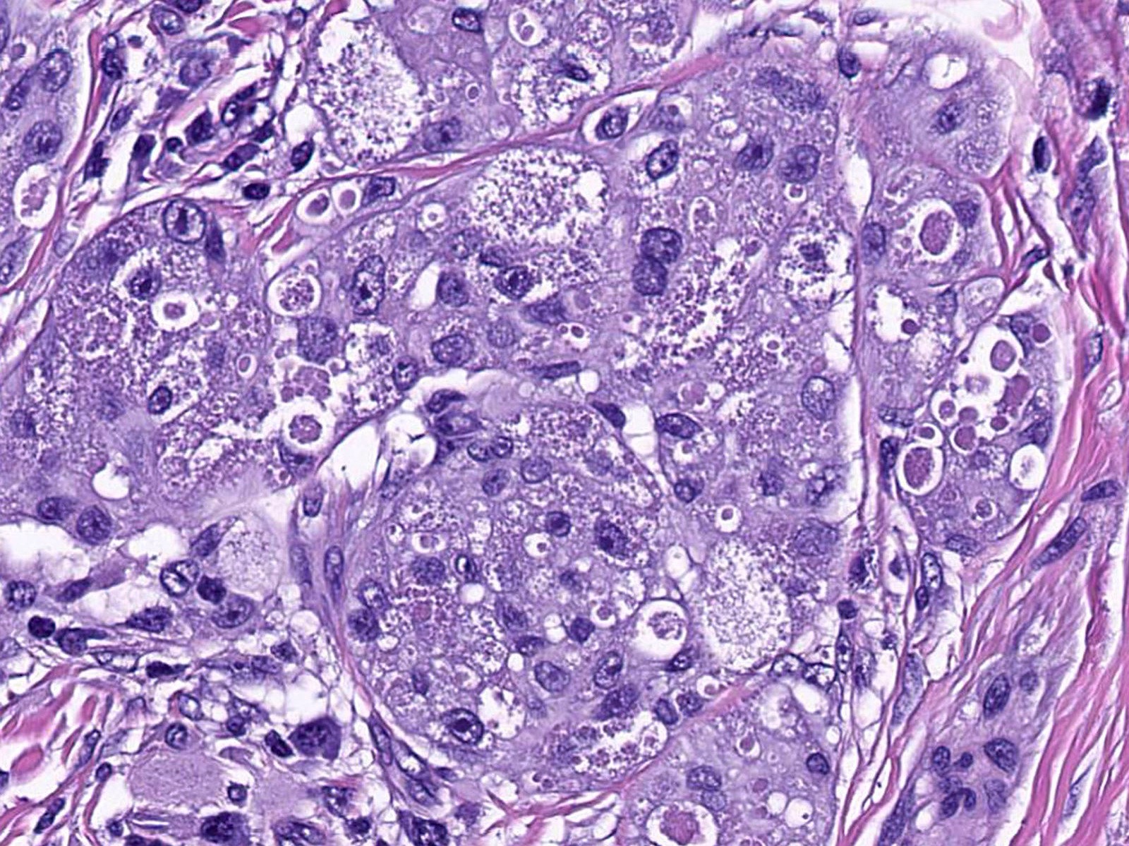
Pustulo-ovoid bodies
Infiltrative array

Accentuation around arrector pili

Abundant granular cytoplasm
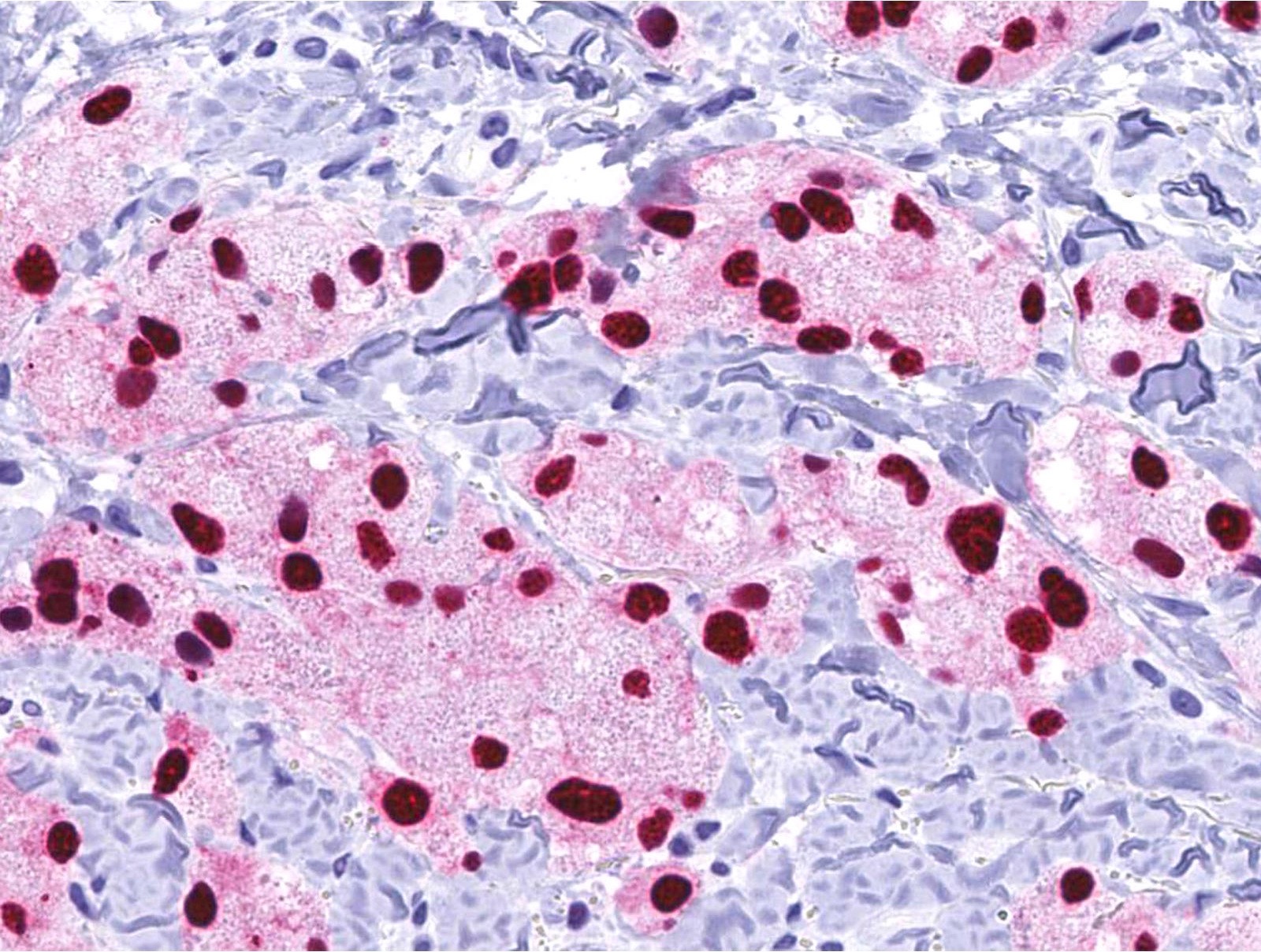
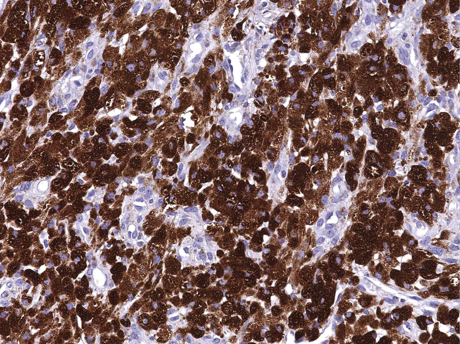
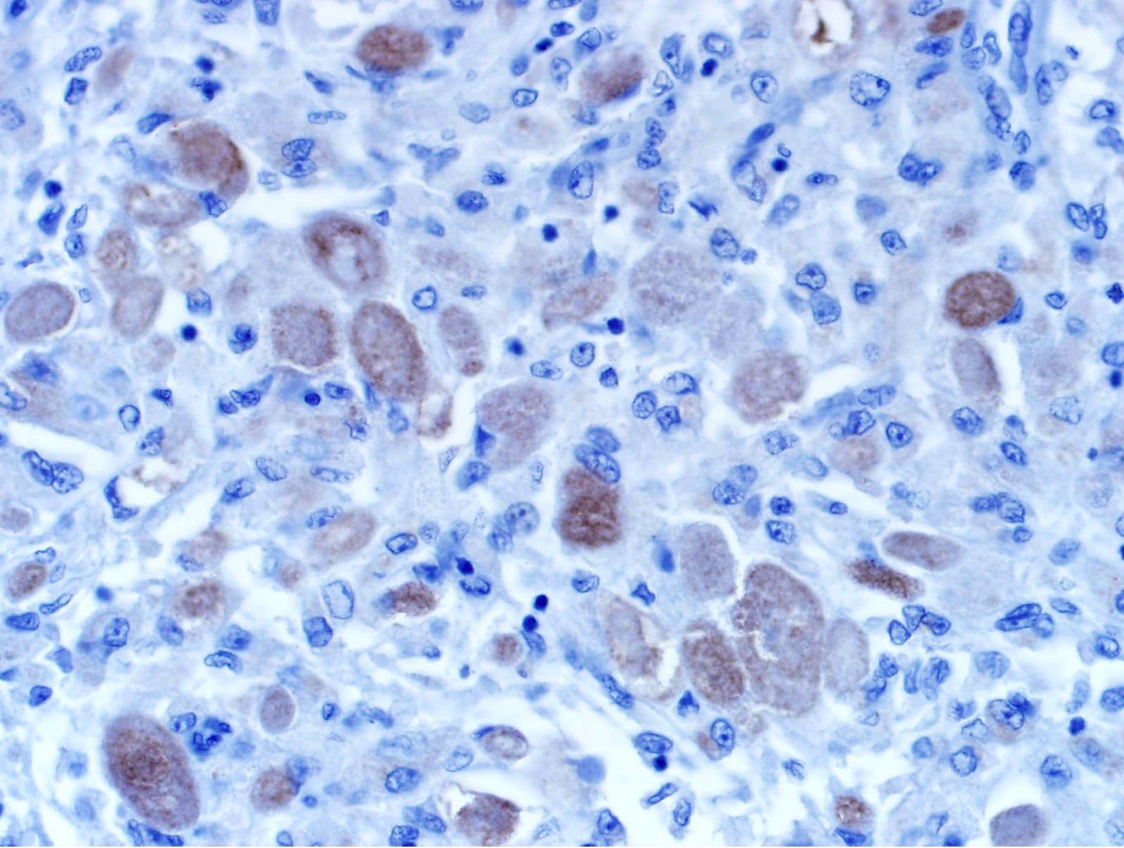
SOX10
Deep dermal tumor

Tumor surrounds nerve
Granular pink cytoplasm


Nonneural granular cell tumor
Nonneural granular cell tumor
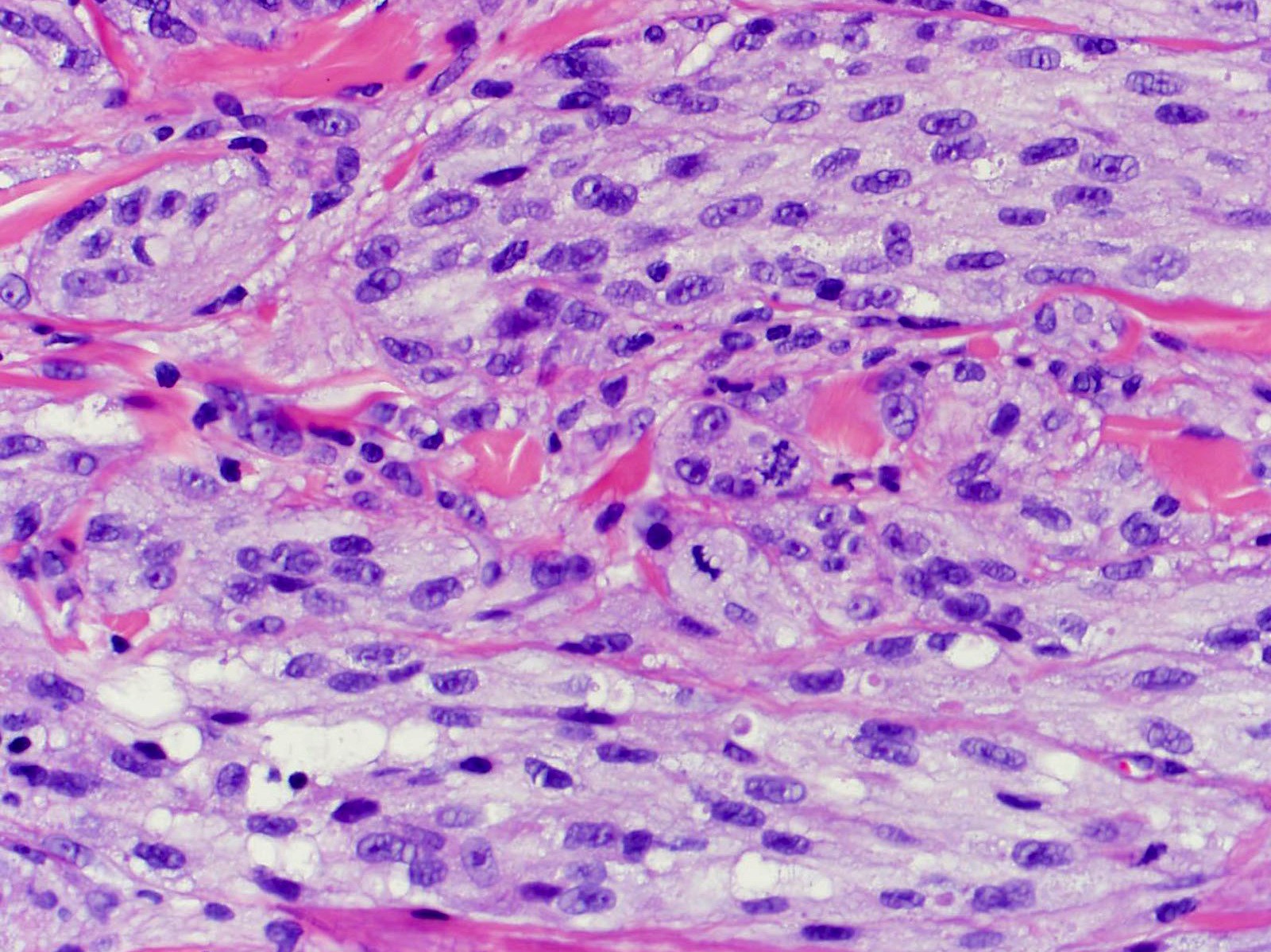
Atypical granular cell tumor
Positive stains
- PASD, Sudan Black B, S100 (except for nonneural granular cell tumors), SOX10, NSE
- Variable staining for vimentin, CD68, NKI-C3 (CD63), MITF, CD56
- A subset of nonneural granular cell tumors express ALK
- References: Cancer 1982;49:1624, Histopathology 2012;61:997, Diagn Histopathol 1982;5:205, Cancer 1989;64:1455, Dermatology 1993;186:106, Am J Dermatopathol 2007;29:22, Hum Pathol 2015;46:813, Am J Surg Pathol 2018;42:1133
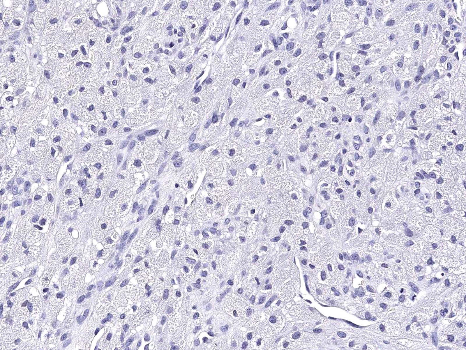
Negative stains
- HMB45, MelanA, AE1 / AE3, EMA, desmin
- Nonneural granular cell tumor is negative for S100
- References: Am J Dermatopathol 2007;29:22, Am J Surg Pathol 1991;15:48
Electron microscopy description
- Membrane bound cytoplasmic granules containing lysosomes of varying sizes (J Cutan Pathol 1981;8:277, Oral Surg Oral Med Oral Pathol 1977;44:227, Ultrastruct Pathol 2018;42:304)
- Larger secondary lysosomes can be seen
- Some membrane bound granules contain aggregates of virus-like particles
- Some lesional cells contain long spacing collagens (Luse bodies)
- Variably abundant basal lamina material may be seen
Molecular / cytogenetics description
- Inactivating ATP6AP1 and ATP6AP2 somatic mutations are highly prevalent in conventional granular cell tumors (Nat Commun 2018;9:3533, Genes Chromosomes Cancer 2019;58:37)
- A subset of nonneural granular cell tumors harbor ALK gene fusions (Am J Surg Pathol 2018;42:1133, J Cutan Pathol 2020;47:1026)
Videos
Granular cell tumor
Sample pathology report
- Skin, left antecubital fossa, shave biopsy:
- Granular cell tumor (granular cell schwannoma) (see comment)
- Comment: The lesional cells are positive with an immunostain for SOX10 protein, a finding that is typical of this entity.
- Skin, left thigh, shave biopsy:
- Nonneural granular cell tumor (see comment)
- Comment: The biopsy shows a polypoid mass of large, oval cells with ample pallid and both finely and coarsely granular cytoplasm, with some pustulo-ovoid bodies. The lesional cells are S100 negative, MITF negative and NKI-C3 strongly positive. An ALK immunostain shows moderate patchy positivity. A phosphohistone H3 shows that cells in mitosis are exceedingly rare, under 1/mm2. The histopathologic and immunohistochemical findings are consistent with nonneural granular cell tumor (also known as primitive polypoid granular cell tumor).
Differential diagnosis
- Melanocytic neoplasms (J Am Acad Dermatol 2004;50:765):
- Leiomyosarcoma (Am J Dermatopathol 1988;10:234, Am J Dermatopathol 1999;21:307):
- Positive for desmin and alpha smooth muscle type
- Negative for S100 and SOX10
- Atypical fibroxanthoma (AFX) (J Cutan Pathol 2005;32:314, J Cutan Pathol 2010;37:380, Am J Dermpathol 2021;43:831):
- A subset of AFX can demonstrate an infiltrative silhouette and cells with abundant granular cytoplasm
- Most AFX will display elevated mitotic activity with atypical forms, prominent nuclear pleomorphism and arise in the setting of marked solar elastosis
- Negative for S100 and SOX10
- Granular AFX may show positive ALK staining that is not reflective of an underlying gene fusion
- Dermatofibroma (benign fibrous histiocytoma) (Dermatology 1999;199:54):
- Xanthoma:
- Reticulohistiocytoma:
- Adult type rhabdomyoma:
- Can be composed of sheets of polygonal cells with abundant eosinophilic, variably granular cytoplasm
- Usually demonstrates cross striations
- Positive for desmin, actin - muscle specific and myogenin
- Negative for S100
- Alveolar soft part sarcoma:
Board review style question #1
- A 40 year old woman presents with a nodule on her trunk. The lesion is biopsied and shows a dermal based proliferation of cells with abundant eosinophilic granular cytoplasm. Which of the following statements is true?
- The abundant granular cytoplasm is indicative of numerous membrane bound cytoplasmic granules containing aggregates of mitochondria
- An S100 or SOX10 stain is negative in most tumors
- Perineural accentuation of cellularity is a feature of malignancy
- These tumors can display infiltrative architecture and can sometimes involve the subcutaneous fat
Board review style answer #1
D. These tumors can display infiltrative architecture and can sometimes involve the subcutaneous fat. This statement is true since granular cell tumors often show infiltrative borders and can occasionally involve the subcutis. No studies have demonstrated an increased aggressive behavior of granular cell tumors that exhibit these features.
Comment Here
Reference: Granular cell tumor
Comment Here
Reference: Granular cell tumor
Board review style question #2
- Which of the following statements about granular cell tumors is true?
- A subset are negative for S100 and other neural markers and can have a polypoid appearance
- Atypical features include association with pseudocarcinomatous hyperplasia and accentuation around peripheral nerves
- NF1 is commonly mutated in conventional granular cell tumors
- Tumors never demonstrate metastatic potential
Board review style answer #2
A. A subset are negative for S100 and other neural markers and can have a polypoid appearance. These so called nonneural granular cells tumors are negative for neural markers (e.g., S100 and SOX10). In addition, a subset has been shown to harbor ALK gene fusions and are positive by ALK immunohistochemistry.
Comment Here
Reference: Granular cell tumor
Comment Here
Reference: Granular cell tumor
