

Adrenal gland & paraganglia
Neuroblastic tumors
Ganglioneuroma
Authors: Carmen Perrino, M.D., Debra L. Zynger, M.D.


Last author update: 1 December 2014
Last staff update: 8 April 2024 (update in progress)
Copyright: 2002-2024, PathologyOutlines.com, Inc.
PubMed Search: Ganglioneuroma adrenal
Table of Contents
Definition / general | Epidemiology | Sites | Etiology | Clinical features | Diagnosis | Laboratory | Radiology description | Radiology images | Prognostic factors | Case reports | Treatment | Gross description | Gross images | Microscopic (histologic) description | Microscopic (histologic) images | Virtual slides | Cytology description | Cytology images | Positive stains | Negative stains | Electron microscopy description | Molecular / cytogenetics description | Differential diagnosisCite this page: Perrino C, Zynger DL. Ganglioneuroma. PathologyOutlines.com website. https://www.pathologyoutlines.com/topic/adrenalganglioneuroma.html. Accessed May 12th, 2024.
Definition / general
- Mature, benign neoplasm that originates from neural crest cells of sympathetic ganglia or adrenal medulla
- On a spectrum, from least → most differentiated: neuroblastoma → ganglioneuroblastoma → ganglioneuroma
Epidemiology
- At all sites, usually age ≥ 7 years old
- Patients with adrenal ganglioneuroma usually 4th to 5th decade (BMC Res Notes 2014;7:791)
- Patients with ganglioneuroma of retroperitoneum or posterior mediastinum usually children (BMC Res Notes 2014;7:791)
- M=F
- Familial disposition and associated with Turner syndrome, multiple endocrine neoplasia type 2 (MEN2) (BMC Res Notes 2014;7:791)
Sites
- Located along distribution of sympathetic nervous system
- Most commonly in posterior mediastinum, followed by retroperitoneum (especially presacral space)
- May occur in many locations, including adrenal gland (~20-30% of cases), cervical and parapharyngeal area, urinary bladder, prostate, bone, pancreas, skin, orbit, paratesticular area, appendix, gastrointestinal tract (Lack: Tumors of the Adrenal Glands and Extraadrenal Paraganglia, Vol.8, 2007)
Etiology
- Clonal proliferation of cells of neural crest origin
Clinical features
- Usually asymptomatic
- May have non-specific signs/symptoms such as abdominal pain, palpable abdominal mass
- Rarely, catecholamine synthesis may cause hypertension
Diagnosis
- Tumor comprised of admixture of ganglion cells and Schwannian stroma/cells
Laboratory
- Usually non-functional
- Occasionally increased vanilmandelic acid and homovanillic acid in urine
Radiology description
- Ultrasound: well-defined, homogeneous, hypoechogenic mass (BMC Res Notes 2014;7:791)
- Computed tomography (CT): circumscribed and well-defined, hypodense, homogeneous or slightly heterogeneous lesion, surrounds peripheral blood vessels without compression or occlusion, may have fine/punctate calcifications (~40-60%), poorly enhanced by contrast medium (<40 Hounsfield units) (BMC Res Notes 2014;7:791)
- Magnetic resonance imaging (MRI): (BMC Res Notes 2014;7:791)
- T1: homogeneous, signal intensity lower than liver
- T2: heterogeneous, signal intensity greater than liver; no absolute change in signal intensity on chemical shift imaging
- Gadolinium administration: delayed and progressive, non-intense enhancement
Radiology images
Images hosted on other servers:
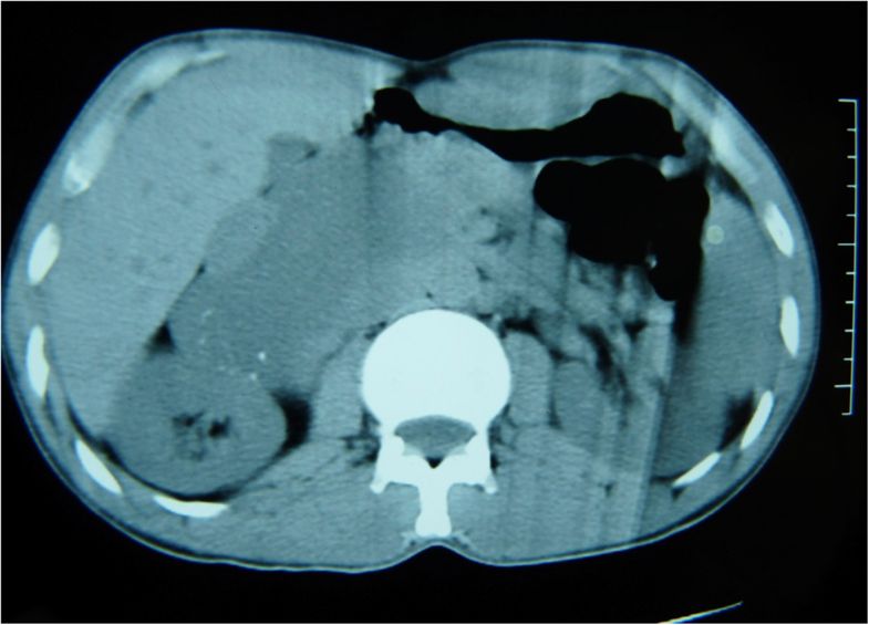
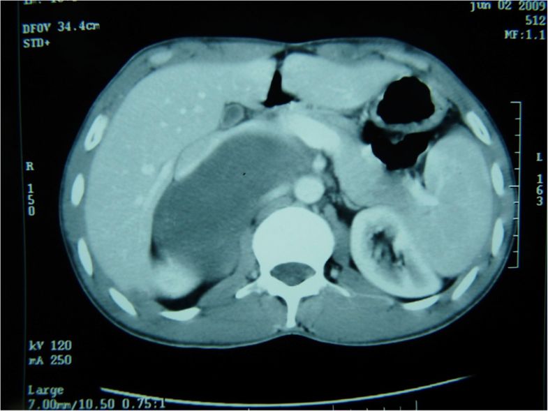
Various images
Prognostic factors
- Benign tumor, good prognosis following adrenalectomy, long term follow-up is recommended (J Med Case Rep 2014;8:131)
- Rarely malignant transformation of Schwannian stroma to malignant peripheral nerve sheath tumor (MPNST), may occur de novo or following abdominal radiation (Clin Endocrinol (Oxf) 2014;80:342)
Case reports
- 13 year old girl with ganglioneuroma and hereditary spherocytosis (Turk J Pediatr 2012;54:187)
- 15 year old girl with incidental adrenal mass (World J Surg Oncol 2012;10:64)
- 18 year old man with ganglioneuroma presenting as an adrenal incidentaloma (J Med Case Rep 2014;8:131)
- 21 year old woman with clinical acute pyelonephritis (Arq Bras Endocrinol Metabol 2012;56:270)
- 28 year old man with adrenal ganglioneuroma (BMC Res Notes 2014;7:791)
- 33 year old woman with laparoscopic extirpation of giant adrenal ganglioneuroma (J Minim Access Surg 2014;10:45)
- 37 year old woman with composite pheochromocytoma-ganglioneuroma of the adrenal gland (Urol Ann 2013;5:115)
- 41 year old man with large adrenal ganglioneuroma (Intern Med 2012;51:2365)
- 62 year old man with ganglioneuroma of the adrenal gland (Case Rep Oncol 2012;5:487)
- 69 year old woman with coexistence of renal cell carcinoma of clear cell type with sarcomatoid cell type component and adrenal mature ganglioneuroma with myelolipoma (Rom J Morphol Embryol 2014;55:425)
Treatment
- Adrenalectomy (J Med Case Rep 2014;8:131)
Gross description
- Well circumscribed
- May have true capsule or fibrous capsule
- Size varies, 8 cm to > 15 cm
- Firm, resilient texture
- Gray-white to tan-yellow
- May have trabecular or whorled appearance
Gross images
Contributed by @SueEPig on Twitter
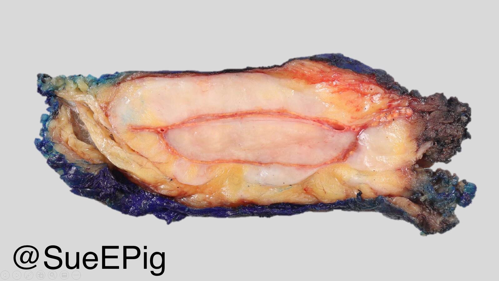
Adrenal ganglioneuroma
Images hosted on other servers:

Adrenal ganglioneuroma

Adrenal ganglioneuroma, cut surface
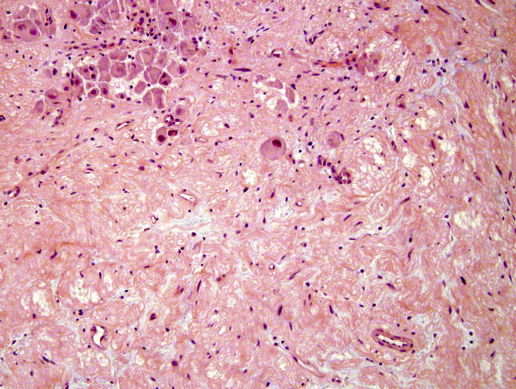
Microscopic (histologic) description
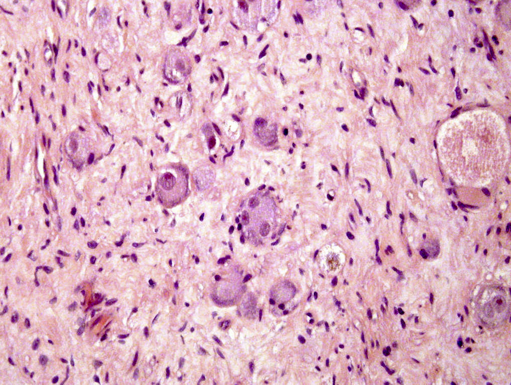
- Admixture of ganglion cells and Schwann cells
- Ganglion cells
- Mature to mildly dysmorphic
- Mature: compact, eosinophilic cytoplasm with distinct cell borders, single eccentric nucleus, prominent nucleolus
- Dysmorphic: single or multiple pyknotic nuclei
- Vary in distribution and number, may be quite sparse
- May contain finely granular, gold to brown pigment (lipofuscin or neuromelanin)
- Mature to mildly dysmorphic
- Schwann cells
- May ensheath neuritic processes
- May be arranged in small intersecting fascicles which are separated by loose myxoid stroma
- Two histologic subtypes:
- Mature = every ganglion cell is mature
- Maturing = minor component of scattered collections of differentiating neuroblasts or maturing ganglion cells
- Unlike intermixed subtype of ganglioneuroblastoma, these immature foci do not form distinct microscopic nests
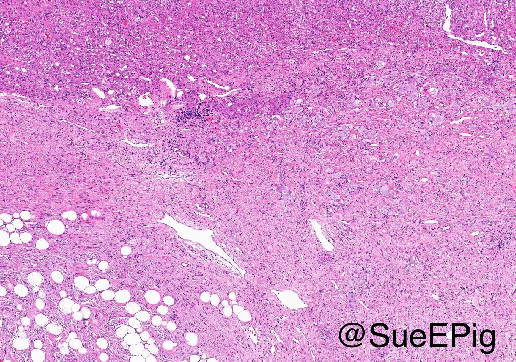
- Background may include lobules of mature adipose tissue (especially at periphery of lesion), mast cells, chronic inflammation, dense collagenized stroma
- Mild variation in cellularity is permitted
- No significant atypia, mitoses, or necrosis should be present
- Masculinizing ganglioneuroma: admixture of ganglioneuroma and Leydig cells with crystalloids of Reinke or strands/clusters of cells resembling adrenal cortical cells
- Composite tumor: rare; usually ganglioneuroma and pheochromocytoma (ìcomposite pheochromocytomaî)
Microscopic (histologic) images
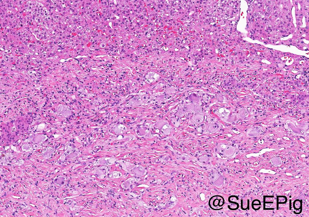
Contributed by Carmen Perrino, M.D., Debra Zynger, M.D. and @SueEPig on Twitter
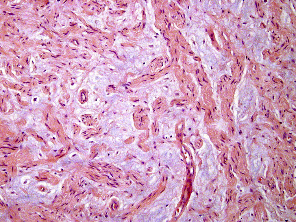
Prominent myxoid background
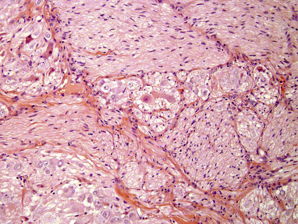
Scattered mature ganglion cells
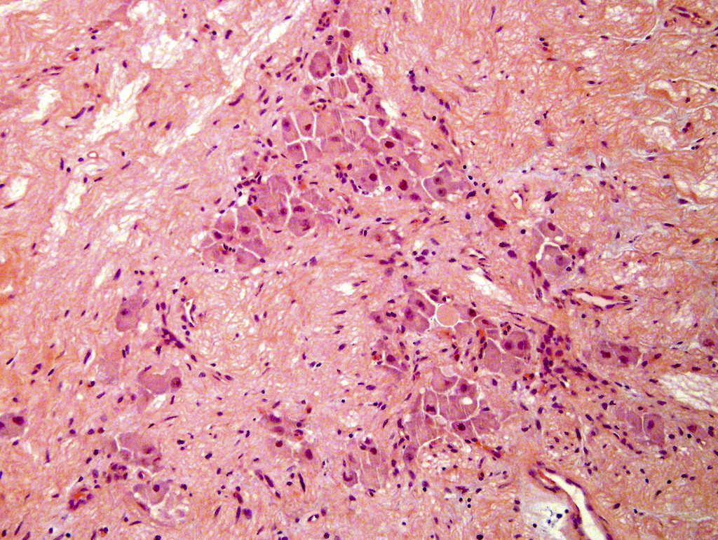
Cluster of mature ganglion cells
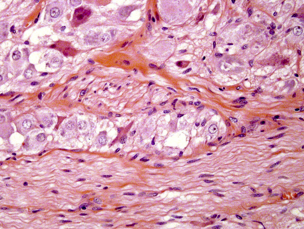
Intervening stroma
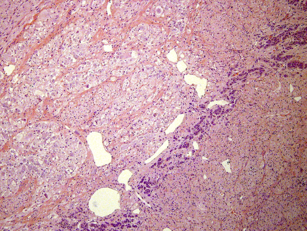
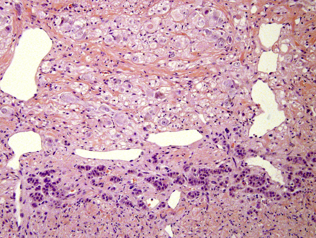
Ganglioneuroma (top) and residual normal adrenal cortical cells (bottom)
Mature ganglion cells admixed with stroma
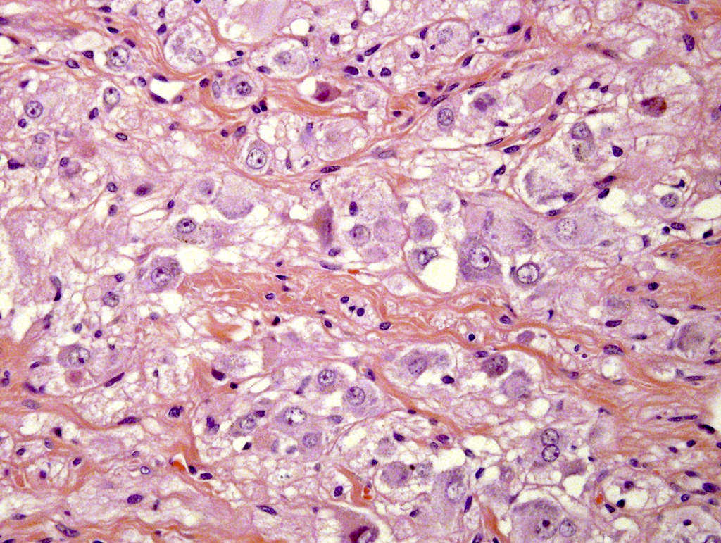
Mature ganglion cells


Adrenal ganglioneuroma
Virtual slides
Images hosted on other servers:


Adrenal gland ganglioneuroma

Pheochromocytoma
with focus of
ganglioneuroma
Cytology description
- Biphasic with large, polyhedral ganglion cells and fibrillary stroma with spindle-shaped cells with cigar-shaped nuclei (J Cytol 2014;31:57)
Cytology images
Images hosted on other servers:

Schwann cells and admixed ganglion cells (insets), neck

Schwann cells (a, c) and ganglion cells (b, d), neck
Positive stains
- Schwann cells/stroma: S100, synaptophysin, neurofilament (NF) protein (Dabbs: Diagnostic Immunohistochemistry, 3rd Edition, 2010, J Med Case Rep 2014;8:131, Am J Pathol 1990;136:375)
- Ganglion cells: S100, synaptophysin, chromogranin A, NF protein, glial fibrillary acidic protein (GFAP), PGP 9.5, type IV collagen, vasoactive intestinal peptide (VIP) (Dabbs: Diagnostic Immunohistochemistry, 3rd Edition, 2010, Endocr Relat Cancer 2003;10:99, Am J Pathol 1990;136:375)
Negative stains
- EMA, cytokeratin, HMB45, WT1, CD99, CD45, desmin, myogenic markers (myogenin, MyoD1) (Dabbs: Diagnostic Immunohistochemistry, 3rd Edition, 2010)
Electron microscopy description
- Mixture of neural bundles and normal appearing ganglion cells with eccentric nuclei and large numbers of cytoplasmic organelles
Molecular / cytogenetics description
- Ganglioneuromas are not usually associated with genetic abnormalities (Clin Endocrinol (Oxf) 2014;80:342)
- RET mutational analysis negative in 4 cases (1 ganglioneuroma, 3 composite ganglioneuroma and pheochromocytomas) (J Clin Endocrinol Metab 2010;95:3118)
- Some still believe that there may be a causative role for an activating RET protooncogene mutation in the pathogenesis of adrenal ganglioneuromas (Clin Endocrinol (Oxf) 2014;80:342)
- RET protooncogene expressed at low levels in 1 of 3 ganglioneuromas (J Mol Med (Berl) 1996;74:617)
- 2 pediatric patients (one with MEN2A, one with MEN2B) with ganglioneuromas and no evidence of pheochromocytoma (J Clin Endocrinol Metab 2005;90:4383)
- MEN2A and MEN2B have rarely been associated with composite adrenal ganglioneuroma and pheochromocytoma (Clin Endocrinol (Oxf) 2014;80:342)
- Loss of heterozygosity (LOH) studies showed that chromosome 1 (1p34-36) and p53 (17p13.1), prognostic markers for neuroblastoma, were not detected in ganglioneuroblastoma (Endocr Relat Cancer 2003;10:99)
Differential diagnosis
- Adrenal cortical adenoma
- Carcinoma
- Composite pheochromocytoma
- Ganglioneuroblastoma
- Neuroblastoma
- Neurofibroma: especially if ganglion cells are sparse
