Testis & paratestis
Nonneoplastic
Hernia sac with mesothelial entrapment
Authors: Jason Tretter, Debra L. Zynger, M.D.
Editorial Board Member: Bonnie Choy, M.D.
Deputy Editor-in-Chief: Maria Tretiakova, M.D., Ph.D.


Last author update: 9 February 2022
Last staff update: 9 February 2022
Copyright: 2003-2025, PathologyOutlines.com, Inc.
PubMed Search: hernia entrapment pathology
Table of Contents
Definition / general | Essential features | Epidemiology | Pathophysiology | Clinical features | Prognostic factors | Treatment | Gross description | Microscopic (histologic) description | Microscopic (histologic) images | Positive stains | Negative stains | Molecular / cytogenetics description | Differential diagnosis | Practice question #1 | Practice answer #1 | Practice question #2 | Practice answer #2Cite this page: Tretter J, Zynger D. Hernia sac with mesothelial entrapment. PathologyOutlines.com website. https://www.pathologyoutlines.com/topic/testisherniasacepid.html. Accessed September 17th, 2025.
Definition / general
- Entrapment of mesothelial cells within hernia sac fibroadipose tissue
Essential features
- Small glands, clusters, cords or individual mesothelial cells within the hernia sac
- Usually no fat, muscle, stromal invasion or necrosis
- Lacks grossly visible proliferation (Virchows Arch A Pathol Anat Histopathol 1989;415:283)
Epidemiology
- 6% of pediatric hernia sacs contained mesothelial proliferation (J Urol 2011;186:1620)
- Mean age in adults is 44.5 years (Am J Surg Pathol 2014;38:54)
- Incidence in adults unknown
Pathophysiology
- Persistent serosal injury causes reactive hyperplasia of the mesothelial lining and submesothelial fibrosis, entrapping mesothelial cells beneath the mesothelial lined surface (Mod Pathol 2005;18:S131, Diagn Pathol 2011;6:78)
Clinical features
- Similar clinical presentation as a patient with a hernia sac without mesothelial entrapment (usually hydrocele, also hematocele, cyst adjacent to epididymis, hemorrhagic epididymal cyst, pyocele) (Am J Surg Pathol 2014;38:54)
Prognostic factors
- No aggressive behavior has been reported (Am J Surg Pathol 2014;38:54)
Treatment
- Similar treatment as a hernia without mesothelial entrapment, i.e. excision of hernia sac (J Urol 2011;186:1620)
Gross description
- No grossly visible proliferation; same findings as hernia sac without mesothelial proliferation (Am J Surg Pathol 2014;38:54)
Microscopic (histologic) description
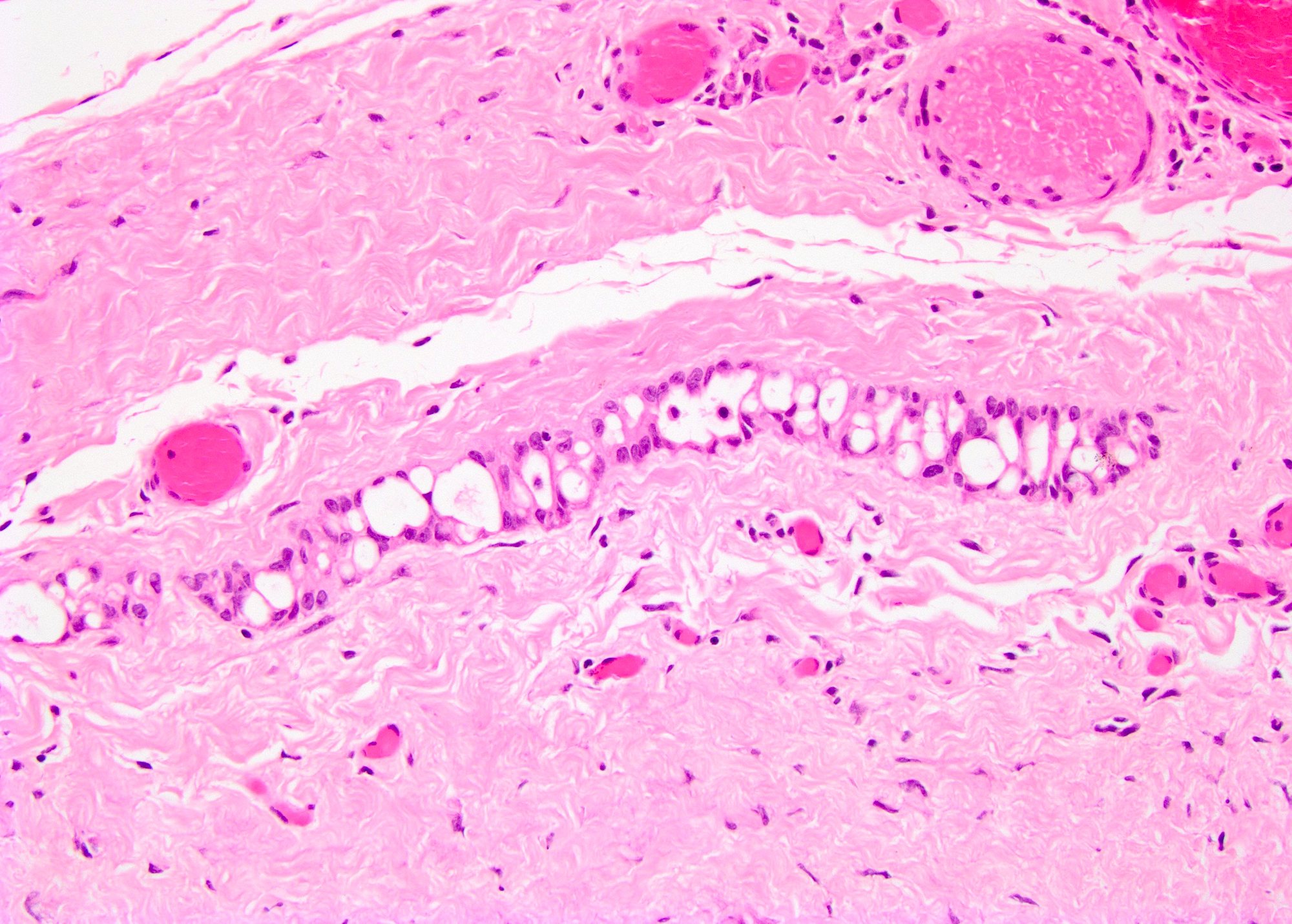
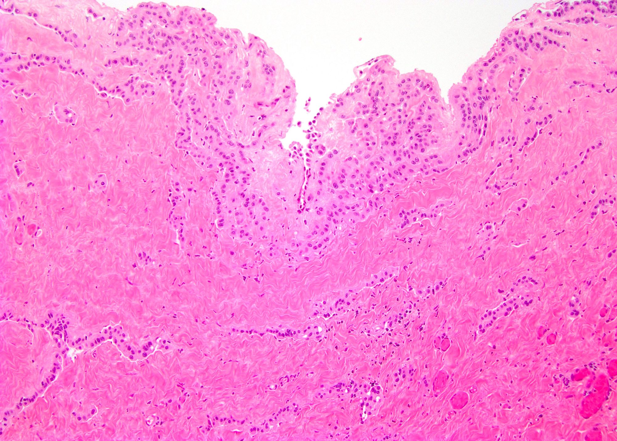
- No surface proliferation; surface often partially or completely denuded (Am J Surg Pathol 2014;38:54)
- Mesothelial proliferation within fibrotic stroma (Am J Surg Pathol 2014;38:54)
- Widely spaced proliferation of mesothelial cells predominately oriented parallel to the surface mesothelium (Am J Surg Pathol 2014;38:54)
- Most commonly form small glands and cords; also small nests and individual appearing cells (Am J Surg Pathol 2014;38:54)
- Proliferation has a well defined edge of pseudoinvasion within the fibrous stroma (Am J Surg Pathol 2014;38:54)
- Usually does not penetrate into muscle (Semin Diagn Pathol 2003;20:272)
- Reactive appearing mesothelial cells with large nuclei and prominent nucleoli (Semin Diagn Pathol 2003;20:272)
- No to low mitotic activity (Am J Surg Pathol 2014;38:54)
- Background of fibrosis and chronic inflammation (Am J Surg Pathol 2014;38:54)
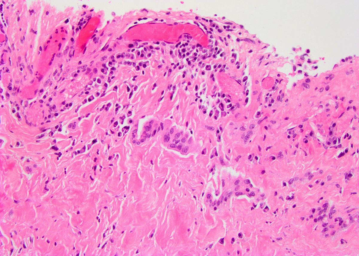
Microscopic (histologic) images
Contributed by Debra L. Zynger, M.D.

Entrapped glands
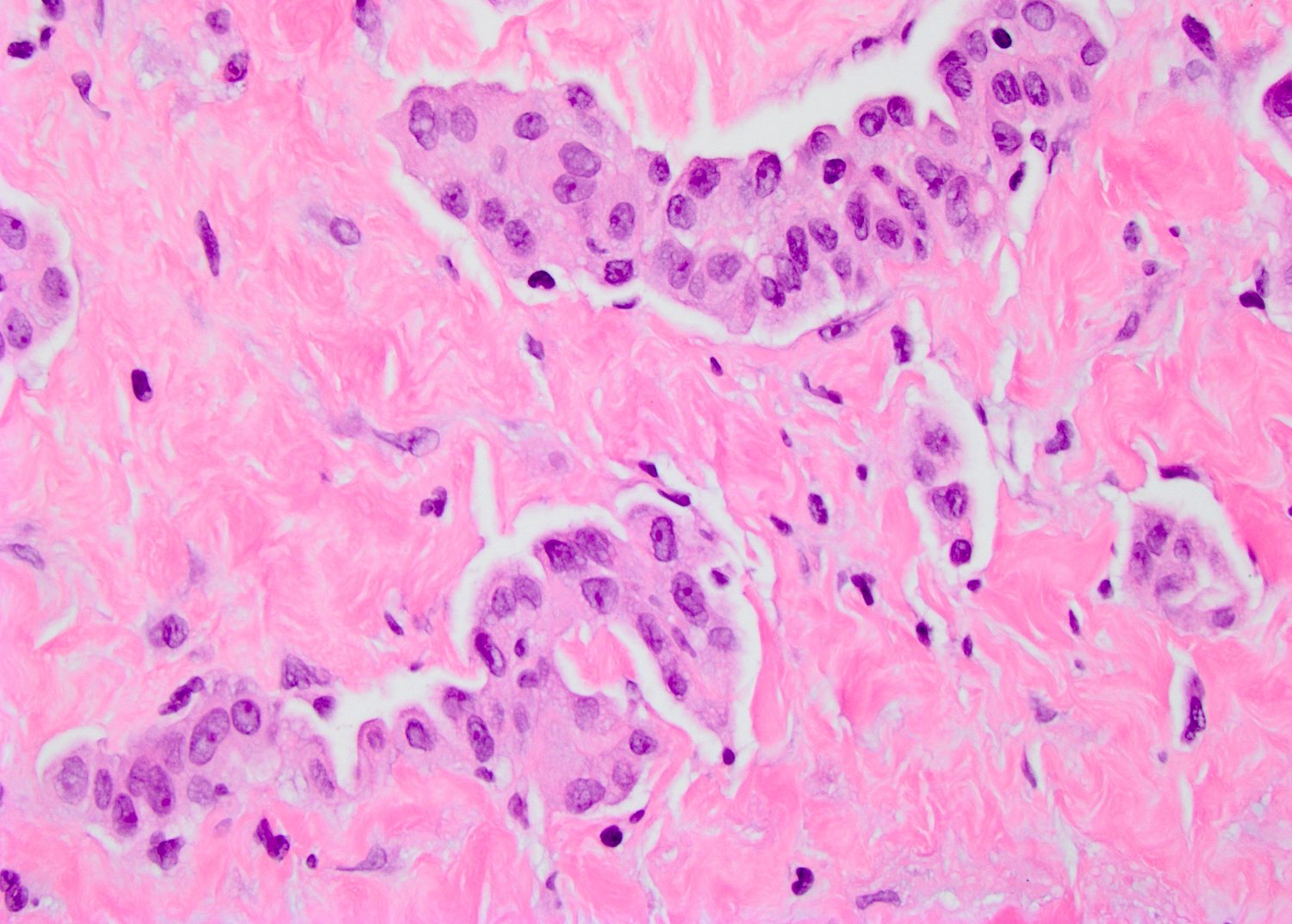
Cords, glands and small nests
Gland formation
Entrapped mesothelium

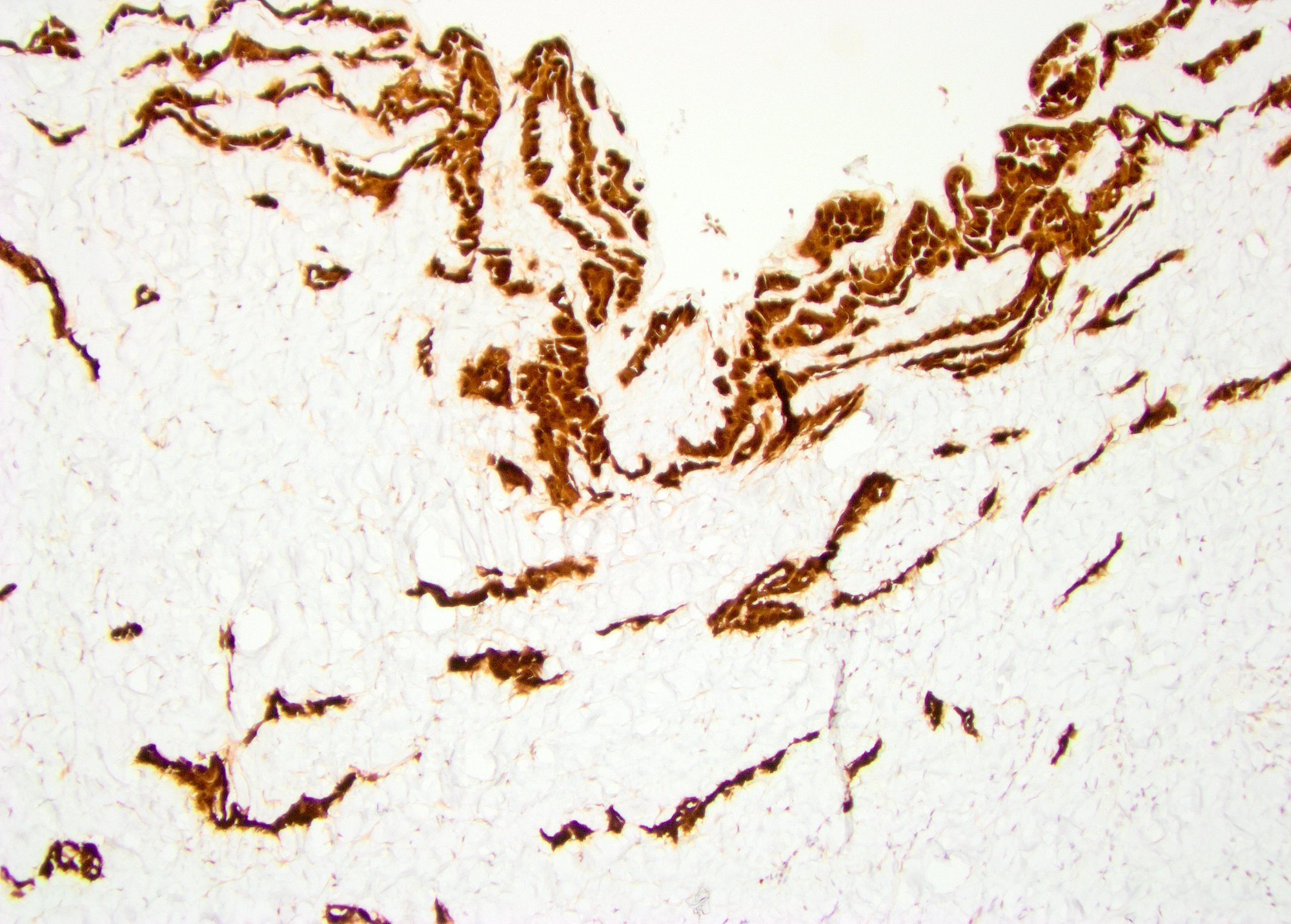
WT1
Calretinin
Positive stains
- Keratins, AE1 / AE3
- CK7
- Mesothelial markers: Calretinin, CAIX, CK5/6, D2-40, CD141, WT1 (Mod Pathol 2005;18:S131, Diagn Cytopathol 2009;37:748, Virchows Arch 2012;460:89)
Negative stains
- p53 (Am J Surg Pathol 2014;38:54)
- Ki67 (6% in reactive mesothelial cells) (Am J Surg Pathol 2014;38:54)
- GLUT1 (0% - 27% in reactive mesothelial cells) (Am J Surg Pathol 2014;38:54)
- CK20, p63, PAX8
Molecular / cytogenetics description
- 9p21 not deleted (Am J Surg Pathol 2014;38:54)
Differential diagnosis
- Mesothelioma
- Grossly visible lesion, usually exophytic in this location (Diagn Pathol 2011;6:78)
- Forms a mass with more tightly packed glands
- True invasion
- Haphazard arrangement of glands rather than in a linear arrangement
- More nuclear atypia and mitotic activity
- Adenocarcinoma:
- Forms a mass
- Desmoplastic reaction
- Back to back glands
- Mitotic activity (Mod Pathol 2005;18:S131)
- Mesothelial markers usually negative
- Adenomatoid tumor:
- Forms a mass lesion near epididymis
- Positive for mesothelial markers (Semin Diagn Pathol 2003;20:272)
- Histiocytic inflammation:
- Keratin negative (Am J Surg Pathol 2014;38:54)
Practice question #1
- Which of the following is a feature of a hernia sac with mesothelial entrapment?
- Glands parallel to surface
- GLUT1 and p53 positivity
- Homozygous for 9p21 deletion
- Invasion into muscle
- Prominent mitotic activity
Practice answer #1
Practice question #2
- A 48 year old man presents with a hydrocele with the findings in the above picture. What is your diagnosis?
- Adenomatoid tumor
- Diffuse malignant mesothelioma
- Hernia sac with mesothelial entrapment
- Histiocytic inflammation
- Metastatic adenocarcinoma
Practice answer #2
C. Hernia sac with mesothelial entrapment
Comment Here
Reference: Hernia sac with mesothelial entrapment
Comment Here
Reference: Hernia sac with mesothelial entrapment
