

Thyroid & parathyroid
Other uncommon thyroid carcinomas
SETTLE
Last author update: 1 July 2016
Last staff update: 7 December 2023
Copyright: 2003-2024, PathologyOutlines.com, Inc.
PubMed Search: SETTLE thyroid


Table of Contents
Definition / general | Essential features | Terminology | Case reports | Treatment | Gross description | Microscopic (histologic) description | Microscopic (histologic) images | Cytology description | Positive stains | Negative stains | Differential diagnosisCite this page: Wei S. SETTLE. PathologyOutlines.com website. https://www.pathologyoutlines.com/topic/thyroidsettle.html. Accessed April 26th, 2024.
Definition / general
- Spindle Epithelial Tumor with Thymus-Like Differentiation
- Rare initially indolent tumor of neck in young patients (4 - 59 years old, median age 18 years), with delayed (after 5 years) metastases to lymph nodes or lungs, indolent even with metastasis (Head Neck 2015;37:746)
Essential features
- Low grade biphasic tumor with metastatic potential which occurs in young patients
Terminology
- Terminology first used in 1991 (Hum Pathol 1991;22:349)
- Synonym: Thyroid spindle cell tumor with mucinous cysts
Case reports
- 6 year old boy with circumscribed tumor with slightly gritty whorled appearance (Hum Pathol 2003;34:190)
- 20 year old woman with enlarged thyroid (Arch Pathol Lab Med 2006;130:405)
- 59 year old man with enlarged thyroid for adult life and recent rapid enlargement of thyroid (Mod Pathol 2000;13:1150)
Treatment
- Thyroidectomy, radiotherapy and chemotherapy are effective in cases with unresectable tumor or metastasis
Gross description
- Circumscribed or infiltrative, lobulated, white / tan cut surface
Microscopic (histologic) description
- Encapsulated or infiltrative tumor separated by sclerotic stroma
- Biphasic pattern: spindle cells and epithelial structures (cords, tubules, papillae or glandular formation)
- Rare monomorphic variant can have spindle cells or glandular only (Histopathology 1998;33:71)
- No or rare lymphocytes
- Mitotic activity or focal necrosis are rare
Microscopic (histologic) images
AFIP images
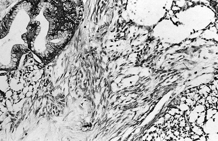
Spindled cells with mesenchymal appearance
Cytology description
- Tumor with minimal epithelial component was moderately cellular with single and loosely grouped spindle cells in homogeneous metachromatic material resembling amyloid (Diagn Cytopathol 2007;35:113)
Positive stains
Negative stains
Differential diagnosis
- Synovial sarcoma: translocation t(x;18) (SSX; SS18 / SYT), increased mitotic figures, no / weak cytokeratin staining, no sclerosing stroma, no epithelial component
- Medullary carcinoma: not biphasic, amyloid+, calcitonin+, chromogranin+
- Anaplastic carcinoma: pleomorphic spindle cells, increased mitotic figures, necrosis
- Ectopic thymoma: with TdT+ lymphocytes
- Malignant teratoma: with other immature tissue

