

Lung
Other carcinomas
Large cell
Authors: Emily O. Symes, M.D., Aliya N. Husain, M.D.
Editorial Board Member: Jefree J. Schulte, M.D.


Last author update: 6 January 2023
Last staff update: 20 January 2023
Copyright: 2003-2024, PathologyOutlines.com, Inc.
PubMed Search: Large cell undifferentiated carcinoma of the lung

Table of Contents
Definition / general | Essential features | Terminology | ICD coding | Epidemiology | Sites | Clinical features | Diagnosis | Radiology description | Prognostic factors | Case reports | Treatment | Gross description | Gross images | Microscopic (histologic) description | Microscopic (histologic) images | Virtual slides | Cytology description | Positive stains | Negative stains | Molecular / cytogenetics description | Sample pathology report | Differential diagnosis | Additional references | Board review style question #1 | Board review style answer #1 | Board review style question #2 | Board review style answer #2Cite this page: Symes EO, Husain AN. Large cell. PathologyOutlines.com website. https://www.pathologyoutlines.com/topic/lungtumorlargecell.html. Accessed May 6th, 2024.
Definition / general
- Resected tumors that lack morphologic or immunohistochemical differentiation toward small cell carcinoma, adenocarcinoma or squamous cell carcinoma (J Thorac Oncol 2015;10:1240)
- Null or unclear immunophenotype based on IHC analysis with TTF1, p40 and neuroendocrine markers (J Thorac Oncol 2019;14:1213)
Essential features
- Malignant, poorly differentiated epithelial neoplasm of lung composed of large, atypical cells
- No morphologic or immunohistochemical evidence of glandular, squamous or neuroendocrine differentiation
- Diagnosis of exclusion; diagnosis can only be made on resected tumors
Terminology
- Synonymous with large cell undifferentiated carcinoma and large cell anaplastic carcinoma
- Previously, large cell carcinoma included variants such as basaloid carcinoma, large cell neuroendocrine carcinoma, lymphoepithelioma-like carcinoma, clear cell carcinoma and large cell carcinoma with rhabdoid phenotype
- As of the 2015 WHO classification, large cell carcinoma is a diagnosis of exclusion, with reclassification of the former large cell carcinoma subtypes into different categories (J Thorac Oncol 2015;10:1243)
ICD coding
Epidemiology
- M > F
- Average age of 65 years
- Associated with cigarette smoking
- Becoming increasingly rare due to immunohistochemical and molecular studies that support the reclassification of entities formerly diagnosed as large cell carcinoma (Sci Transl Med 2013;5:209ra153)
Sites
- Peripheral lung
- 50% with connection to large airways
Clinical features
- Cough, chest pain, shortness of breath
- Typically larger than 5 cm
Diagnosis
- Diagnosis of exclusion
- Small biopsy specimens and cytology specimens should not be used for diagnosis
- According to the 2021 WHO, only resection specimens with exclusion of a differentiated component, via thorough sampling, can be classified as large cell carcinoma (J Thorac Oncol 2022;17:362)
- NSCC, NOS (nonsmall cell carcinoma, not otherwise specified) should be used in small biopsies or cytology specimens
Radiology description
- Usually large, lobulated or well marginated, peripheral, heterogeneously enhancing mass, often containing central necrosis (Ghosh: Handbook of Imaging in Pulmonary Disease, 1st Edition, 2021)
- Rapid growth is common with metastasis (chest wall, thoracic lymph nodes, distant) at presentation
- PET CT preferred over CT for identification of full extent of disease and metastases
Prognostic factors
- Generally poor prognosis with limited treatment options (J Thorac Oncol 2019;14:1213)
Case reports
- 52 year old man with respiratory failure and obstructing endobronchial mass (Cureus 2018;10:e2202)
- 58 year old man with prolonged cough and hemoptysis (Thorac Cancer 2021;12:1141)
- 80 year old woman with shoulder pain and vertebral compression fracture (Respir Med Case Rep 2020;31:101197)
Treatment
- Dependent on stage
- Surgical excision, chemotherapy or radiation therapy
Gross description
- Usually peripheral lung and unifocal; large, circumscribed with well defined borders and bulging, lobulated, homogeneous gray-white "fish flesh" cut surface
- Internal necrosis and hemorrhage are common (Fletcher: Diagnostic Histopathology of Tumors, 5th Edition, 2020)
Gross images
Contributed by Debra L. Zynger, M.D.
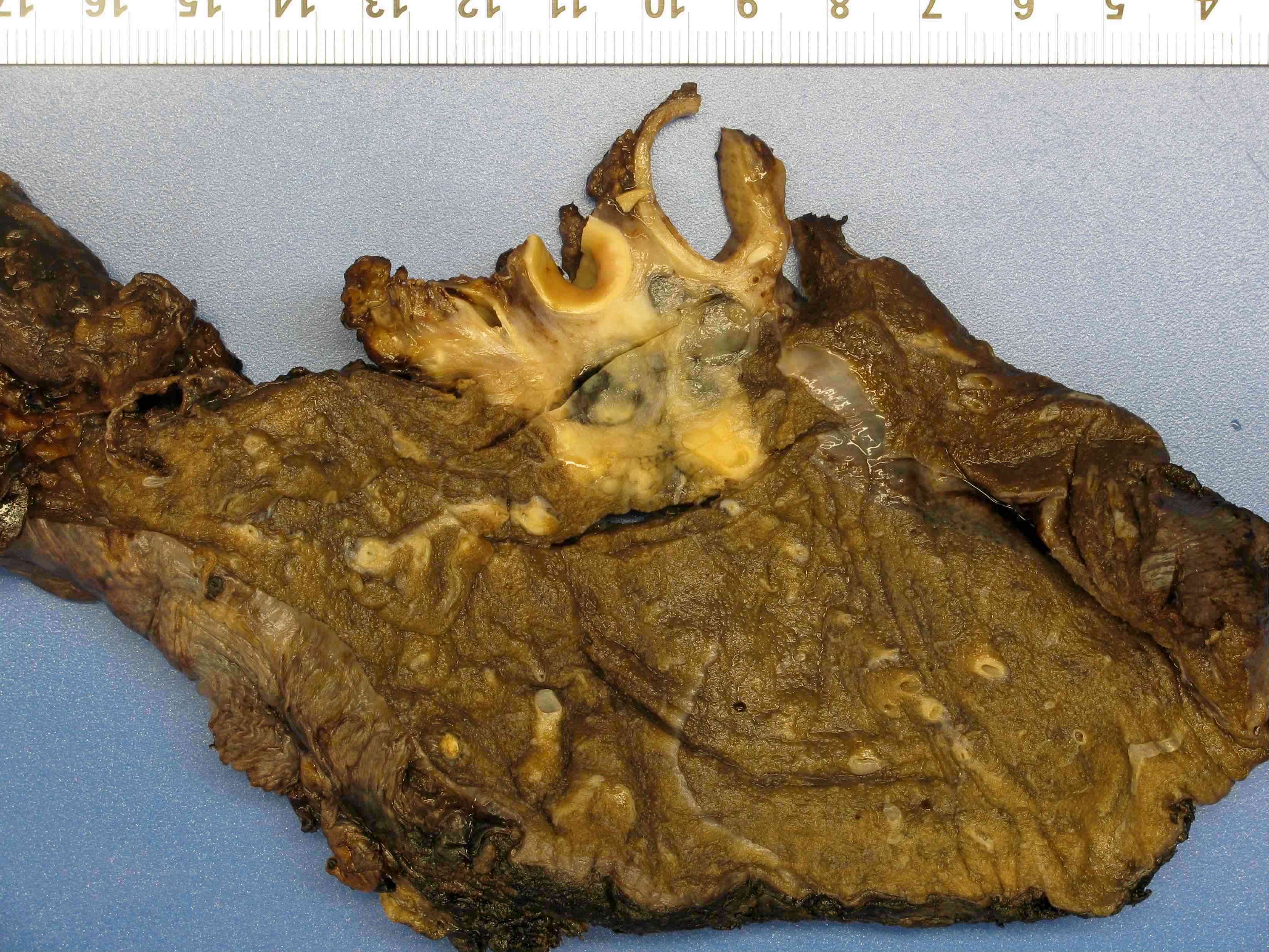
Right lung mass
Microscopic (histologic) description
- Sheets or solid nests of round to polygonal cells
- Moderately abundant cytoplasm, well defined cell borders
- Vesicular nuclei, prominent nucleoli
- Foci of central necrosis and hemorrhage may be present
- No glandular, squamous or neuroendocrine differentiation is present (Fletcher: Diagnostic Histopathology of Tumors, 5th Edition, 2020)
- Reference: Clin Chest Med 2020;41:67
Microscopic (histologic) images
Contributed by Emily O. Symes, M.D.
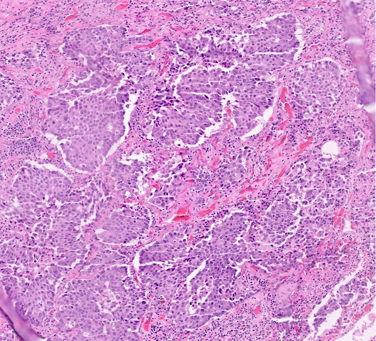
Sheets of tumor cells

Necrosis and lymphovascular invasion

Extensive necrosis
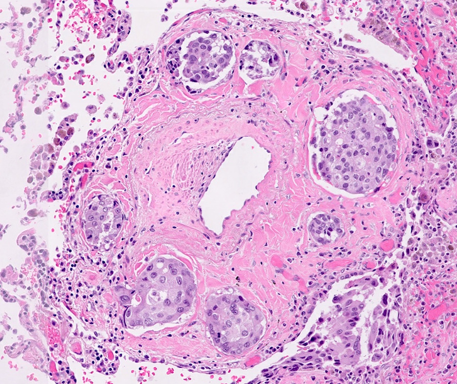
Lymphovascular invasion
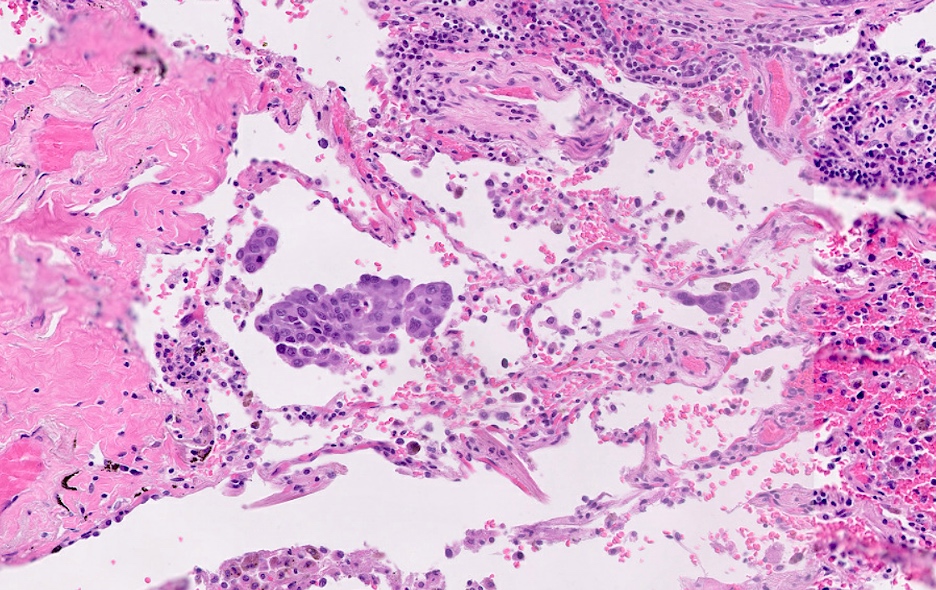
Spread through air spaces (STAS)
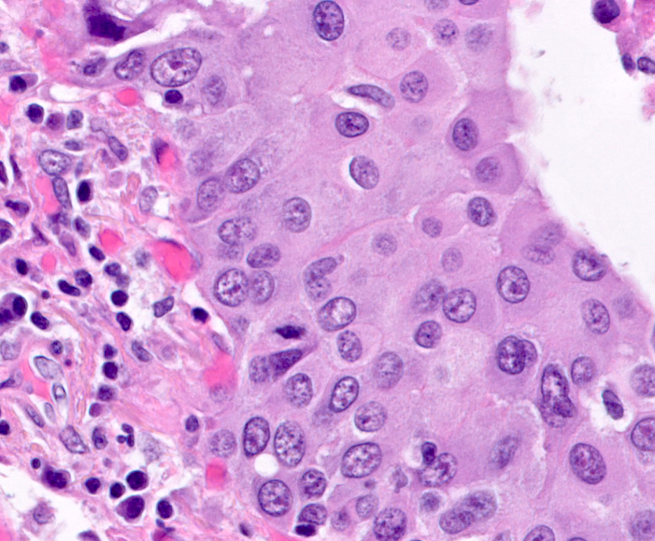
High power tumor cells

TTF1
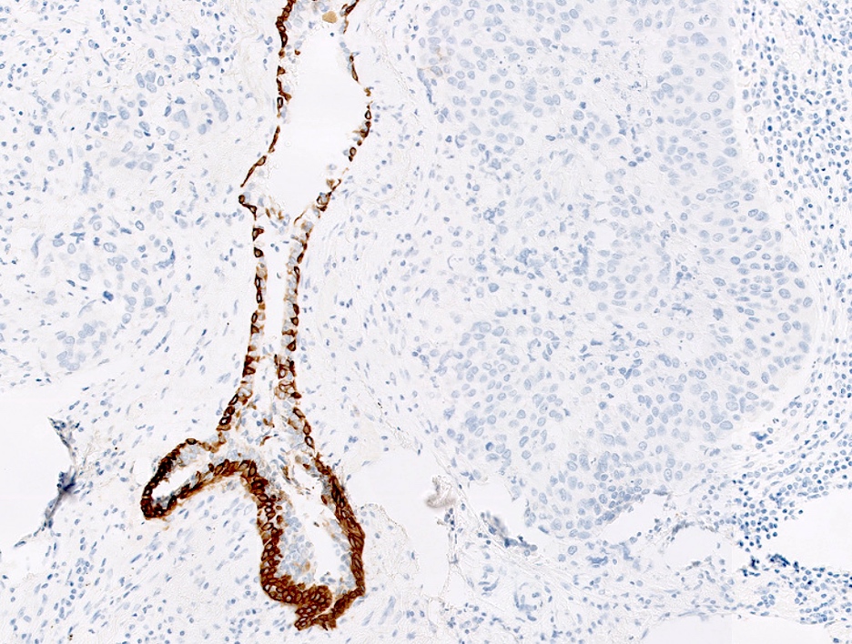
CK5/6

p40
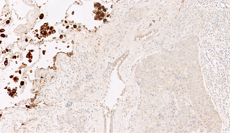
Napsin A

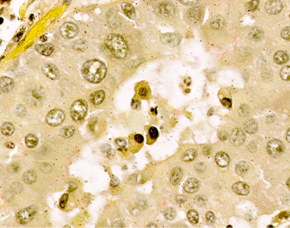
PDL1

Mucin stain
Virtual slides
Images hosted on other servers:


Undifferentiated large cell carcinoma


Undifferentiated large cell carcinoma
Cytology description
- Large pleomorphic cells with a moderate to abundant amount of cytoplasm, vesicular nuclei, prominent nucleoli
Positive stains
- Broad spectrum and low molecular weight cytokeratins, such as CAM5.2
Negative stains
- TTF1, Napsin, mucicarmine, p40, p63, synaptophysin, chromogranin, CD56
Molecular / cytogenetics description
- Molecular studies show that some null phenotype large cell carcinomas contain mutations seen in conventional adenocarcinoma (e.g., KRAS, NRAS, MET, STK11) and squamous cell carcinoma (e.g., PIK3CA amplification and mutation); this indicates that tumors classified as large cell carcinomas could represent poorly / undifferentiated nonsmall cell lung cancer (NSCLC) (J Thorac Oncol 2019;14:1213, Sci Transl Med 2013;5:209ra153)
Sample pathology report
- Lung, left, resection:
- Large cell carcinoma (1.1 cm) (see comment)
- Comment: Immunohistochemical studies are performed with appropriate controls. The tumor is negative for TTF1, Napsin A, CK5/6 and p40. There is no morphologic evidence of differentiation to adenocarcinoma or squamous cell carcinoma. The histologic features are not those of neuroendocrine carcinoma. Given these findings, the tumor is best classified as large cell carcinoma. PDL1 immunostain shows weak expression in 20% of tumor cells.
Differential diagnosis
- Basaloid squamous cell carcinoma:
- Expresses squamous markers (i.e., p40)
- Large cell neuroendocrine carcinoma:
- Expresses neuroendocrine markers (i.e., chromogranin, synaptophysin, CD56)
- Solid adenocarcinoma:
- Metastatic carcinoma:
Additional references
Board review style question #1
Large cell carcinoma of the lung will generally stain for which of the following immunohistochemical markers?
- CAM5.2
- Chromogranin
- p40
- SOX10
- TTF1
Board review style answer #1
Board review style question #2
Which one of the following special stains is shown in the image above?
- CK5/6
- Chromogranin
- TTF1
- Mucin
- p40
Board review style answer #2
D. Mucin. Tumor cells are negative for mucin immunostaining but show degenerative vacuoles, which can mimic mucin on H&E.
Comment Here
Reference: Large cell carcinoma
Comment Here
Reference: Large cell carcinoma
