Ovary
Sex cord stromal tumors
Pure stromal tumors
Steroid cell tumor
Editorial Board Member: Kyle Devins, M.D.
Deputy Editor-in-Chief: Gulisa Turashvili, M.D., Ph.D.


Last author update: 3 April 2024
Last staff update: 3 April 2024
Copyright: 2002-2025, PathologyOutlines.com, Inc.
PubMed search: Ovarian steroid cell tumor
Table of Contents
Definition / general | Essential features | Terminology | ICD coding | Epidemiology | Sites | Pathophysiology | Etiology | Clinical features | Diagnosis | Laboratory | Radiology description | Radiology images | Prognostic factors | Case reports | Treatment | Clinical images | Gross description | Gross images | Frozen section description | Frozen section images | Microscopic (histologic) description | Microscopic (histologic) images | Virtual slides | Cytology description | Cytology images | Positive stains | Negative stains | Molecular / cytogenetics description | Videos | Sample pathology report | Differential diagnosis | Additional references | Practice question #1 | Practice answer #1 | Practice question #2 | Practice answer #2Cite this page: Amlashi FG, Kalir T. Steroid cell tumor. PathologyOutlines.com website. https://www.pathologyoutlines.com/topic/ovarytumorsteroidcelltumornos.html. Accessed September 25th, 2025.
Definition / general
- Ovarian stromal tumor composed of steroid cells with malignant potential
Essential features
- Mostly unilateral
- Half of cases have androgenic manifestations
- Microscopic features include diffuse sheets of polygonal to rounded tumor cells with moderate to abundant cytoplasm (Am J Surg Pathol 1987;11:835)
Terminology
- Steroid cell tumor, not otherwise specified (NOS)
ICD coding
- ICD-O
- ICD-11: 2C73.Y & XH4L39 - other specified malignant neoplasms of the ovary & steroid cell tumor, malignant
Epidemiology
- Rare (< 1% of ovarian tumors)
- Average age in general: 43 year old
- Average age in von Hippel-Lindau: 27 years old (Int J Clin Exp Pathol 2022;15:332)
- In an analysis of 63 cases, patients with clinically malignant tumors were 16 years older than patients with benign tumor (Am J Surg Pathol 1987;11:835)
Sites
- Ovary
- Extraovarian origin (very rare) (Int J Gynecol Pathol 2019;38:151)
Pathophysiology
- Tumors are presumed to be of ovarian stromal cell origin
Etiology
- Dysregulation in the hypoxia signaling pathway (Endocr Relat Cancer 2023;30:e230179)
- VHL aberration in von Hippel-Lindau (Int J Clin Exp Pathol 2022;15:332)
- Genomic instability, MDM1 / CDK4 coamplification, ATRX rearrangement, MDM2 / CDK4 copy number gain, BAP1 mutation in malignant tumors
- In nonmalignant tumors: mutation in FH, CTTNB1, CASP10 and P53 (Am J Surg Pathol 2023;47:1398)
Clinical features
- Androgenic (~50%), estrogenic changes including rare examples of isosexual pseudoprecocity (~10%), occasional progestogenic changes, Cushing syndrome or elevated cortisol levels (uncommon)
- Rarely with hypercalcemia, erythrocytosis, ascites or acute heart failure
- Reported in von Hippel-Lindau (both types I and II), with the most common presentation including abnormal uterine bleeding, amenorrhea and infertility (Int J Clin Exp Pathol 2022;15:332)
Diagnosis
- Histologic examination
Laboratory
- In patients with androgenic changes, Cushing syndrome or both: elevated urinary levels of 17-ketosteroids and 17-hydroxycorticosteroids, serum testosterone and androstenedione
- In Cushing syndrome: elevated free cortisol in the blood or urine
Radiology description
- Ultrasound: hypoechoic / isoechoic, homogenous or heterogeneous texture, characterized by abundant blood flow signals (Oncol Lett 2022;24:370)
- Computed tomography (CT): low density area, consistent with lipid content within the mass (AJR Am J Roentgenol 2007;188:W393, Magn Reson Med Sci 2019;18:251)
- Magnetic resonance imaging (MRI): intermediate signal intensity on T2 weighted images and avid contrast enhancement (indicating hypervascularity of the tumor); signal loss on out of phase MR imaging, consistent with lipid content within the mass (AJR Am J Roentgenol 2007;188:W393, Magn Reson Med Sci 2019;18:251)
- Overall CT and MRI findings are variable, owing to the amount of lipid component and fibrous stroma (AJR Am J Roentgenol 2007;188:W393, Magn Reson Med Sci 2019;18:251)
Radiology images
Images hosted on other servers:


Enhancing adnexal mass

Fat containing pelvic mass


Homogeneous isoechoic mass

Solid adnexal mass
Prognostic factors
- Clinically malignant in 25 - 43% of cases (Am J Surg Pathol 1987;11:835)
- Patients with clinically malignant tumor were on average 16 years older than patients with benign tumors in one series (Am J Surg Pathol 1987;11:835)
- No malignant steroid cell tumors have been reported in patients in their first 2 decades
- Rare tumors have recurred up to 19 years postoperatively (Am J Surg Pathol 1987;11:835)
- Extraovarian spread of tumor is present at the time of operation in a small minority of cases; 3 cases with Cushing disease with extensive intra-abdominal spread of tumor (Int J Gynecol Pathol 1987;6:40)
- Features associated with malignancy (Am J Surg Pathol 1987;11:835)
- 2+ mitoses per 10 high power fields (92% malignant)
- Necrosis (86% malignant)
- 7 cm or larger (78% malignant)
- Intratumoral hemorrhage (77% malignant)
- Grade 2 or 3 nuclear atypia (64% malignant)
- Aggressive behavior with 4 or more malignant features (Am J Surg Pathol 2023;47:1398)
Case reports
- 16 year old girl with history of von Hippel-Lindau (VHL) syndrome and a 3 cm well defined nodule in right ovary (Int J Gynecol Pathol 2020;39:473)
- 21 and 23 year old women with malignant and benign steroid cell tumors, NOS (J Ovarian Res 2013;6:53)
- 28 year old woman with acute heart failure, virilization and 7 cm right adnexal mass (J Obstet Gynaecol Res 2020;46:1211)
- 35 year old woman with virilization and a pelvic mass (Arch Pathol Lab Med 2006;130:113)
- 56 and 74 year old women with mixed steroid cell tumor and fibroma in extraovarian and ovarian tissue, respectively (Int J Gynecol Pathol 2019;38:151)
- 64 year old woman with collision signet ring stromal tumor and steroid cell tumor of the ovary (Int J Gynecol Pathol 2017;36:261)
Treatment
- Unilateral salpingo-oophorectomy if fertility sparing is desired; otherwise, total hysterectomy and bilateral salpingo-oophorectomy
- Adjuvant chemotherapy has been used for cases with malignant type; however, there is no consensus (Case Rep Oncol 2020;13:358, Case Rep Obstet Gynecol 2016;2016:6184573)
- Tumor excision, chemotherapy and gonadotropin releasing hormone (GnRH) analog therapy are reported for disease recurrence or progression (BMC Endocr Disord 2022;22:265, Gynecol Oncol Rep 2023;46:101169)
Clinical images
Images hosted on other servers:

Hirsutism

Male pattern baldness
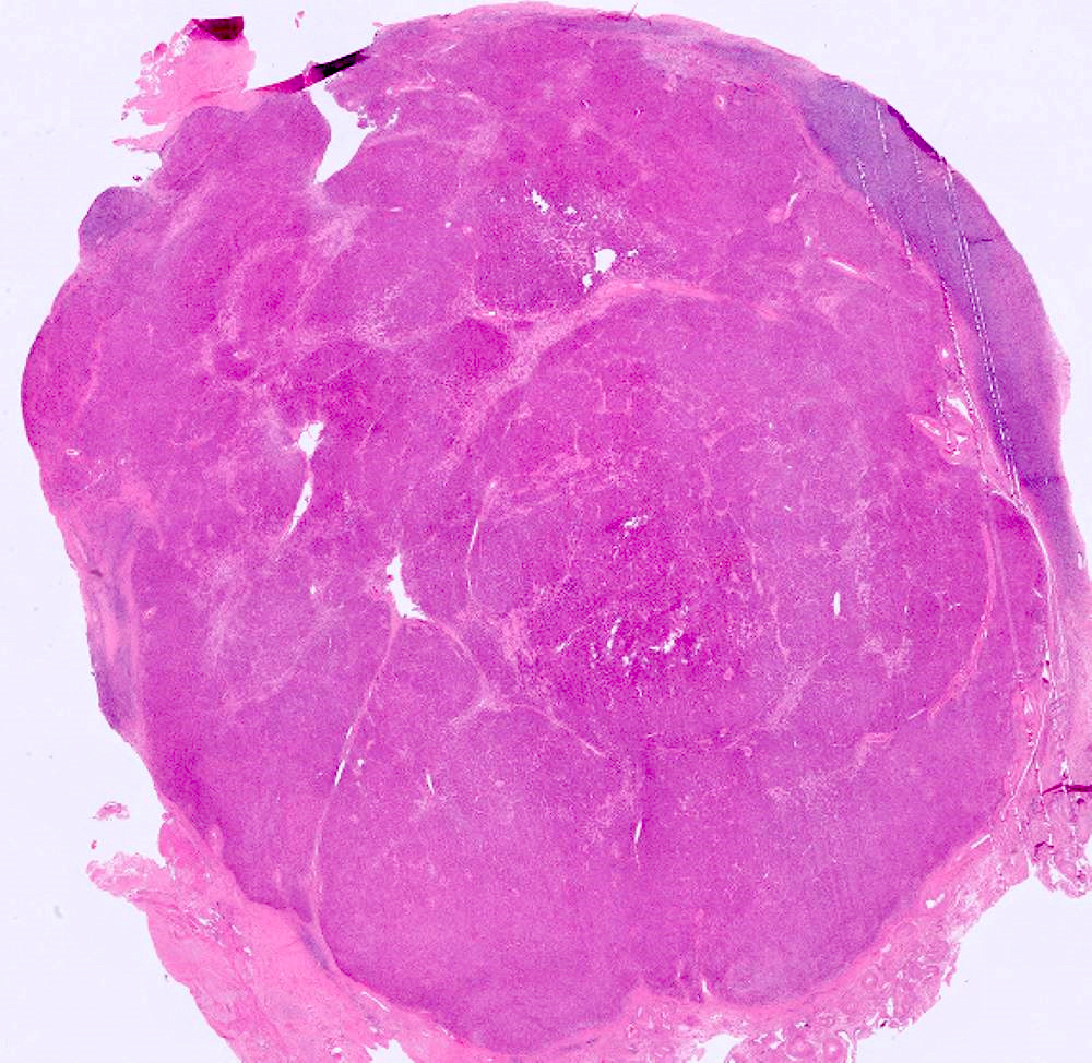
Gross description
- Wide size range (mean size: 8.4 cm) (Am J Surg Pathol 1987;11:835)
- Unilateral (95%)
- Solid, well circumscribed, occasionally lobulated
- Sections from yellow-orange (lipid rich) to red-brown (lipid poor) to dark brown-black (abundant lipochrome pigment)
- Occasionally with necrosis, hemorrhage and cystic degeneration
Gross images
Images hosted on other servers:

Yellow cut surface

Metastatic steroid cell tumor

Lobulated cut surface

Nodular solid tumor

Tan-brown, well circumscribed tumor with hemorrhage
Frozen section description
- Diffuse or nested arrangement of tumor cells
- Cells with abundant eosinophilic or clear, foamy cytoplasm
Frozen section images
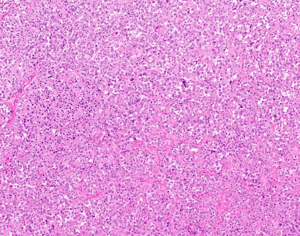
Contributed by Fatemeh Ghazanfari Amlashi, M.D. and Tamara Kalir, M.D., Ph.D.
Monotonous sheets of cells
Abundant cytoplasm
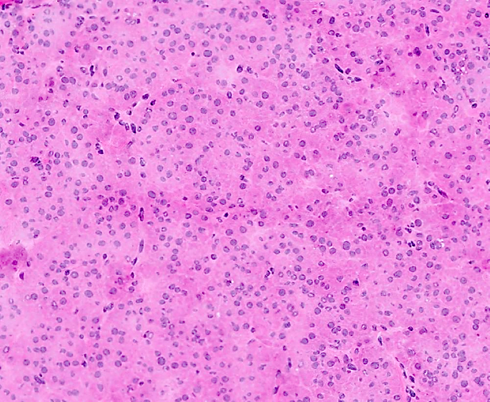
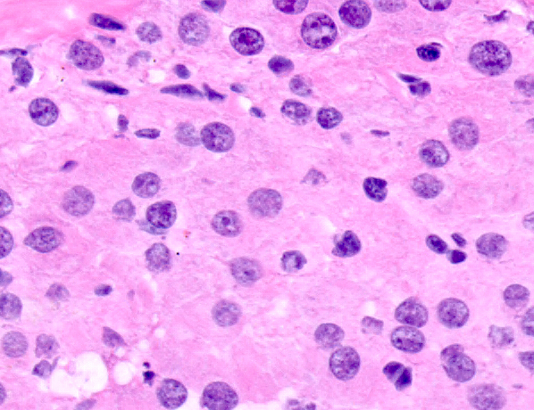
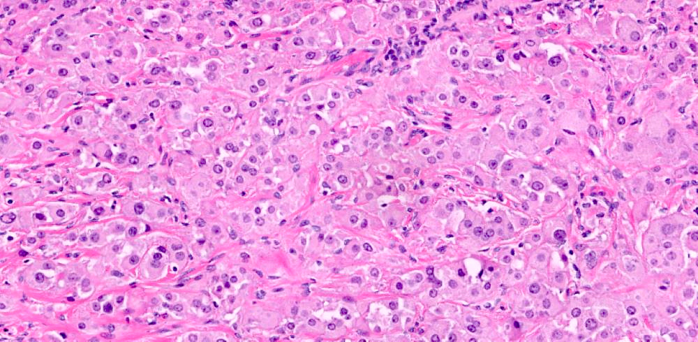
Microscopic (histologic) description
- Architectural pattern: diffuse (most common) but occasionally in nests, cords, pseudoglandular and follicle-like arrangements (Am J Surg Pathol 1987;11:835)
- Stroma: usually sparse (85%) but may be fibrotic or hyalinized, rarely edematous or myxoid, exceptionally with calcification and psammoma body formation
- Polygonal to rounded tumor cells with distinct cell borders, central nuclei and moderate to abundant cytoplasm
- Tumor cell cytoplasm: eosinophilic and granular (lipid poor) to vacuolated and spongy (lipid rich); tumors with a mixture of 2 cell types are also present
- More lipid rich cytoplasm in this tumor compared to other subtypes of steroid cell tumor
- Lipochrome pigment present (40%)
- Rarely, signet ring appearance
- Adipocytic metaplasia and hyaline globule (unusual) (Am J Surg Pathol 2023;47:1398)
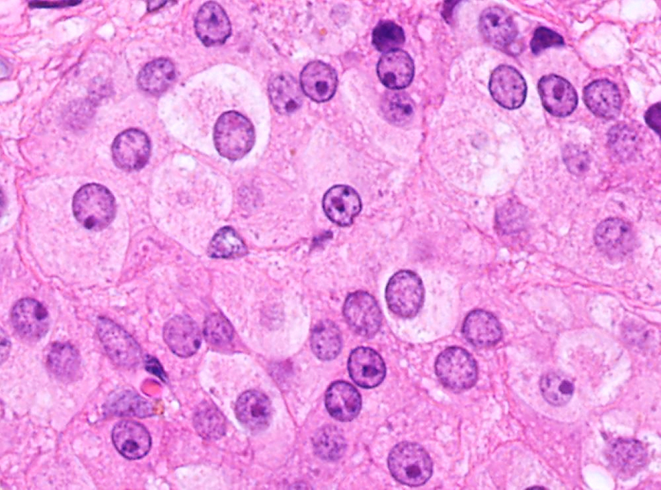
- Usually absent / slight nuclear atypia along with low mitotic activity (< 2 mitotic figures/10 high power fields); those with grades 1 - 3 nuclear atypia associated with increased mitotic activity (up to 15 mitotic figures/10 high power fields)
- Necrosis and hemorrhage are occasionally present, particularly in tumors with significant cytologic atypia
- Metastatic tumor is similar to the primary tumor in some cases but more poorly differentiated in others
- Stromal hyperthecosis may be seen in the adjacent ovarian stroma and contralateral ovary, particularly in small tumors
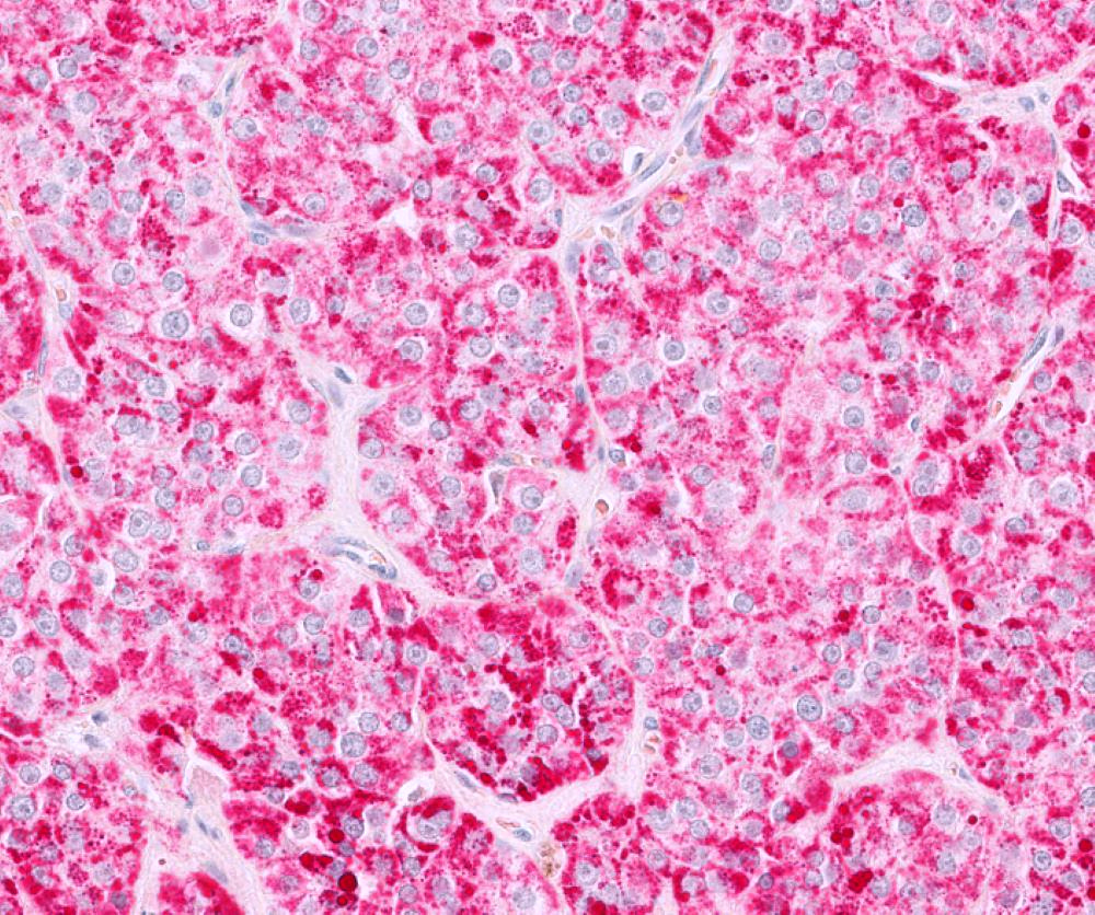
Microscopic (histologic) images
Contributed by Fatemeh Ghazanfari Amlashi, M.D., Tamara Kalir, M.D., Ph.D. and AFIP
Well defined tumor
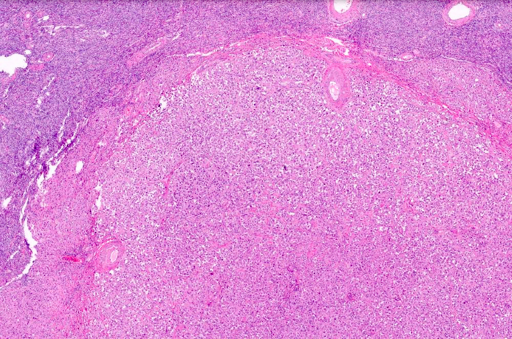
Sparse stroma
Diffuse sheets
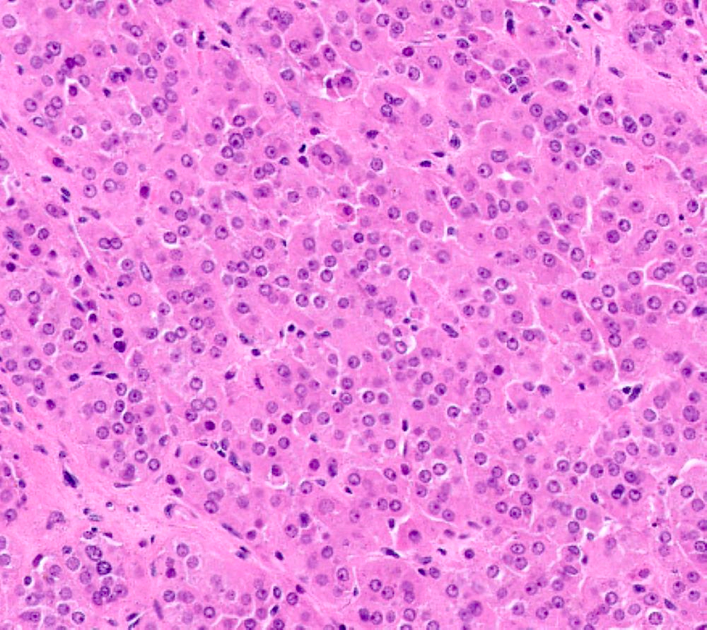
Moderate to abundant cytoplasm
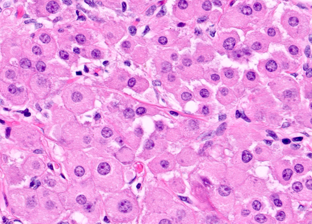
Round to polygonal cells
Distinct cell borders
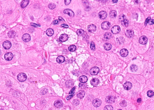
Spongy and eosinophilic cytoplasm
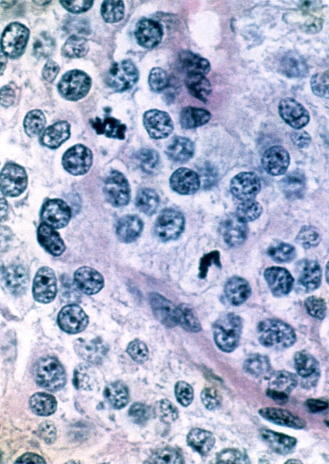
2 atypical mitotic figures
Inhibin
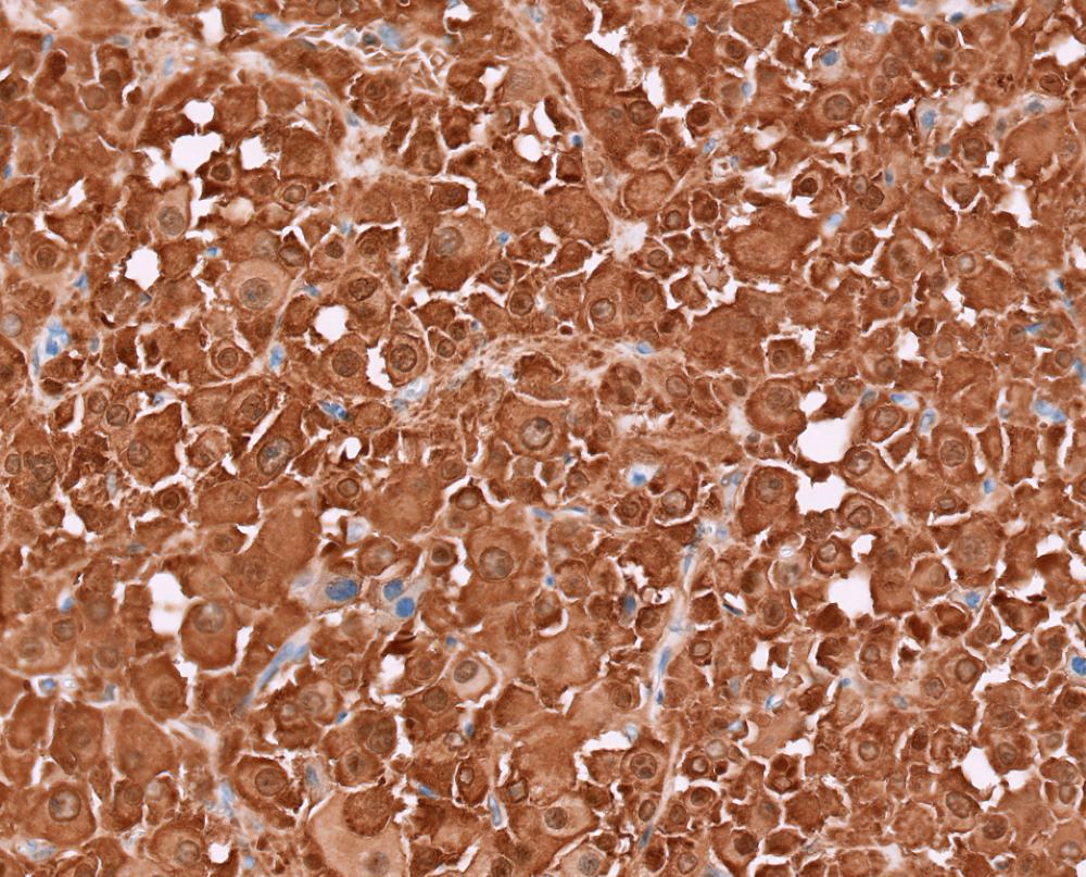
Calretinin
MelanA
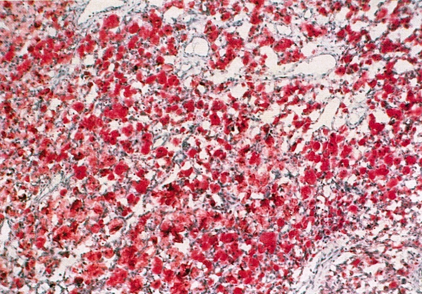
Abundant lipid (oil red O stain)
Virtual slides
Images hosted on other servers:

Lipid rich tumor cells
Cytology description
- Large polygonal to round cells
- Arranged in sheets and attached with vascular stromal tissue fragments (J Cytol 2015;32:284)
- Cells with small central round nuclei with conspicuous nucleoli and abundant granular to pale foamy cytoplasm
- Ascites in malignant steroid cell tumor: isolated tumor cells or clusters with slight overlapping and cannibalism formation (Acta Cytol 2017;61:165)
Cytology images
Images hosted on other servers:

Large polygonal cells
Positive stains
Negative stains
Molecular / cytogenetics description
- Pathogenic variants in several hypoxia related genes, including HIF1A, VHL, SDHB, SRC, IDH2 and FOXO4 (Endocr Relat Cancer 2023;30:e230179)
- Mutation in FH, CTTNB1, CASP10 and P53 (Am J Surg Pathol 2023;47:1398)
- Malignant steroid cell tumor with genomic instability, MDM1 / CDK4 coamplification, ATRX rearrangement, MDM2 / CDK4 copy number gain, BAP1 mutation (Am J Surg Pathol 2023;47:1398)
Videos
Steroid cell tumor
Sample pathology report
- Right ovary and fallopian tube, salpingo-oophorectomy:
- Ovarian steroid cell tumor, not otherwise specified (6 cm size) (see comment)
- Fallopian tube with no significant pathologic change
- Comment: Sections show a well circumscribed neoplasm composed of abundant clear cytoplasm. There is mild nuclear atypia. No mitosis or necrosis identified. Immunostains show the tumor cells are positive for inhibin, calretinin and negative for CD10, supporting the diagnosis.
- Left ovary and fallopian tube, salpingo-oophorectomy:
- Ovarian steroid cell tumor, not otherwise specified (12 cm size) (see comment)
- Fallopian tube with no significant pathologic change
- Comment: Sections show a well circumscribed neoplasm composed of sheets of round cells with abundant eosinophilic granular cytoplasm. Intratumoral hemorrhage and focal necrosis is present. Moderate nuclear atypia and mitoses (4/10 high power fields) are seen. The ovarian capsule is intact and uninvolved by the tumor cells. Immunostains show the tumor cells are positive for calretinin and inhibin, supporting the diagnosis.
Differential diagnosis
- Ovarian clear cell carcinoma:
- Periodic acid-Schiff (PAS) positive, diastase sensitive, glycogen rich cytoplasm and eccentric nuclei
- Variety of architectural patterns (Int J Gynecol Pathol 2019;38:S40)
- Almost always accompanied by a variable component of clear and hobnail cells
- Metastatic renal cell carcinoma:
- Glycogen rich cytoplasm and eccentric nuclei
- Prominent sinusoidal vascular framework (Int J Gynecol Pathol 2018;37:525)
- Areas with tubular differentiation
- History of a prior or concurrent renal mass
- No endocrine manifestation
- CD10 positive
- Pregnancy luteoma:
- Third trimester of pregnancy
- Bilateral (33%) (Am J Surg Pathol 2014;38:239)
- Multiple (50%)
- Abundant eosinophilic cytoplasm containing little or no lipid
- Mitotic figures may be numerous, up to 2 - 3/10 high power fields
- Leydig cell tumor:
- Reinke crystalloids (Am J Surg Pathol 2023;47:1398)
- Acellular eosinophilic zone
- Nuclear clustering
- Fibrinoid material within vessel walls
- Located in ovarian hilum
- Sertoli cell tumor (lipid rich):
- At least focal tubular differentiation in adequately sampled tumors
- Absence of lipochrome pigments
- Metastatic melanoma:
- Bilateral (40%)
- No endocrinologic manifestation
- More malignant nuclear features than steroid cell tumors
- MelanA, S100 positive (Am J Surg Pathol 2004;28:771)
- Inhibin, calretinin, SF1 negative
- Oxyphilic struma ovarii:
- Association with other teratomatous elements (Endocr Pathol 2015;26:342)
- Presence of colloid
- Thyroglobulin positive
- Hepatoid yolk sac tumor:
- Elevated serum alpha fetoprotein (Am J Surg Pathol 2022;46:309)
- Glandular lumens may be seen
- AFP positive (Cancer 1982;50:2355)
Additional references
Practice question #1
Which of the following statements is correct regarding steroid cell tumor, not otherwise specified (NOS) of the ovary?
- Inhibin+, calretinin+, MelanA+, PAX8-
- Less than 1% risk of malignancy
- Mostly bilateral
- Various architectural patterns
Practice answer #1
A. Inhibin+, calretinin+, MelanA+, PAX8-. Steroid cell tumor, NOS shows positive inhibin, calretinin and MelanA and negative PAX8. Note that MelanA (A103 clone) can be expressed in steroid cell tumor and the presence of it should not necessarily prompt the diagnosis of melanoma. Answer B is incorrect because steroid cell tumor has a 30% risk of malignancy. Answer C is incorrect because steroid cell tumors are mostly unilateral. Answer D is incorrect because having various architectural patterns is a feature of ovarian clear cell carcinoma.
Comment Here
Reference: Steroid cell tumor
Comment Here
Reference: Steroid cell tumor
Practice question #2
A 40 year old woman presented with 5 cm, solid, well defined adnexal mass. Which of the following features is in favor of steroid cell tumor, NOS?
- Acellular eosinophilic material and nuclear clustering
- Location in hilum
- Reinke crystal
- Sheets of tumor cells with distinct cell borders and clear to foamy cytoplasm
Practice answer #2
D. Sheets of tumor cells with distinct cell borders and clear to foamy cytoplasm. Steroid cell tumor, NOS is mostly seen as sheets of tumor cells with distinct cell borders and can have clear to foamy cytoplasm in lipid rich tumor. Answers A, B and C are incorrect because these are features of Leydig cell tumor.
Comment Here
Reference: Steroid cell tumor
Comment Here
Reference: Steroid cell tumor
