Ovary
Other carcinomas
Carcinosarcoma
Editorial Board Member: Kyle Devins, M.D.
Deputy Editor-in-Chief: Gulisa Turashvili, M.D., Ph.D.


Last author update: 21 January 2025
Last staff update: 21 January 2025
Copyright: 2003-2025, PathologyOutlines.com, Inc.
PubMed Search: Carcinosarcoma ovary
Table of Contents
Definition / general | Essential features | Terminology | ICD coding | Epidemiology | Sites | Pathophysiology | Etiology | Clinical features | Diagnosis | Laboratory | Radiology description | Prognostic factors | Case reports | Treatment | Gross description | Gross images | Microscopic (histologic) description | Microscopic (histologic) images | Positive stains | Negative stains | Molecular / cytogenetics description | Sample pathology report | Differential diagnosis | Additional references | Practice question #1 | Practice answer #1 | Practice question #2 | Practice answer #2Cite this page: Ammann L, Kaseb H. Carcinosarcoma. PathologyOutlines.com website. https://www.pathologyoutlines.com/topic/ovarytumormmt.html. Accessed September 21st, 2025.
Definition / general
- Biphasic, malignant tumor with high grade epithelial and sarcomatous components
Essential features
- Rare and aggressive ovarian neoplasm (Gynecol Oncol 2016;142:248, Curr Treat Options Oncol 2023;24:1667)
- Carcinoma variant rather than a true mixed epithelial and mesenchymal tumor (WHO, 2020) (Histopathology 2022;80:762)
- Histology consists of both high grade carcinomatous and sarcomatous elements
- High morbidity and mortality rate, with median overall survival of < 2 years (Curr Treat Options Oncol 2023;24:1667)
- Stage is the best predictor of outcome and most patients present at advanced stage
Terminology
- Ovarian carcinosarcoma (OCS)
- Previously called malignant mixed Müllerian tumor (MMMT) (Curr Treat Options Oncol 2023;24:1667)
ICD coding
- ICD-O: 8980/3 - carcinosarcoma, NOS
Epidemiology
- Rare, OCS accounts for < 5% of ovarian malignancies (Curr Treat Options Oncol 2023;24:1667)
- Occurs mainly in postmenopausal, low parity women; median age at diagnosis is typically between 60 and 70 years old (Histopathology 2000;37:427)
- Higher prevalence in Black individuals (African Americans)
- ~90% of OCS cases exhibit malignant spread (Int J Gynecol Cancer 2014;24:S55)
Sites
- More common in other sites such as uterus; rarely involves ovaries, fallopian tubes or cervix
- High rates of lymph node metastasis and vascular invasion
- > 90% of carcinosarcomas spread beyond the ovaries and 33% of cases are associated with peritoneal effusion (Front Oncol 2023;13:1278300)
Pathophysiology
- 3 different theories proposed: conversion theory, the collision theory and the combination theory
- Conversion theory
- This is the most accepted theory as shown by studies indicating most carcinosarcomas are monoclonal (Int J Gynecol Pathol 2003;22:368)
- Epithelial origin with epithelial - mesenchymal transition
- Carcinomatous portion arises first, over time differentiating into the sarcomatous portion (Gynecol Oncol 2016;142:248)
- Cells in a carcinomatous component continuously transform into sarcomatous cells during the growth of carcinosarcoma (Anticancer Res 2014;34:7351)
- Supported by the concordant p53 abnormalities seen in both the epithelial and sarcomatous components
- Collision theory
- No longer widely accepted
- States that carcinosarcomas consist of 2 juxtaposed independent tumors: 1 tumor of epithelial origin and 1 of mesenchymal origin merge to form a single carcinosarcoma (Gynecol Oncol 2016;142:248, Int J Gynecol Pathol 2003;22:368)
- Combination theory
- Common stem cell (cells which can give rise to all cell types) gives rise to both the epithelial and mesenchymal components (Cochrane Database Syst Rev 2013;2013:CD006246)
- Majority of carcinosarcomas are monoclonal, making this theory also likely (Int J Gynecol Pathol 2003;22:368)
- Conversion theory
Etiology
- Almost all are sporadic
Clinical features
- Clinical manifestations are nonspecific and related to tumor size and extraovarian extension (J Ovarian Res 2020;13:129)
- May be asymptomatic if lower stage (J Ovarian Res 2020;13:129)
- Symptomatic cases (typically advanced stage) may present with (Gynecol Oncol 2012;125:271)
- Pelvic or abdominal pain / mass
- Early satiety
- Bloating
- Abdominal distention
- Ascites
- Other gastrointestinal complaints
- Tend to manifest at later stages of disease (Curr Opin Obstet Gynecol 2006;18:20)
- Endometriosis significantly more common in ovarian carcinosarcoma than high grade serous carcinoma (Jpn J Radiol 2021;39:357)
Diagnosis
- Like other ovarian tumors, the diagnosis is frequently only rendered after surgical resection
Laboratory
- Many patients may present with elevated CA-125, few with elevated CA-153 levels (Cancer 1998;82:1731, J Ovarian Res 2020;13:129, Cancer 2004;100:2148)
- May be used in the follow up of patients
- However, CA-125 is significantly lower in OCS than in high grade serous carcinoma
- Alpha fetoprotein (AFP) may be elevated in some cases (Gynecol Oncol 2000;77:203)
Radiology description
- Pelvic ultrasound and computed tomography (CT): large, solid heterogeneous pelvic mass, with / without ascites (Ultrasound Obstet Gynecol 2022;59:241)
- Magnetic resonance imaging (MRI) demonstrates stained glass appearance, hemorrhage and necrosis more common in ovarian carcinosarcoma than in high grade serous carcinoma (Jpn J Radiol 2021;39:357)
Prognostic factors
- Prognosis is poor; median overall survival time ranging from 8 to 26 months (Gynecol Oncol 2016;142:248)
- Worst survival among carcinosarcomas of the female genital tract
- Most important prognostic factor: pathological / clinical staging at the time of diagnosis (Gynecol Oncol 2016;142:248)
- At the time of diagnosis, the FIGO stage is usually III - IV (Crit Rev Oncol Hematol 2019;134:46)
- Adverse prognostic factor: presence of sarcomatous component outside the ovary (Am J Surg Pathol 2012;36:831)
- Majority of cases present with extraovarian spread at diagnosis
- Risk factors
- Older age
- Suboptimal surgical resection (Gynecol Oncol 2016;142:248)
- Presence of lymph node metastasis (Curr Treat Options Oncol 2023;24:1667)
Case reports
- 41 year old woman undergoing fertility treatment via in vitro fertilization (Int J Surg Case Rep 2023;104:107937)
- 48 year old woman with cutaneous metastasis of carcinomatous component (Diagn Pathol 2022;17:76)
- 52 year old woman with bilateral OCS (Clin Case Rep 2021;9:e05160)
- 55 year old woman with Cowden syndrome (Gan To Kagaku Ryoho 2022;49:783)
- 77 year old woman with OCS growing into an inguinal hernia sac (Surg Today 2003;33:797)
Treatment
- Mainstay of treatment is multidisciplinary management, including cytoreductive surgery followed by platinum based chemotherapy (usually carboplatin - paclitaxel) (Int J Gynecol Cancer 2014;24:S55)
- Use of ifosfamide / cisplatin combination chemotherapy compared to a carboplatin / taxol combination correlates with better overall survival (Gynecol Oncol 2006;100:128)
- Use of paclitaxel / platinum chemotherapy versus other chemotherapies is optimal for nonprogression and longer survival (Gynecol Obstet Invest 2011;72:208)
- Anthracycline, alkylating agent and cisplatin demonstrated a complete or partial response rate but also high toxicity (Int J Gynecol Cancer 2009;19:1142)
- Similar overall survival to ovarian high grade serous carcinoma when treated in the same manner with optimal cytoreduction and platinum / taxane combination chemotherapy (Int J Clin Oncol 2018;23:329)
Gross description
- Typically, unilateral solid or solid and cystic mass with friable, necrotic / hemorrhagic cut surface (Am J Surg Pathol 2012;36:831)
- Typically unilateral, only 10% bilateral (Clin Case Rep 2021;9:e05160)
- Typically large mass; mean size: 13.6 cm (Am J Surg Pathol 2012;36:831)
- Often spread beyond the ovary and over peritoneal surfaces (Crit Rev Oncol Hematol 2019;134:46)
Gross images
Contributed by Mona Kandil, M.D., Ph.D.

Ovary

Uterus
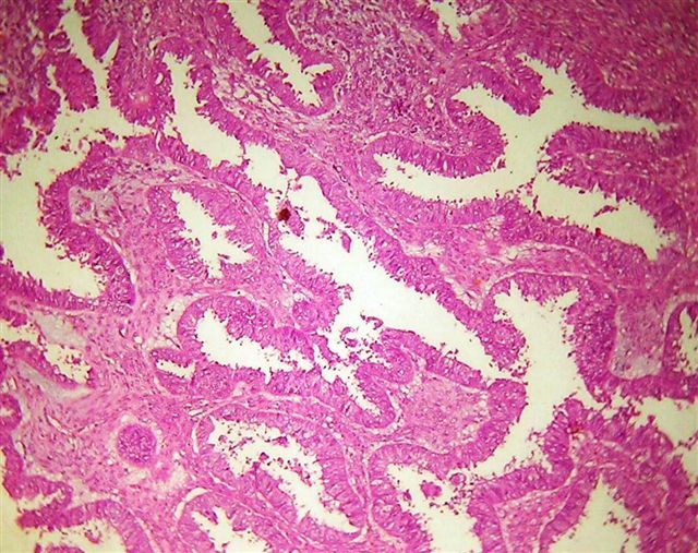
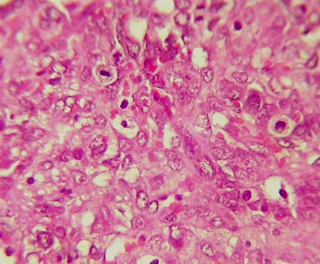
Microscopic (histologic) description
- High grade malignant carcinomatous and sarcomatous elements admixed
- Carcinomatous element
- Most common carcinomatous element is high serous adenocarcinoma; the second most common is endometrioid (Am J Surg Pathol 2012;36:831)
- Serous > endometrioid > clear cell > squamous > mixed > undifferentiated carcinoma
- Can also be mucinous, mixed high grade serous and endometrioid carcinoma, mixed high grade serous and clear cell carcinoma, or undifferentiated adenocarcinoma (Gynecol Oncol 2016;142:248)
- Most common carcinomatous element is high serous adenocarcinoma; the second most common is endometrioid (Am J Surg Pathol 2012;36:831)
- Sarcomatous element: can be homologous (50%) or heterologous (50%)
- Homologous
- Typically nonspecific malignant mesenchymal cells but may resemble fibrosarcoma, leiomyosarcoma or other sarcomas native to Müllerian organs (Clin Case Rep 2021;9:e05160)
- Heterologous
- Specific malignant elements of a non-Müllerian tissue including cartilaginous, osteosarcomatous or rhabdomyoblastic elements (J Ovarian Res 2020;13:129)
- Incidence of sarcomatous types: chondrosarcoma > rhabdomyosarcoma > osteosarcoma and liposarcoma > neuroectodermal elements
- Most often only 1 type of heterologous sarcoma
- Homologous
- Lymphovascular invasion common
- High grade cytologic atypia and brisk mitotic activity
- Intracytoplasmic hyaline globules in sarcomatous component are frequently seen
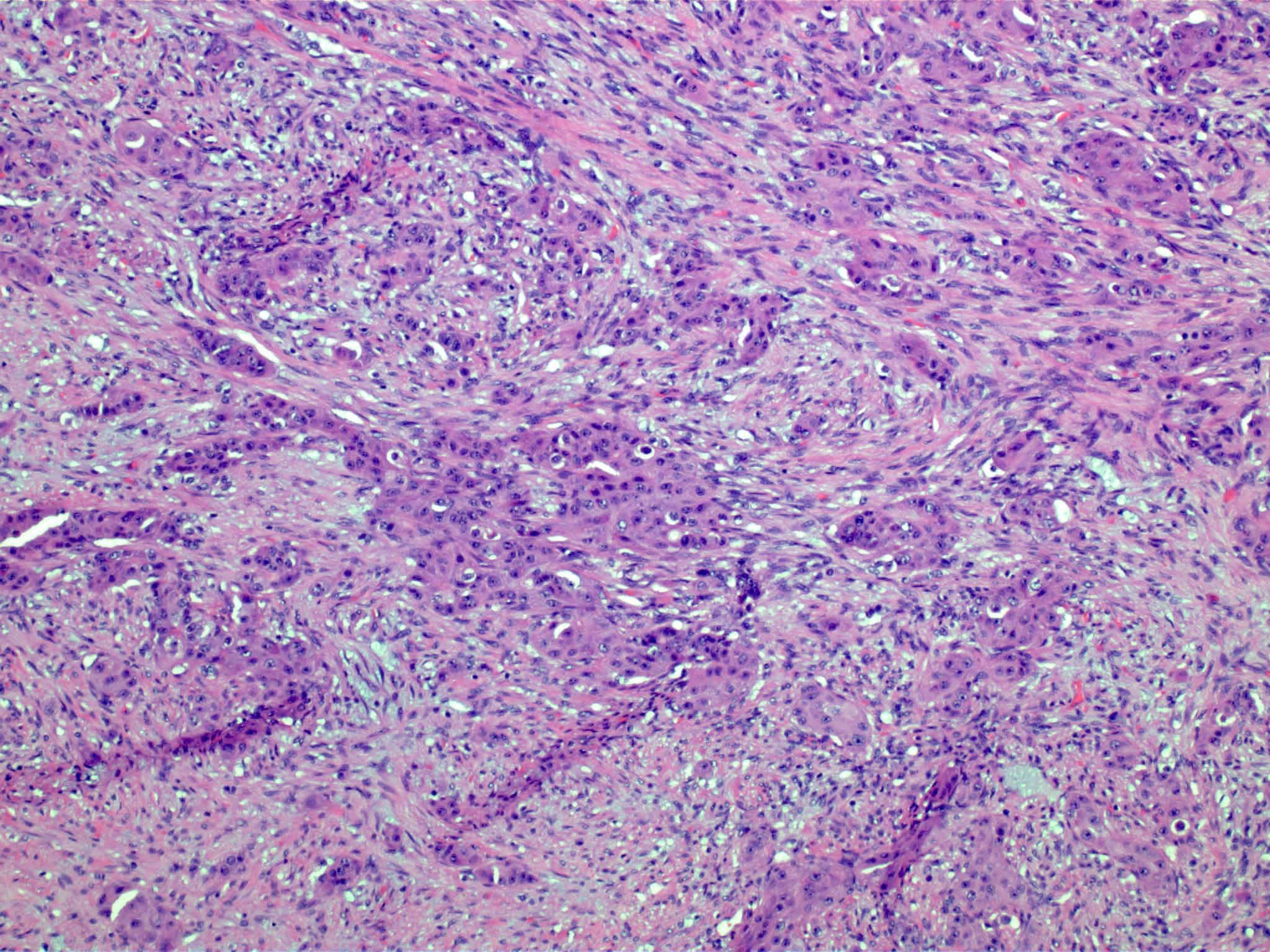
Microscopic (histologic) images
Contributed by Hatem Kaseb, M.D., Ph.D., M.P.H. and Mona Kandil, M.D., Ph.D.


Carcinomatous component
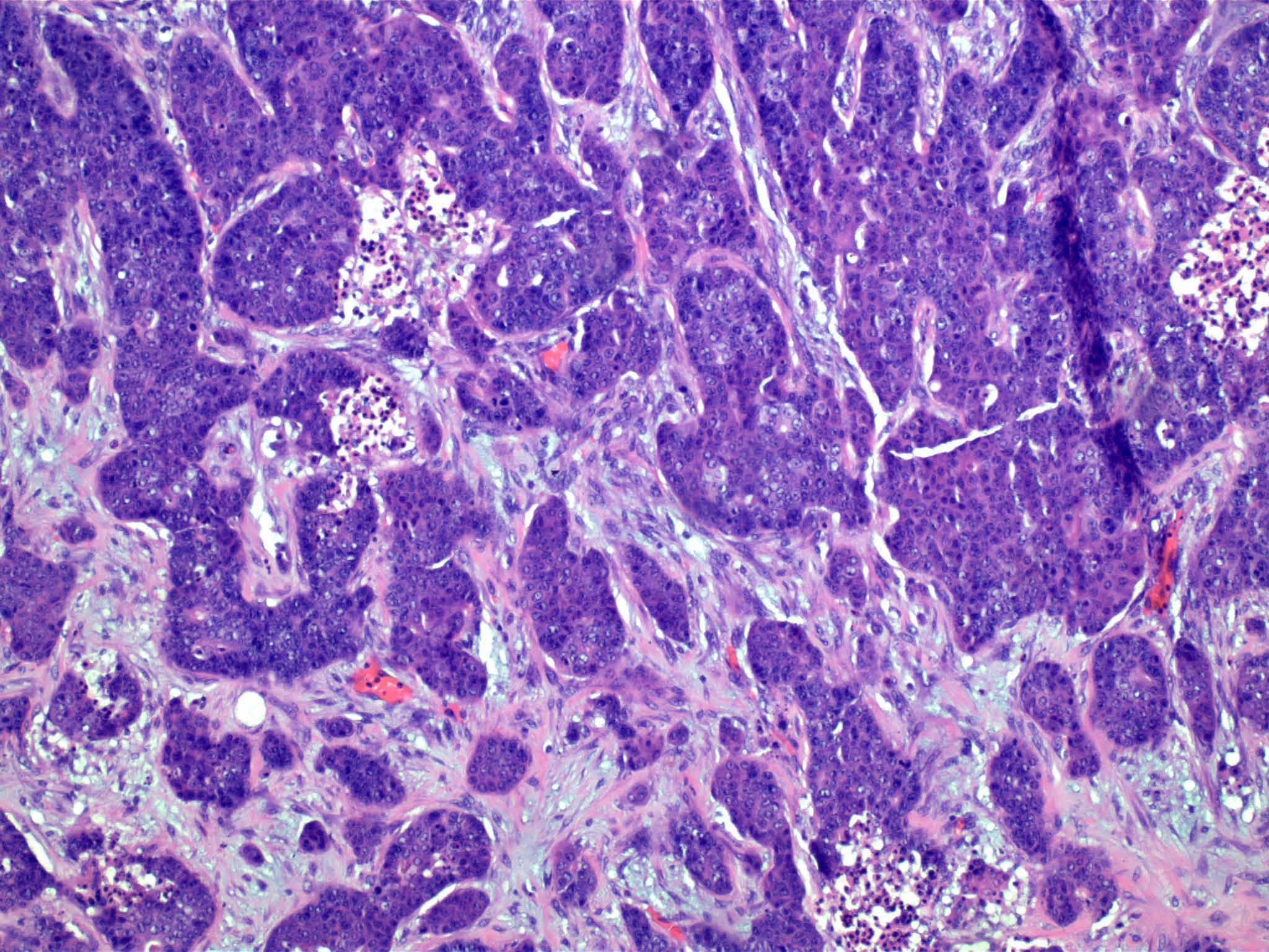


Carcinomatous component
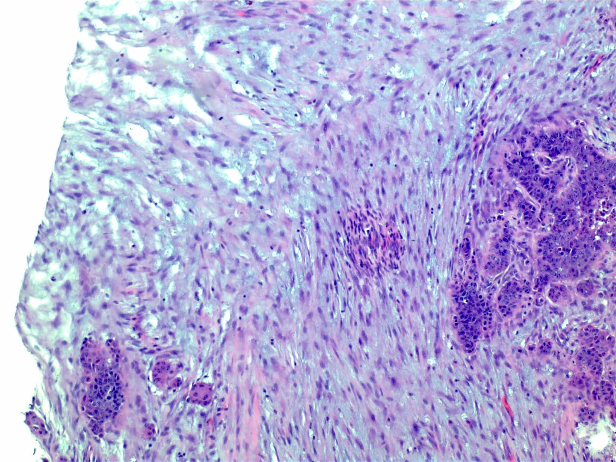
Carcinomatous and sarcomatous components

Sarcomatous component
Malignant stroma
High grade malignant glands
High grade tumor cells with mitotic figures
Positive stains
- Both carcinomatous and sarcomatous elements are usually positive for p53 abnormal expression and p16 (Histopathology 2000;37:427)
- Both carcinomatous and sarcomatous elements may be positive for
- Tumor cell droplets: PAS+ diastase resistant
- Sarcomatous component
- Variable: CEA, EMA, keratins (CAM 5.2 and AE1 / AE3)
- Vimentin (J Cancer Res Ther 2015;11:1022)
- SALL4, glypican 3 (Patholog Res Int 2012;2012:569609)
- Myogenin, myoD1, muscle specific actin (HHF35) and desmin if rhabdomyoblastic elements (Histopathology 2000;37:427)
- Chondrosarcoma / osteosarcoma areas: S100 protein
- CD99, FLI1 and GFAP may be positive in neuroectodermal component
- SATB2 positive in osteosarcoma and sarcoma, NOS
- Carcinomatous component
- Chromogranin, NSE, CD56 and synaptophysin may be positive
- May show positivity for ER / PR (J Cancer Res Ther 2015;11:1022)
- HER2 / neu
- Variable positivity in OCS (Am J Clin Oncol 2023;46:572)
- Typically expressed in epithelial component (Cancer Sci 2003;94:986)
- Ki67
- Higher expression of Ki67 in carcinomatous element, indicating more aggressive portion of the tumor (Virchows Arch 2008;452:259)
- Immunohistochemical loss of mismatch repair (MMR) proteins may occur (Nat Commun 2014;5:5006)
Negative stains
- AFP, CDX2, desmin, CD34 (epithelioid sarcoma) (Appl Immunohistochem Mol Morphol 2000;8:293)
Molecular / cytogenetics description
- Shown to have mutations in genes commonly associated with uterine and ovarian carcinomas, such as TP53, KRAS and PIK3CA (most commonly observed mutations); others reported include CDKN1B, CTNNB1, FBXW7, PPP2R1A, CDH4, PTEN, PIK3CA, CCNE1, TERT, ARID1A, ARID1B, SPOP, BAZ1A, MLL3 and BCOR (Proc Natl Acad Sci U S A 2016;113:12238, Nat Commun 2014;5:5006)
- TP53 (Am J Clin Oncol 2023;46:572, Histopathology 2000;37:427, Proc Natl Acad Sci U S A 2016;113:12238, Obstet Gynecol 1994;83:118)
- Mutations in TP53 are the most common in OCS, found in > 50% of cases
- Often associated with tumorigenesis, advanced stage and worse survival, though not necessarily prognostic
- Commonly deleted and frequently present in the serous epithelial component (> 50%)
- PIK3CA
- ~40% of OCS exhibit PIK3CA missense mutations; these mutations show no prognostic significance (Nat Commun 2014;5:5006, Histopathology 2000;37:427, J Cancer Res Ther 2015;11:1022, Front Oncol 2023;13:1278300)
- EGFR or KIT
- Overexpression of EGFR or KIT proteins may occur in OCS (Cancer Sci 2003;94:986)
- Mismatch repair (MMR)
- Alterations or losses in MMR proteins are observed in some OCS, with defects more common in uterine than ovarian carcinosarcomas (Nat Commun 2014;5:5006, Am J Clin Oncol 2023;46:572)
- HER2 / Neu, MYC and VEGFA
- Amplification of HER2, MYC and VEGFA genes may be present, potentially impacting tumor growth and progression (Am J Clin Oncol 2023;46:572)
- BRCA1 / BRCA2
- BRCA1 / BRCA2 mutations and associated deficiencies in DNA double strand break repair have been reported, especially in cases where the carcinomatous component is serous carcinoma (Am J Clin Oncol 2023;46:572)
- Tumor mutational burden (TMB)
- TMB in OCS and ovarian tumors is typically low (Am J Clin Oncol 2023;46:572)
Sample pathology report
- Uterus, total hysterectomy and bilateral salpingo-oophorectomy:
- Ovarian carcinosarcoma with a homologous sarcomatous component (see comment)
- Comment: There is a malignant biphasic cell proliferation composed of carcinomatous elements (serous carcinoma) intimately admixed with a sarcomatous element. The constellation of morphological features supports the diagnosis of carcinosarcoma (malignant mixed Müllerian tumor). These are considered malignant epithelial tumors, which behave like a high grade carcinoma.
Differential diagnosis
- Immature teratoma (children):
- Presence of young, immature neuroectodermal elements
- Usually women < 20 years old
- Endometrioid carcinoma with spindle cells:
- Ovaries must be uninvolved or clear metastasis
- Tubal histologic pattern
- Pure carcinomas with desmoplastic stroma:
- Resembles sarcomatous elements
- Sarcomatous components exhibit high grade nuclear atypia and frequent mitotic figures
- Pure carcinoma with desmoplastic stroma is an epithelial tumor with a fibrotic stromal reaction (versus biphasic in OCS)
- Sarcomatoid carcinoma:
- Combination of glandular and spindle components
- Widespread expression of keratin markers
- Dedifferentiated carcinoma:
- Low grade endometrioid carcinoma adjacent to noncohesive poorly differentiated tumor cells
- Occasional rhabdoid morphology
- Mesodermal adenosarcoma with or without sarcomatous overgrowth or mesodermal adenosarcoma with endometrioid carcinoma:
- Benign glandular elements mixed with a sarcomatous stroma
- Presence of periglandular cuffs of cellular stroma
- Lacks high grade malignant epithelial features
- Metastatic tumor from nongynecologic site (e.g., gastric adenocarcinoma [Krukenberg tumor]):
- Highly cellular fibromatous stroma in Krukenberg tumors
- Signet ring cell morphology is common in this disorder but rare in gynecologic primaries (OCS)
- Other tumors showing multilineage differentiation:
- Examples include poorly differentiated Sertoli-Leydig cell tumor with heterologous elements, Müllerian adenosarcoma and immature teratoma
- Do not typically show high grade carcinomatous components (may have benign or low grade neoplastic epithelial elements)
- Relevant immunohistochemical stains for sex cord and germ cell tumors aid in diagnosis
- Ovarian metastasis from a uterine primary:
- Presence of a large tumor in the uterine cavity in patients with both endometrial and ovarian tumors suggests an endometrial primary
Additional references
Practice question #1
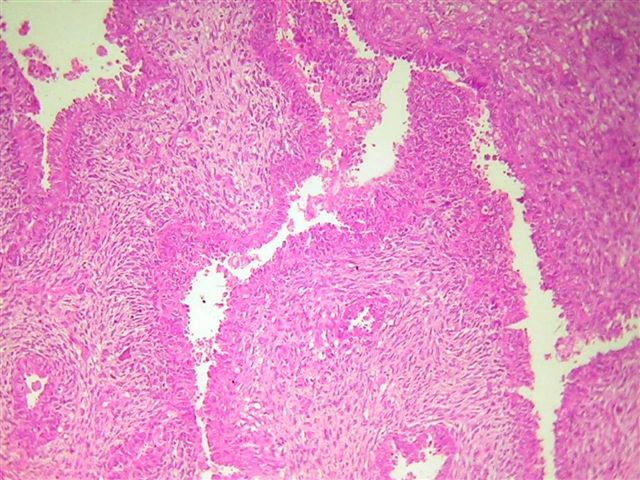
A 55 year old woman presents with an abdominal mass, which is represented in the microscopic image above. She denies any accompanying symptoms. Which of the following immunohistochemical staining results will fit this patient profile?
- CK7+, vimentin-
- CK7-, vimentin-
- CK7+, vimentin+
- CK7-, vimentin+
Practice answer #1
C. CK7+, vimentin+. In ovarian carcinosarcoma, the carcinomatous component (demonstrated here on the right) will be positive for CK7, while the sarcomatous component (on the left) will be positive for vimentin. Answer B is incorrect because at least one component should be positive for CK7 and the other for vimentin. Answers A and D are incorrect because as mentioned above, the carcinomatous component is positive for CK7 and the sarcomatous component is typically positive for vimentin, therefore, the tumor overall should show positivity for CK7 and vimentin in at least one component.
Comment Here
Reference: Carcinosarcoma
Comment Here
Reference: Carcinosarcoma
Practice question #2
A 65 year old woman presents with a 15 cm solid cystic mass of the ovary. An excisional biopsy is performed. Microscopic images of 2 distinct areas are shown above. Which of the following genes is the most likely to be mutated?
- EGFR
- HER2
- KRAS
- TP53
Practice answer #2
D. TP53 is the most common mutation (> 50%) in carcinosarcomas of the ovary.
Answer B is incorrect because HER2 is amplified in only a small fraction of ovarian carcinosarcoma cases. Answer C is incorrect because KRAS is commonly mutated in ovarian carcinosarcoma cases; however, TP53 is the most commonly reported mutation in ovarian carcinosarcoma. Answer A is incorrect because EGFR is rarely mutated / overexpressed in ovarian carcinosarcoma cases.
Comment Here
Reference: Carcinosarcoma
Comment Here
Reference: Carcinosarcoma
