
Pancreas
Other tumors
Sclerosing epithelioid mesenchymal neoplasm
Authors: Aslihan Yavas, M.D., Olca Basturk, M.D.
Deputy Editor-in-Chief: Raul S. Gonzalez, M.D.


Last author update: 15 November 2022
Last staff update: 15 November 2022
Copyright: 2019-2024, PathologyOutlines.com, Inc.
PubMed Search: Sclerosing epithelioid mesenchymal neoplasm

Table of Contents
Definition / general | Essential features | Epidemiology | Sites | Clinical features | Laboratory | Prognostic factors | Case reports | Gross description | Gross images | Microscopic (histologic) description | Microscopic (histologic) images | Positive stains | Negative stains | Electron microscopy description | Molecular / cytogenetics description | Molecular / cytogenetics images | Sample pathology report | Differential diagnosis | Board review style question #1 | Board review style answer #1 | Board review style question #2 | Board review style answer #2Cite this page: Yavas A, Basturk O. Sclerosing epithelioid mesenchymal neoplasm. PathologyOutlines.com website. https://www.pathologyoutlines.com/topic/pancreassclerosingepit.html. Accessed May 13th, 2024.
Definition / general
- A novel indolent pancreatic neoplasm with unique morphologic / immunophenotypic features and a distinct methylation pattern, different from both epithelial and mesenchymal neoplasms described in other anatomical sites (Mod Pathol 2020;33:456)
- First described in 2020 (Mod Pathol 2020;33:456)
Essential features
- Indolent
- Well circumscribed tumor with a geographic pattern of epithelioid cell nests alternating with spindle cell fascicles
- Vimentin (diffuse), CD99 (diffuse) and cytokeratin (AE1 / AE3 and CK18, dot-like) expression
- Lack of abnormalities in any of key genetic drivers
- Mesenchymal transcriptomic features and distinct methylation signature
Epidemiology
- Mean age: 43 years (Mod Pathol 2020;33:456)
- F:M = 3:1 (Mod Pathol 2020;33:456)
Sites
- Mostly occurs in head and neck of the pancreas (Mod Pathol 2020;33:456)
Clinical features
- Nonspecific symptoms
Laboratory
- Serum tumor markers (CA19.9 and CEA) within normal limits
Prognostic factors
- None known; exhibits indolent behavior (Mod Pathol 2020;33:456)
Case reports
- 31 year old woman with suspected solid pseudopapillary neoplasm of the pancreatic tail (Pancreas 2021;50:e47)
Gross description
- Well circumscribed and solid, usually confined to the pancreatic parenchyma
- White-tan and firm cut surface
- Median tumor size: 1.8 cm (Mod Pathol 2020;33:456)
Gross images
Images hosted on other servers:

Well circumscribed solid lesion
Microscopic (histologic) description
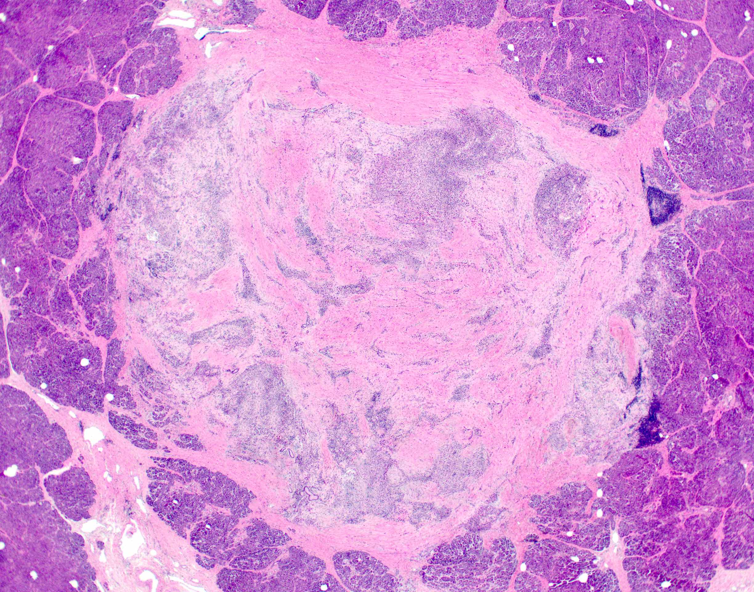
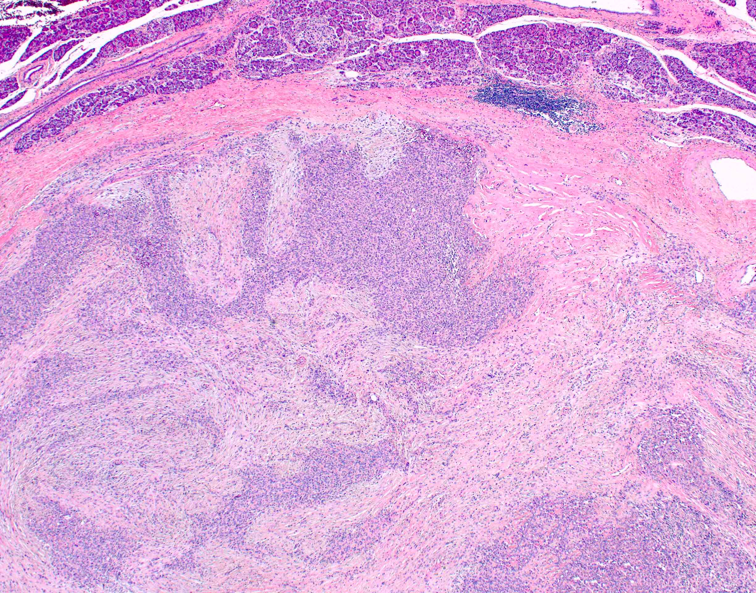
- Solid neoplasms associated with extensive fibrosis and dense lymphoid aggregates at the periphery (Mod Pathol 2020;33:456)
- Entrapped pancreatic parenchyma may be seen at the periphery or within the tumor
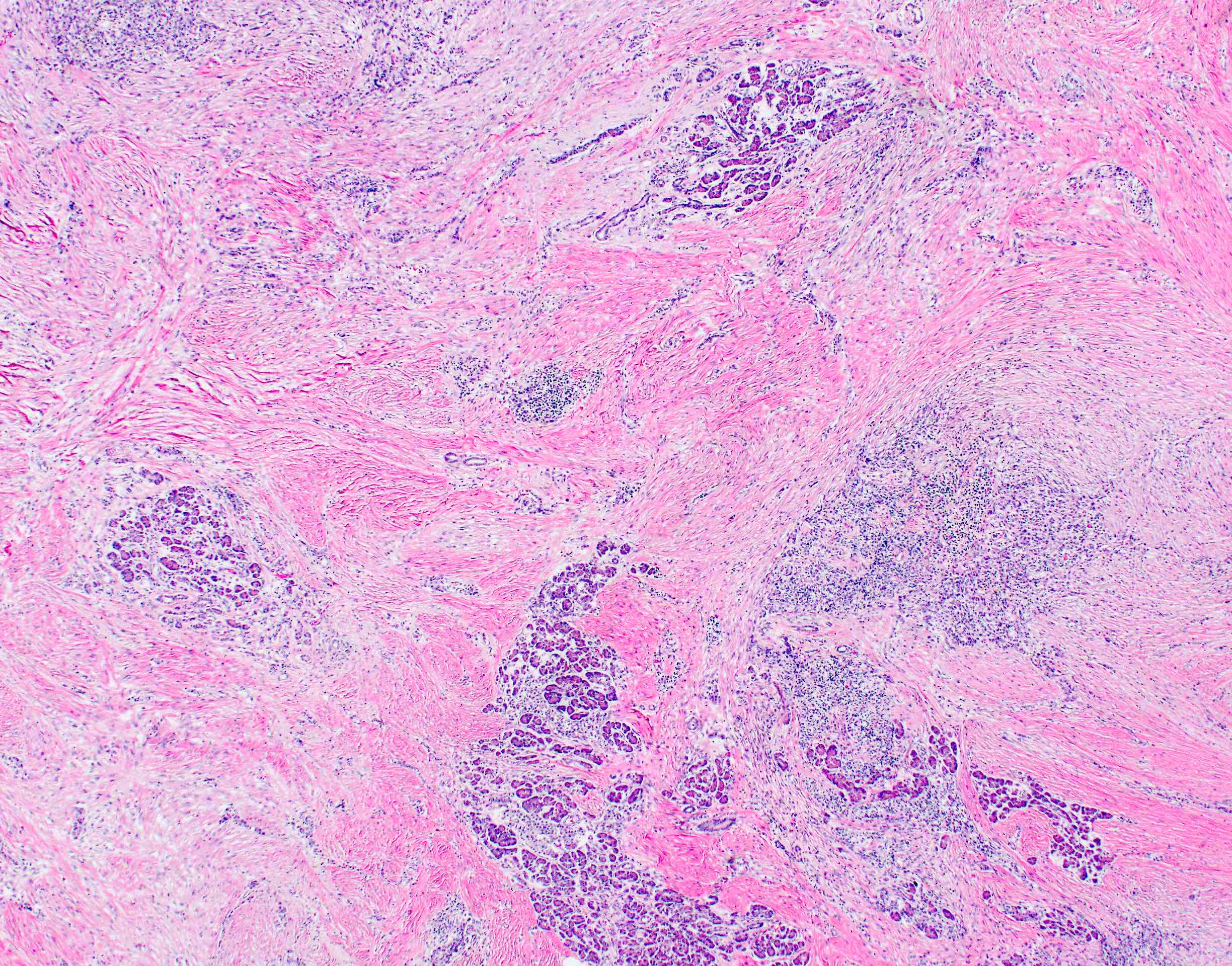
- Density of tumor cells varies significantly, not only among tumors but also throughout each tumor, creating a geographic appearance of hypercellular and hypocellular areas with collagenous stroma
- No true epithelial structures such as glands, papillae or pseudopapillae (Mod Pathol 2020;33:456)
- Most cells are epithelioid to spindled with scant cytoplasm and round to oval nuclei with open chromatin
- Others display more irregular, hyperchromatic nuclei
- Occasional mitotic figures may be seen
Microscopic (histologic) images
Contributed by Olca Basturk, M.D.
Overall appearance
Well circumscribed lesion
Peripheral dense lymphoid aggregates
Entrapped pancreatic parenchyma
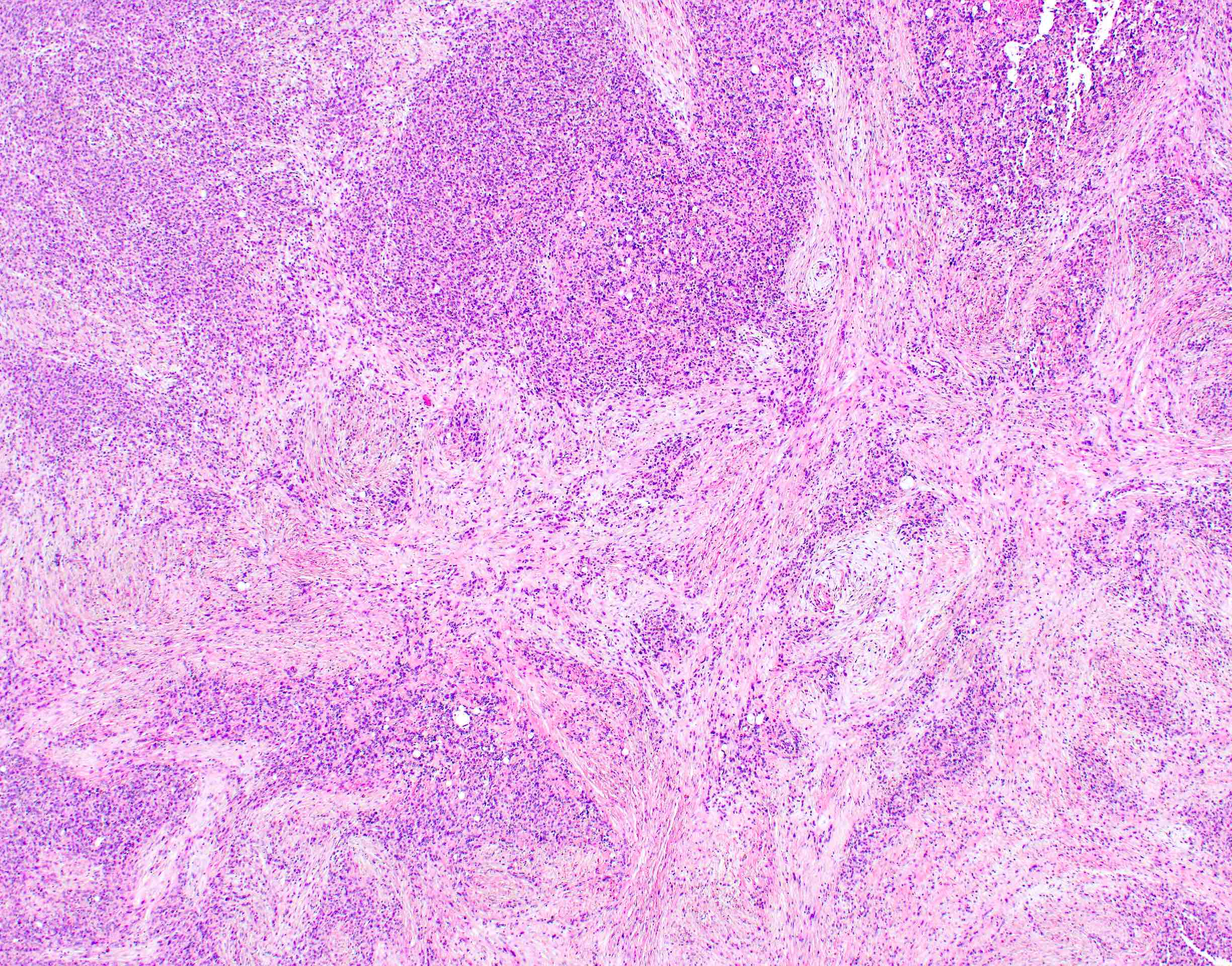
Hypercellular and hypocellular areas
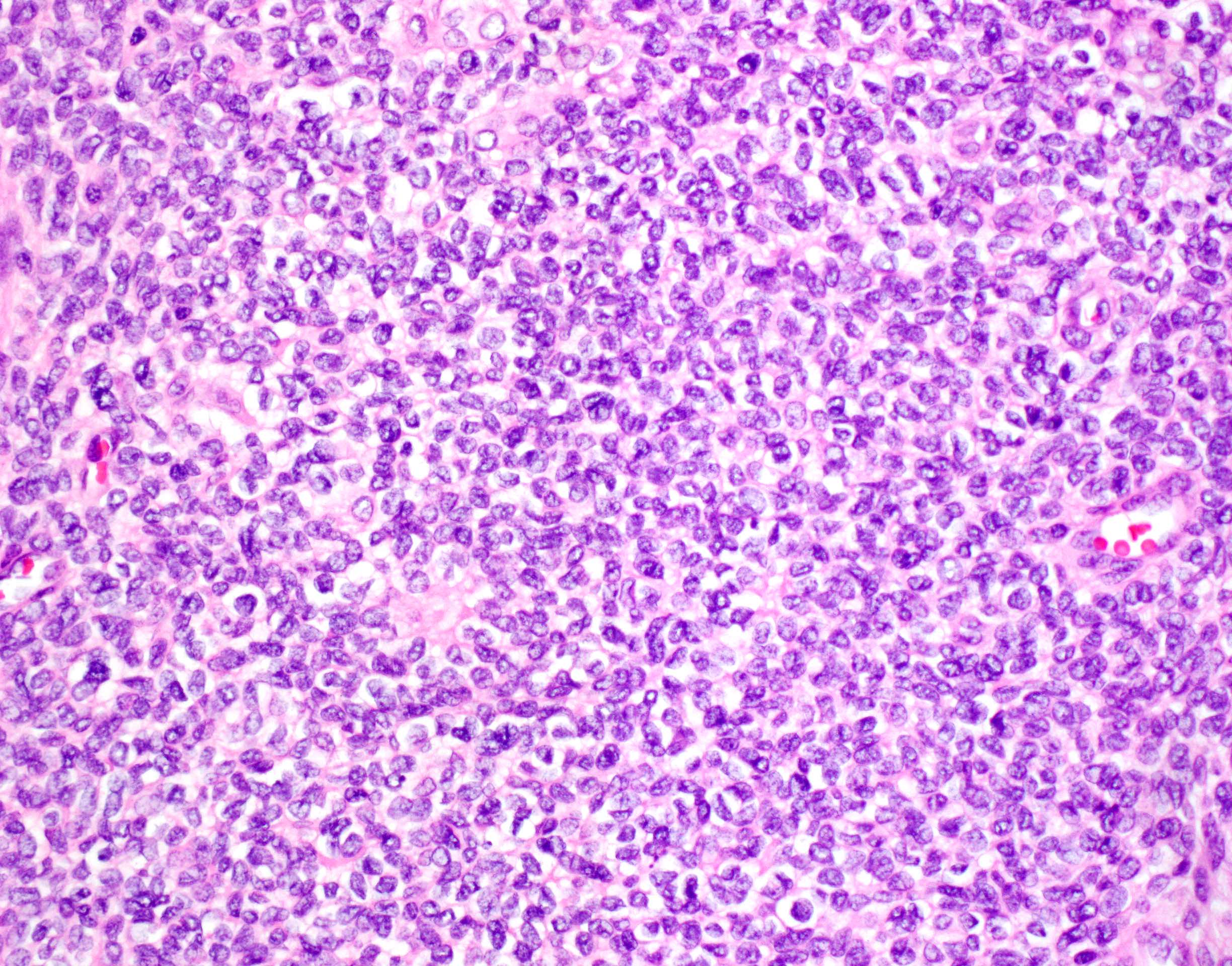
Sheets of epithelioid cells
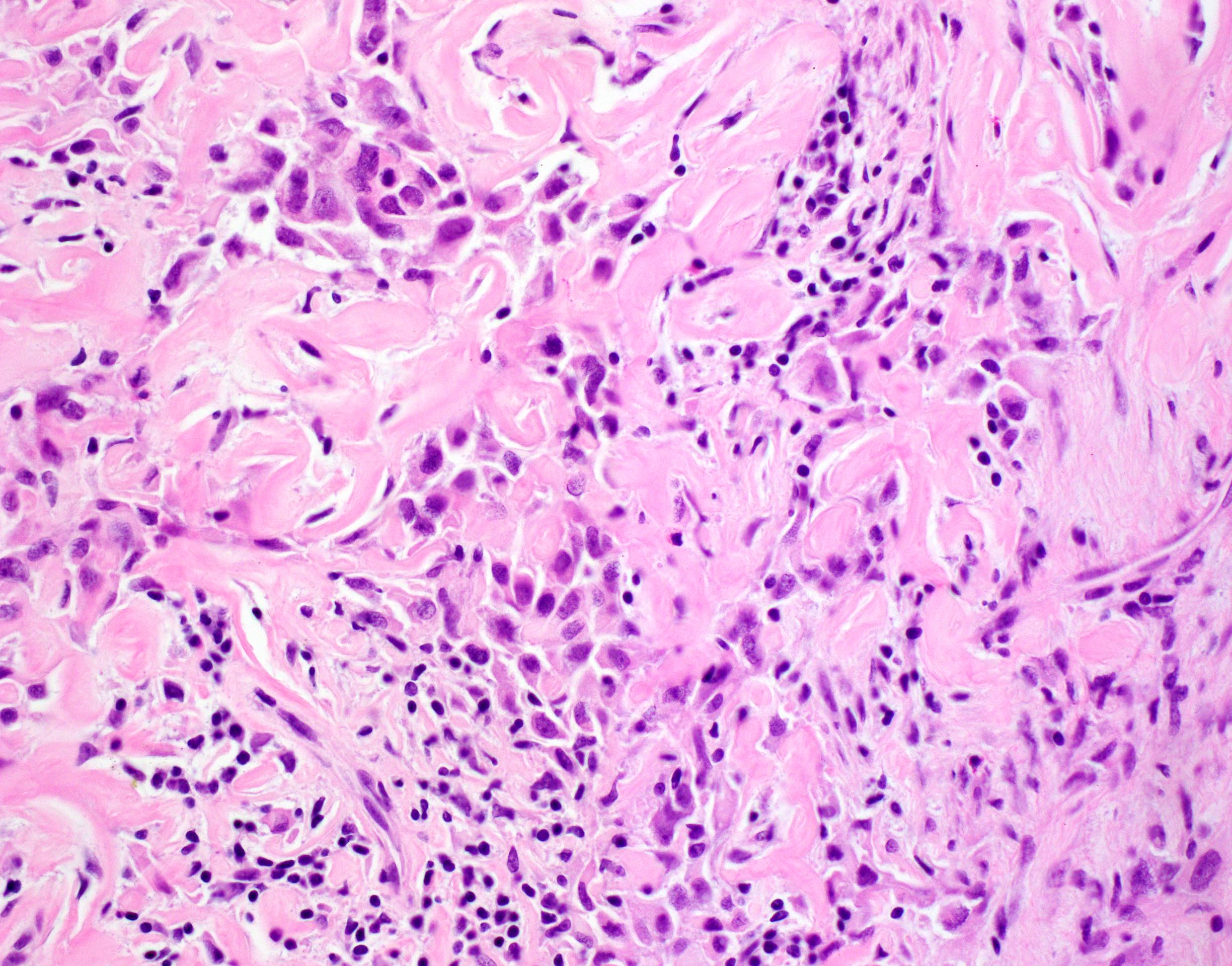
Atypical spindle cells
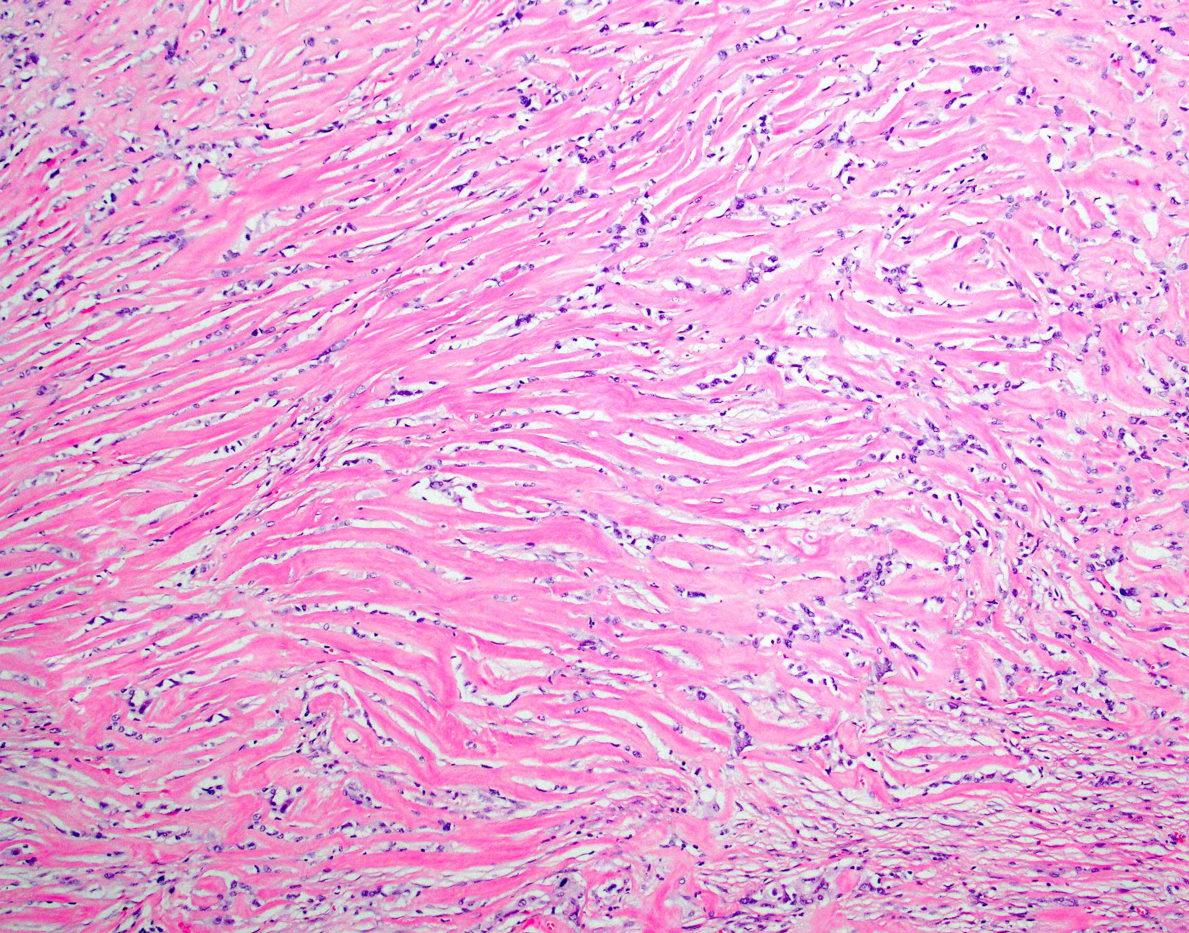
Dense hyaline fibrosis
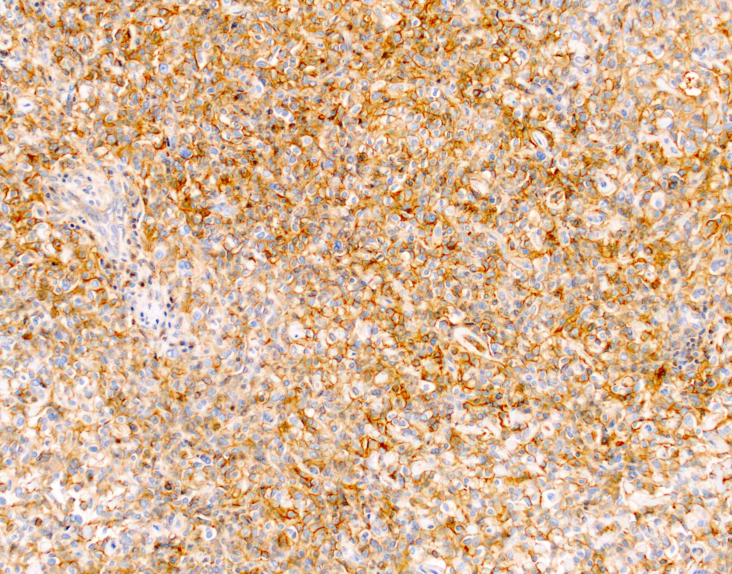
Membranous CD99 expression
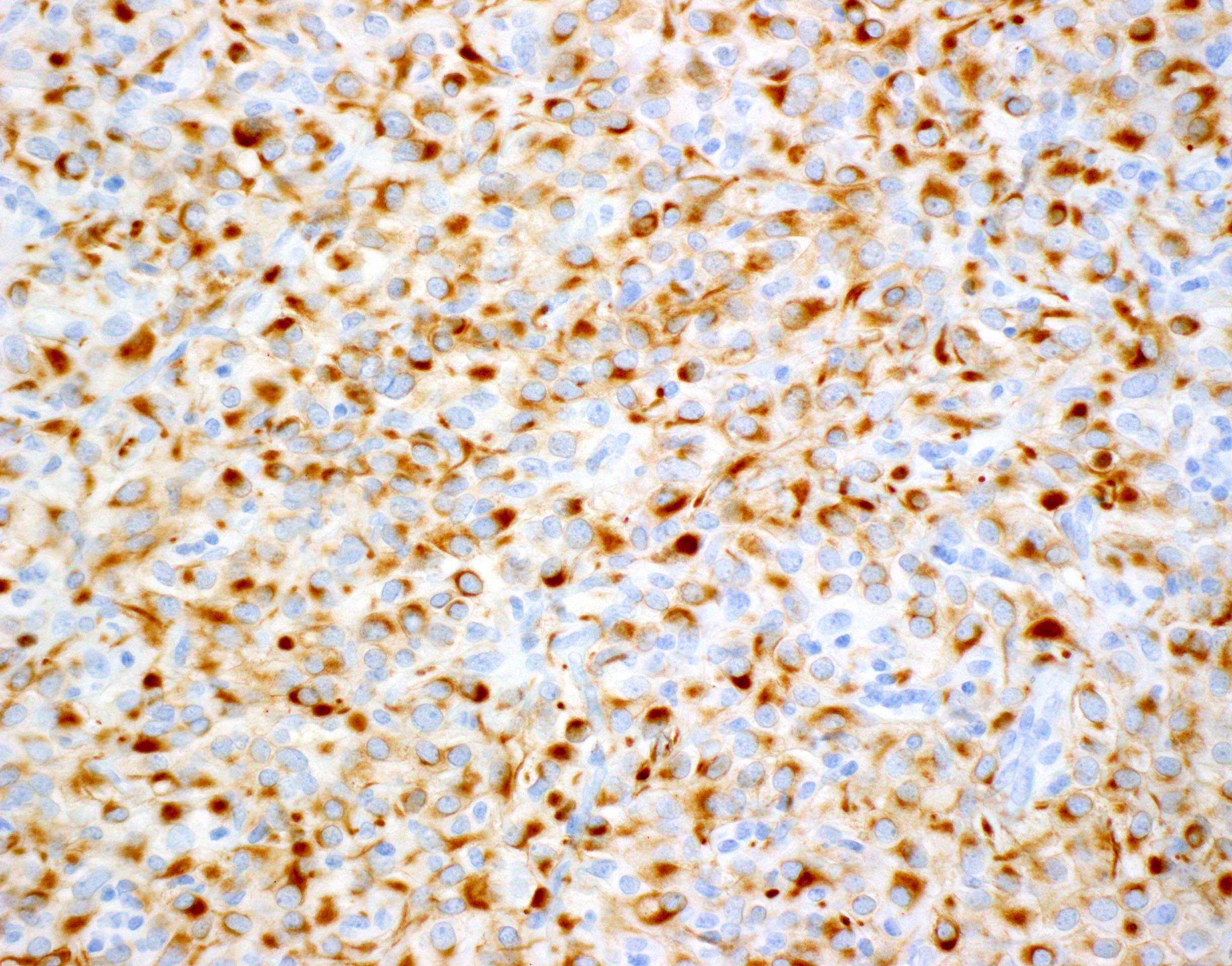
Dot-like CK18 expression
Positive stains
- Vimentin, CD99, AE1 / AE3, CK18 (dot-like) (Mod Pathol 2020;33:456)
Negative stains
- Synaptophysin, chromogranin, abnormal beta catenin, progesterone receptor, CD10, trypsin, chymotrypsin, MUC4, desmin, myogenin, INI1 (retained), ERG, CD31, CD34, S100, HMB45, MelanA, CD117, DOG1, TTF1, HepPar1, BCL2, ALK, STAT6, CD21, CD35, CD45 (Mod Pathol 2020;33:456)
Electron microscopy description
- Nonspecific findings; occasional desmosomes, perinuclear tonofilaments and abundant rough endoplasmic reticulum (Mod Pathol 2020;33:456)
Molecular / cytogenetics description
- No recurrent somatic mutations, amplifications or deletions in any known oncogenes or suppressor genes (Mod Pathol 2020;33:456)
- Activation or enrichment of pathways related to the extracellular matrix, tight junctions, adherens junctions and TGFβ signaling by Gene Set Enrichment Analysis (Mod Pathol 2020;33:456)
- A unique cluster by unsupervised analysis of the methylation profiles (Mod Pathol 2020;33:456)
Molecular / cytogenetics images
Images hosted on other servers:

Genomic and transcriptomic characterization
Sample pathology report
- Pancreas and duodenum, pancreaticoduodenectomy:
- Sclerosing epithelioid mesenchymal neoplasm (see comment)
- Comment: The tumor is confined to the pancreatic parenchyma and measures 2.2 cm in greatest dimension. Adjacent pancreas reveals atrophy. Surgical margins are free of tumor. 16 benign lymph nodes are identified (0/16).
Differential diagnosis
- Solid pseudopapillary neoplasm:
- Pseudopapillae, areas of macrophages, hyaline globules and nuclear grooves
- Abnormal beta catenin and nuclear progesterone receptor expression
- CTNNB1 mutations
- Sarcomatoid carcinoma:
- Larger infiltrative tumors with considerable pleomorphism, necrosis and proliferative activity
- Glandular component may be present
- KRAS, TP53, SMAD4, CDKN2A mutations
- Sclerosing epithelioid fibrosarcoma:
- MUC4 expression
- EWSR1-CREB3L1, FUS-CREB3L21 or FUS-CREB3L2 fusions
- Solitary fibrous tumor:
- Inflammatory myofibroblastic tumor:
- Perivascular epithelioid cell neoplasm (PEComa):
- Gastrointestinal stromal tumor:
Board review style question #1
Which one of the following stains is positive in sclerosing epithelioid mesenchymal neoplasm of the pancreas in the figure shown above?
- CD34
- CD99
- DOG1
- MUC4
- S100
Board review style answer #1
Board review style question #2
Which one of the following microscopic features is seen in sclerosing epithelioid mesenchymal neoplasms of the pancreas?
- No entrapped pancreatic parenchyma is seen within the tumors.
- They are composed of epithelioid cells only.
- They are predominantly hypocellular tumors.
- They have well circumscribed borders.
- True epithelial structures such as glands, papillae and pseudopapillae are common.
Board review style answer #2
D. They have well circumscribed borders.
Comment Here
Reference: Sclerosing epithelioid mesenchymal neoplasm
Comment Here
Reference: Sclerosing epithelioid mesenchymal neoplasm
